Introduction
Cognitive functions including visuospatial ability, executive function, comprehension, and semantic memory are necessary to complete the clock drawing test (CDT; Rouleau et al., Reference Rouleau, Salmon, Butters, Kennedy and Mcguire1992; Royall et al., Reference Royall, Cordes and Polk1998; Shulman, Reference Shulman2000; Leyhe et al., Reference Leyhe2009). Previously, we investigated the neural correlates of CDT performance using the free-drawn method and the pre-drawn method, with measurements using magnetic resonance imaging (MRI) voxel-based morphometry (Matsuoka et al., Reference Matsuoka2011). Brain damage in the bilateral posterior temporal lobes was shown to affect performance on the pre-drawn method, whereas brain damage in the right posterior temporal lobe affected the free-drawn method. Thus, the study suggested that the different clock drawing methods evaluated different cognitive functions (Matsuoka et al., Reference Matsuoka2011).
In addition to the use of different methods, the CDT can be evaluated quantitatively or qualitatively. More than a dozen different quantitative scoring systems for the CDT have been published (Shulman, Reference Shulman2000). Quantitative evaluation is easy to score; is relatively independent of culture, language, and education; and has achieved widespread clinical use for cognitive screening. We demonstrated that distinct brain regions might be associated with CDT performance using different quantitative scoring systems (Matsuoka et al., Reference Matsuoka2011). These results suggested that the different quantitative scoring systems required different cognitive functions.
On the other hand, qualitative evaluation is useful for screening for dementia and in differentiating between dementia subtypes (Kitabayashi et al., Reference Kitabayashi, Ueda, Narumoto, Nakamura, Kita and Fukui2001; Blair et al., Reference Blair, Kertesz, Mcmonagle, Davidson and Bodi2006; Lee et al., Reference Lee, Kim, Hong, Lee, Oh and Cheong2008b). While conceptual and spatial and/or planning deficits were frequent in patients with Alzheimer's disease (AD), graphic difficulties were frequent in patients with mild to moderate vascular dementia (Kitabayashi et al., Reference Kitabayashi, Ueda, Narumoto, Nakamura, Kita and Fukui2001). This result suggested that the neural correlates of the different qualitative errors were distinct.
Few studies have investigated the relationship between qualitative deficits on the CDT and brain regions. Suhr et al. (Reference Suhr, Grace, Allen, Nadler and Mckenna1998) showed that qualitative scoring could be used to differentiate among stroke patients with different local lesions. In the study, the location of the lesions was roughly divided based on laterality, cortical or subcortical, and anterior or posterior. Therefore, the brain regions associated with the qualitative deficits on the CDT have not been fully investigated. Qualitatively, the CDT can be separated into three components: clock face representation, layout of the numbers, and position of the hands. The Rouleau CDT is designed to assess the accuracy of each of these components (Rouleau et al., Reference Rouleau, Salmon, Butters, Kennedy and Mcguire1992). The purpose of this study was to identify neural correlates of each component of the Rouleau CDT using single photon emission computed tomography (SPECT) in patients with AD.
Methods
Participants
Ninety-five right-handed drug-naïve patients with AD (33 males, 62 females; mean age ± SD, 74.8 ± 8.2 years old) were recruited from the Center for Diagnosis of Dementia at the Kyoto Prefectural University of Medicine. All patients had been assessed comprehensively by a geriatric psychiatrist, received their information including medical and social histories, physical and neurological examinations, neuropsychological tests, laboratory data, and brain imaging. Cognitive impairments were assessed by clinical examination and neuropsychological tests, and functional status was confirmed by family members or other available caregivers. The diagnosis was made according to the National Institute of Neurological and Communicative Disease and Stroke–Alzheimer's Disease and Related Disorders Association (NINCDS-ADRDA) criteria for probable AD (McKhann et al., Reference Mckhann, Drachman, Folstein, Katzman, Price and Stadlan1984). Patients were excluded from the study if they had a significant history of psychiatric or neurological disorders (other than AD), including stroke, head injury, epilepsy, psychiatric disorders, alcohol abuse, or a serious medical condition. None of the patients had been prescribed anti-dementia or other psychoactive drugs because our center examines patients who have not been diagnosed with dementia. The ethics committee of the Kyoto Prefectural University of Medicine approved the study and informed consent was obtained from all participants.
Neuropsychological tests
All patients with AD were evaluated using the CDT and the Mini-Mental State Examination (MMSE; Folstein et al., Reference Folstein, Folstein and Mchugh1975). For the CDT, patients were presented with a blank sheet of paper and a pencil and given the following instructions: “I would like you to draw a clock, put in all the numbers, and set the hands at 20 past 8.” The free-drawn clock drawings were scored using the Rouleau CDT on a 10-point scale (Rouleau et al., Reference Rouleau, Salmon, Butters, Kennedy and Mcguire1992), with higher scores reflecting better CDT performance. This 10-point scale (R total) is designed to assess the accuracy of the clock face representation (R1, maximum 2 points), the layout of the numbers (R2, maximum 4 points), and the position of the hands (R3, maximum 4 points). Two raters (T.M. and A.O.) blinded to the clinical data independently scored all the drawings. The inter-rater reliability was examined by calculation of intraclass correlation coefficients (ICCs) based on the rating of all patients by the two raters. The inter-rater ICCs for the R total, R1, R2, and R3 scores were all >0.942. All drawings were reassessed after at least four weeks by one rater (T.M.) and the intra-rater ICCs for the four scores were all >0.955.
Image acquisition
All patients with AD underwent brain perfusion SPECT within one month after they underwent MMSE and CDT. SPECT was performed by intravenous injection of 185 MBq of N-isopropyl-p-[123I]iodoamphetamine (I-123-IMP) (Nihon Mediphysics, Hyogo, Japan) in participants seated at rest with their eyes open. SPECT imaging commenced 22 minutes after the injection and continued for 16 minutes. A triple-head gamma camera (PRISM IRIX, Picker International, Cleveland, OH, USA) equipped with low-energy, high-resolution, parallel collimators was used. Projection data from each camera were obtained in a 128 × 128 format for 40 steps of 120° at 8 seconds per step (voxel size: 2 × 2 × 2 mm).
Image analysis was performed using Statistical Parametric Mapping (SPM) 8 (Wellcome Department of Cognitive Neurology, University College, London) in Matlab 7.5 (Mathworks Inv., Sherborn, MA, USA). The acquired SPECT images were converted from DICOM to Analyze format using MRIcro (http://www.mricro.com) and transferred to SPM8. After confirmation of no significant artifacts using MRI or computed tomography (CT), all SPECT images were anatomically normalized using the I-123-IMP template (Fujifilm RI Pharma, Tokyo, Japan) matched to the Montreal Neurological Institute (MNI) template. The normalized images were smoothed using a 12-mm full-width half-maximum isotropic Gaussian kernel. To examine regional differences, the images were scaled to a mean global cerebral blood flow of 50 mL/100 g/min.
Statistical analyses
A multiple regression analysis was performed to detect a relationship between each score (R total, R1, R2, and R3) and regional cerebral blood flow (rCBF) in the patients with AD. Age, gender, and education were included as covariates. The X, Y, and Z coordinates provided by SPM were used to approximate the MNI brain space. Statistical thresholds were set to a family-wise error (FWE)-corrected p value of 0.05 at the voxel level in all analyses.
Results
Characteristics of patients
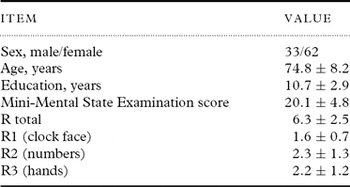
Table 1 shows the characteristics of the patients with AD. Although the MMSE score ranged from 5 to 28, the mean MMSE score was 20.1 ± 4.8, and the cognitive dysfunction in most of the patients with AD ranged from mild to moderate. The R1 score (maximum 2 points) was relatively high, whereas R2 (maximum 4 points) and R3 (maximum 4 points) were relatively low.
Clinical characteristics of patients (n = 95) with Alzheimer's disease

Except for sex, data are shown as the mean ± standard deviation.
Multiple regression analysis
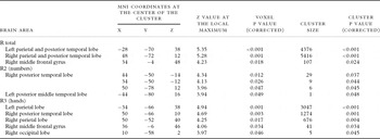
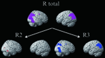
The multiple regression analysis showed a positive correlation between R total and rCBF in the bilateral parietal and posterior temporal lobes and the right middle frontal gyrus (Table 2). R1 was not significantly positively correlated with rCBF, R2 was significantly positively correlated with rCBF in the right posterior temporal lobe and the left posterior middle temporal lobe, and R3 was significantly positively correlated with rCBF in the bilateral parietal lobes, the right posterior temporal lobe, the right middle frontal gyrus, and the right occipital lobe (Figure 1 and Table 2).
Results of the multiple regression analysis

Regions in which rCBF showed a positive correlation with each CDT score (purple, R total; red, R2; blue, R3) for statistical thresholds set to an FWE-corrected p value of 0.05 at the voxel level.

Discussion
In this study, we found that performances on two different components of the CDT, numbers and hands, were associated with rCBF in various brain regions, including the parietal, temporal, and frontal lobes. These brain regions showed both overlap and differences between the CDT components. The results support that the analysis of the components of the CDT is useful for the assessment of localization of brain damage and differential diagnosis.
Regarding the performance on the drawing of the clock hands, we identified a correlation with the right middle frontal gyrus, in addition to those with the bilateral parietal lobes and right posterior temporal lobe, which have been repeatedly reported in previous studies (Lee et al., Reference Lee2008a; Matsuoka et al., Reference Matsuoka2011). Ino et al. (Reference Ino, Asada, Ito, Kimura and Fukuyama2003) showed that drawing the hands of a clock corresponding to the time presented acoustically was related to brain areas including the bilateral posterior parietal lobes and bilateral dorsal premotor areas. Seidl et al. (Reference Seidl2012) showed that performance on the Shulman CDT, using the pre-drawing method and assessing only the numbers and hands, was associated with impairment in the bilateral hippocampus and right globus pallidus. The dorsolateral prefrontal circuit including the dorsolateral prefrontal and globus pallidus plays a role in the organization of information to facilitate a response (Bonelli and Cummings, Reference Bonelli and Cummings2007. In addition, a spatial working memory task activates a dorsal pathway distributed between the parieto-occipital and dorsal prefrontal cortex, predominantly in the right hemisphere (Ventre-Dominey et al., Reference Ventre-Dominey2005). Other studies have suggested involvement of the fronto-parietal network in visuomotor control (Wise et al., Reference Wise, Boussaoud, Johnson and Caminiti1997) and attention control (Neufang et al., Reference Neufang2011). Therefore, the present results suggest that the performance on the drawing of the clock hands in the CDT requires executive function, spatial working memory, and visuomotor and attention control, which are supported by the fronto-parietal network. Moreover, the present results show that the right posterior temporal lobe, involved in visual knowledge (Owen et al., Reference Owen, Milner, Petrides and Evans1996; Kellenbach et al., Reference Kellenbach, Brett and Patterson2001) and spatial attention (Chen et al., Reference Chen, Marshall, Weidner and Fink2009), also plays an important role for the drawing of the clock hands.
The performance on the drawing of the clock numbers was mainly affected by dysfunction of the right posterior temporal lobe. In previous lesion studies, impaired spatial organization, including impaired number placement and/or omission of numbers, was much more frequent in participants with right hemisphere lesions (Suhr et al., Reference Suhr, Grace, Allen, Nadler and Mckenna1998; Tranel et al., Reference Tranel, Rudrauf, Vianna and Damasio2008). Moreover, a SPECT study showed that a reversed CDT phenomenon, which is an error in the layout of the numbers, was associated with right fronto-temporal dysfunction (Brugnolo et al., Reference Brugnolo2010). The right posterior temporal lobe plays a role in retrieval of visual knowledge (Owen et al., Reference Owen, Milner, Petrides and Evans1996; Kellenbach et al., Reference Kellenbach, Brett and Patterson2001) and spatial attention (Chen et al., Reference Chen, Marshall, Weidner and Fink2009), and the performance on the drawing of the clock numbers in the CDT might reflect these cognitive functions.
The importance of the performance on the drawing of the numbers and hands in the CDT is still uncertain. Some studies have shown that the assessment of numbers and hands has a higher discriminatory power than the assessment of numbers only (Brodaty and Moore, Reference Brodaty and Moore1997; Storey et al., Reference Storey, Rowland, Basic and Conforti2001; Schramm et al., Reference Schramm, Berger, Muller, Kratzsch, Peters and Frolich2002), whereas other studies indicate that only a number assessment was useful to detect cognitive impairment in patients with dementia and mild cognitive impairment (Lee et al., Reference Lee, Kim, Hong, Lee, Oh and Cheong2008b; Jouk and Tuokko, Reference Jouk and Tuokko2012). In this study, we identified both common and distinct brain regions associated with performance on the drawing of the numbers and hands. Therefore, a combination of these assessments can cover broader brain regions than the assessment of each component alone.
Finally, the total score for the CDT was significantly positively correlated with rCBF in the bilateral parietal and posterior temporal lobes and the right middle frontal gyrus. Previous studies have shown that the Rouleau CDT is associated with the right parietal lobe (Lee et al., Reference Lee2008a; Matsuoka et al., Reference Matsuoka2011), the right posterior inferior temporal lobe (Matsuoka et al., Reference Matsuoka2011), the left hippocampus (Takahashi et al., Reference Takahashi2008; Matsuoka et al., Reference Matsuoka2011), and the left posterior temporal lobe (Ueda et al., Reference Ueda2002). Therefore, the total score on the Rouleau CDT appears mainly to reflect the function of the bilateral posterior temporal lobes and the right parietal lobe, including visuospatial ability and semantic memory.
There were a large number of participants in the study and all were drug-naïve. However, the study still has some limitations. First, we failed to show a region that was significantly correlated with performance on the drawing of the clock face. This may be because the cognitive dysfunction in most of the patients ranged from mild to moderate and most patients were able to draw an acceptable clock face, leading to a skewed data distribution. However, this suggests that the drawing of the clock face has no influence on the performance of numbers and hands. Second, we used the CDT instructions “set the hands at 20 past 8” although the original Rouleau CDT used the instructions “set the hands for 10 after 11.” It has been considered that “10 after 11” may be more sensitive to frontal dysfunction including stimulus bound responses as the “10” has both concrete and abstract representations on the clock. For “20 past 8,” the participant must recode the “20” as “4.” Moreover, some patients with frontal lobe deficits set the minute hand at the “2” because the “2” most closely resembles the “20” (Freedman et al., Reference Freedman, Leach, Kaplan, Winocur, Shulman and Delis1994). In this study, the performance on the drawing of the hands was associated with the right middle frontal gyrus. Therefore, the instructions “set the hands at 20 past 8” may also be sensitive to frontal dysfunctions. Third, the data for the healthy participants lacked. Fourth, we performed multiple regression analyses using only functional images. Fifth, the assessment of the vascular lesions might be insufficient although the patients with apparent cerebral vascular disease were excluded using MRI or CT. Thus, an interpretation of our results should be performed within these limitations and requires caution. Additional studies are necessary to examine the neural correlates of the components of CDT performance.
In summary, various brain regions were associated with different components of the CDT. These results suggest that the assessment of each of these components might be useful for the detection of localization of brain damage.
Conflict of interest
None.
Description of authors’ roles
T. Matsuoka designed the study, analyzed the data, and wrote the paper. J. Narumoto designed the study, supervised the data collection, and assisted with writing the paper. A. Okamura scored the CDT and assisted with writing the paper. S. Taniguchi, Y. Kato, K. Shibata, and K. Nakamura collected the data and assisted with writing the paper. C. Okuyama and K. Yamada collected the image data and assisted with writing the paper. K. Fukui supervised the study and assisted with writing the paper.
Acknowledgments
None.