Introduction
Advances in the external beam radiation therapy resulted in achieving tumour dose escalation and a better precision during treatment delivery. It is crucial to ensure that in each fraction, an accurate dose is delivered to the tumour. Therefore, patient-specific quality assurance (PSQA) is an essential step to ascertain that the equipment is capable of delivering the plan generated in the treatment planning system (TPS) within the acceptable tolerance. Reference Huang, Medlam and Lee1-Reference Low, Toye, Phung and Huston3 Introduction of modern calculation algorithms, such as Monte Carlo, Reference Roche, Crane, Powers and Crabtree4 Acuros XB, Reference Fogliata, Nicolini, Clivio, Vanetti and Cozzi5 Analytical Anisotropic Algorithm (AAA) Reference Sievinen, Ulmer and Kaissl6 etc., have improved the accuracy of radiotherapy dose calculations and demands PSQA for its complexity. These algorithms have also the better capability to account for the heterogeneity in CT set while dose calculation. Reference Chen, Xiao and Li7 Therefore, any artefact-related heterogeneity may produce unwanted results in dose calculation.
Among a few previous studies, Laub et al Reference Laub and Wong8 studied the effect of ion chamber volumes for small field dosimetry and concluded that the use of a diamond detector is suitable. Low et al. Reference Low, Parikh, Dempsey, Wahab and Huq9 also studied the volume effect on dose measures in small field geometry and addressed the issue of larger volume ion chambers in small fields and found more than 10% variation for farmer chamber in small field dosimetry. None of these studies had addressed the role of artefact caused heterogeneity by the central electrode of the ionisation chamber during verification dose calculation using TPS. For the routine PSQA, commonly use CT set of phantom with an ion chamber in place to create verification plans are used. All the advanced dose calculation algorithms are augmented for accurate dose calculation accounting the heterogeneity of the medium. This work describes the effect of artefacts due to the central electrode of the different ion chambers during dose calculation using AAA and Acuros XB algorithm.
Materials and Methods
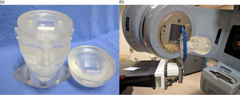
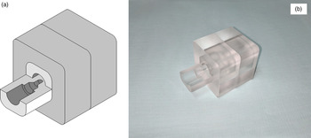
A locally fabricated polymethyl methacrylate head phantom was used in this study. Figure 1a shows the head phantom assembly and Figure 1b shows a phantom positioned on the treatment couch. A cuboid of size 40 × 40 × 40 mm3 was fabricated for inserting the ion chamber detectors into the phantom. Care was taken to align the point of measurement of the detectors at the centre of the cavity. Figure 2 shows the drawing of 40 × 40 × 40 mm3 cuboid with a detector holder and the corresponding machined part. The head phantom set-up and alignment were done using a BrainLab® (Feldkirchen, Germany) Stereotactic Frame Interface as shown in Figure 1b.
(a) Head phantom assembly and (b) it was mounted on the LINAC couch for the measurements.

(a) 40 × 40 × 40 mm3 cuboid 3D model with detector insert and (b) the corresponding machined part.

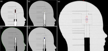
CT scan of the phantom with four commonly used ionisation chamber namely FC65 (IBA Dosimetry (Schwarzenbruck, Germany), Active volume 0.65 cc), CC13 (IBA Dosimetry (Schwarzenbruck, Germany), Active volume 0.13 cc), CC01 (IBA Dosimetry (Schwarzenbruck, Germany), Active volume 0.01 cc) and A14SL (Standard Imaging (Middleton, WI), Active volume 0.015 cc) and with dummy fillet were acquired using CT simulator (GE Optima (580W), GE Healthcare, Chicago, USA) with a slice thickness of 1.25 mm. These CT sets were imported into the Eclipse V13.7.14 (Varian Medical Systems, Palo Alto, CA, USA) TPS for creating the treatment verification plans. Figure 3 shows the sagittal view of the head phantom with all four detectors placed at the isocentre along with the dummy insert.
Sagittal CT view of the head phantom with detectors (a) FC65, (b) A14SL, (c) CC01, (d) CC13 and (e) dummy detector insert (superimposed contours of the active volumes of all four detectors).

Gradient of HU value in the axial cavity volume of the FC65 dosimeter (X and Y are the axial 2D coordinates).

Sensitive volumes of each detector were contoured on the CT set with detectors in place. CT sets of the phantom with detector and dummy fillet were fused to obtain the sensitive volume of the detector on CT set with dummy fillet. The following CT sets were made for dose calculation: 1. CT set with the detector, 2. CT set with dummy fillet having the contour of detectors sensitive volume through the image fusion and 3. CT set with chamber sensitive volume [for FC65 and CC13 only (Group-A)] assigned to air constant parameters to remove the effect of artefacts caused by the central electrode. For small volume, chambers like CC01 and A14SL (Group-B) were not been assigned the air cavity since its volumes are too small to make a significant difference.
The volumetric-modulated arc therapy verification plans were created with co-planar arcs. AAA and Acuros XB were used for dose calculation for each verification plan which is currently available in our institute. Mean doses to different detector volumes were estimated for both the algorithms. Plans were made such that there were no high-dose gradients in chamber-sensitive volume. The quality assurance treatment plans were delivered on the phantom in Varian Clinac iX 6 MV (Varian Medical Systems, Palo Alto, CA, USA) Medical Linear Accelerator (LINAC). The statistical analysis was done with paired t-test.
Results
The results of percentage deviations of the measured and the planned doses, for the stated detectors, using different CT sets were shown in Table 1 to 4. The mean percentage variation was −2.615 (SD: 0.394) for Group-A chambers and the corresponding mean percentage variation was −0.474 (SD: 0.485) for Group-B and was statistically significant with p-value 0.001 using Acuros XB. However, among Group-A dosimeters, the mean percentage variation was not statistically significant (p = 0.711) [FC65: −2.588 (SD: 0.256); CC13: −2.643 (SD: 0.504)]. Similarly, there was no significant variation between Group-B dosimeters [A14SL: −0.432 (SD: 0.392); CC01:−0.517 (SD: 0.575) (p = 0.641)]. If the calculations were done using the CT set with the detector, the mean of percentage deviations between TPS calculated and measured dose found to be significant for the Acuros XB algorithm compared to AAA for Group-A dosimeters. The mean variation was found to be >2.5% for FC65 and CC13.
PSQA-based point dose measurements of cranial VMAT verification plans of 15 patients with FC65 ion chamber using PMMA head phantom

AAA, Analytical Anisotropic Algorithm; VMAT, volumetric-modulated arc therapy; PMMA, polymethyl methacrylate; PSQA, patient-specific quality assurance. *Measured dose (cGy) with chamber–M; TPS calculated dose (cGy) with chamber–A; TPS calculated dose (cGy) with dummy chamber–B; TPS calculated dose (cGy) with assigned air cavity inside the chamber–C.
PSQA-based point dose measurements of cranial VMAT verification plans of 15 patients with CC13 ion chamber using PMMA head phantom

AAA, Analytical Anisotropic Algorithm; VMAT, volumetric-modulated arc therapy; PMMA, polymethyl methacrylate; PSQA, patient-specific quality assurance. *Measured dose (cGy) with chamber–M; TPS calculated dose (cGy) with chamber–A; TPS calculated dose (cGy) with dummy chamber–B; TPS calculated dose (cGy) with assigned air cavity inside the chamber–C.
PSQA-based point dose measurements of cranial VMAT verification plans of 15 patients with A14SL ion chamber using PMMA head phantom

AAA, Analytical Anisotropic Algorithm; VMAT, volumetric-modulated arc therapy; PMMA, polymethyl methacrylate; PSQA, patient-specific quality assurance. *Measured dose (cGy) with chamber–M; TPS calculated dose (cGy) with chamber–A; TPS calculated dose (cGy) with dummy chamber–B.
PSQA-based point dose measurements of cranial VMAT verification plans of 15 patients with CC01 ion chamber using PMMA head phantom

AAA, Analytical Anisotropic Algorithm; VMAT, volumetric-modulated arc therapy; PMMA, polymethyl methacrylate; PSQA, patient-specific quality assurance. *Measured dose (cGy) with chamber–M; TPS calculated dose (cGy) with chamber–A; TPS calculated dose (cGy) with dummy chamber–B.
Discussion
According to Fogliata et al., Reference Fogliata, Nicolini, Clivio, Vanetti and Cozzi5 Acuros XB gives higher accuracy in heterogeneous media. Acuros XB calculated and the measured doses in present phantom with detectors show a significant variation (>2.5%) for FC65 and CC13 ion chambers. The verification plan that uses the phantom CT set with chambers may have heterogeneity caused by artefacts. It is also observed from Table 1 to 4 that the TPS overestimates the dose. The artefacts affect the TPS-calculated dose leading to a comparatively higher deviation with the measured dose for Acuros XB, due to a better accounting of artefact-related heterogeneity. Hoffmann et al., Reference Hoffmann, Jørgensen, Muren and Petersen10 conclude Acuros XB shows the measured dose to be in good agreement (a mean deviation of 1.0 ± 1.9%) with the TPS-calculated dose using Gafchromic film measurements. However, in our study, the mean variation of measured dose against TPS calculated with dummy detector inserts shows a lower variation (≤0.8%) for all detectors using algorithms (AAA and Acuros XB). This emphasises the artefacts in the CT set that cause higher deviations using Acuros XB. The TPS dose results of the dummy detector show a closer value compared to the results obtained by Fogliata et al. and Hoffmann et al. It is observed in A14SL and CC01 chambers, the variations are relatively less as these may not produce any significant artefact in CT. However, the FC65 and CC13 chambers produce significant artefacts causing considerable variations between the calculated (Acuros XB) and measured doses.
A relevant study by B.R. Muir et al. Reference Muir and Rogers11 suggested avoiding making ionisation chambers that use high-Z electrodes to the manufactures, which may cause significant variation in K Q values in the current dosimetric protocols due to the central electrode effect. The gradient in the hounsfield unit (HU) value (Figure 4) from the central anode to the outer graphite wall for the FC65 chamber might be caused by the artefacts from the chamber central anode in the CT set. Therefore, when the sensitive volume is assigned as air constants, it leads to a better agreement with the measured value. The dose variation shows similar to the results obtained as in the dummy chamber. The detector volume and the type of detector electrode materials may also contribute to the artefacts leading to the quantity of dose difference, although the magnitude is small. Suitable artefacts reducing methods may help to resolve the artefacts due to the detector in-homogeneity of the phantom while taking the CT set. Reference Link, Berning and Scherf12-Reference Shaiju, Kumar and Varadarajulu15
It is evident that the percentage variation depends on the detector volume as the dose difference is higher for Group-A when compared with Group-B dosimeters for both the algorithms. In this scenario, it is advisable to use the CT set with dummy detectors to create verification plans. It is also observed that the variation in Acuros XB calculated and measured the dose using A14SL is minimum. It may be attributed that there are minimum artefacts with this detector. AAA calculated dose gives a better correlation with the measured values for all detectors. It might be because of lesser consideration of the artefacts generated heterogeneity due to the detectors, unlike Acuros XB. However, the mean of percentage deviation between measured and calculated doses with dummy chamber using stated algorithms are similar.
Conclusions
The study concludes that the CT set of the detectors in place give more artefacts/heterogeneity with volume, which may cause significant variations in the calculated dose using Acuros XB. The central electrode artefacts related to heterogeneity may be the reason for the dose discrepancy. Therefore, the study suggests using the CT set without detector for calculation of mean dose for larger chambers while the creation of verification plans especially for Acuros XB. The study also put forward to adopt suitable methods for reducing the artefact-related heterogeneities during dose calculations.
Acknowledgements
This research work has been funded by Kerala State Council for Science, Technology, and Environment (KSCSTE), Kerala, India. Sanction Order Number KSCSTE/5712/2017-SRSHS, dated 11/09/2017. We extend our sincere thanks to Dr Thayal Singh Elias (Professor), and Mr. Rojas K. Jose, (Medical physicist), Radiation Physics Division, Regional Cancer Centre, Thiruvananthapuram, India for their valuable support. The Authors have no conflicts of interest to declare.