Implications
Early detection of karyotype abnormalities could aid producers in excluding candidate parents of the next generation. Karyotype analyses have heretofore been undertaken using cytogenetic analysis which is costly to undertake routinely on commercial animals. We describe the statistics that can be generated from now commonly available genotype allele intensity data to detect aneuploidy using a set of dizygotic 53,X female sheep twins as a case study. The approach described is accurate and agnostic to the commercial genotype platforms currently available.
Introduction
Chromosomal abnormalities have been well documented in many species including sheep (Broad et al., Reference Broad, Hayes and Long1997; Raudseep and Chowdhary, Reference Raudseep and Chowdhary2016). Despite this, only two studies have attempted to characterise sheep with X-monosomy (Zartman et al., Reference Zartman, Hinesley and Gnatkowski1981; Baylis et al., Reference Baylis, Wayte and Owen1984). Zartman et al. (Reference Zartman, Hinesley and Gnatkowski1981) reported the first case of X-monosomy in sheep; the 5-year old sheep from New Mexico had apparently never given birth, but was masculine in appearance yet her external genitalia appeared normal. Baylis et al. (Reference Baylis, Wayte and Owen1984) reported on a mosaic X0/XX Cambridge female sheep who, although phenotypically normal, exhibited gonadal dysgenesis.
While cytogenetic analysis to determine the presence of chromosomal abnormalities is possible, the generally low expected frequency of such abnormalities in live animals, coupled the high cost of cytogenetic analysis relative to the value of the animal, contributes to a low uptake of routine cytogenetic screening in many domesticated species, including sheep. The recent availability of low cost genome-wide dense genomic marker panels based on single nucleotide polymorphisms (SNPs) (Boichard et al., Reference Boichard, Chung, Dassonneville, David, Eggen, Fritz, Gietzen, Hayes, Lawley, Sonstegard, Van Tassell, VanRaden, Viaud-Martinez and Wiggans2012; Berry et al., Reference Raudseep and Chowdhary2016), however, provide a rich source of genomic data which could possibly be used in the detection of some chromosomal abnormalities.
Berry et al. (Reference Berry, Wolfe, O’Donovan, Byrne, Sayers, Dodds, McEwan, O’Connor, McClure and Purfield2017) illustrated how the genotype allele intensity values of SNPs from commercially available genotype panels could be used to detect X-monosomy in cattle; such an approach has never been attempted in sheep. Several studies in humans have also concluded that SNP data can be used to detect aneuploidy (Treff et al., Reference Treff, Su, Tao, Levy and Scott2011; Xiong et al., Reference Xiong, Tan, Tan, Gong, Zhang, Lu, Luo, Lu and Lin2014). Previous studies on aneuploidy detection in non-human species using genome-wide SNP array data (e.g. Berry et al. Reference Berry, Wolfe, O’Donovan, Byrne, Sayers, Dodds, McEwan, O’Connor, McClure and Purfield2017), however, confined their analyses to genotypes generated from platforms of just one commercial vendor, namely Illumina (Illumina Inc., San Diego, CA, USA). The benefit of mining SNP data for detecting aneuploidy is that the SNP allele intensity values are generally already available as part of genome-wide enabled selection programmes in many species including sheep (Duchemin et al., Reference Duchemin, Colombani, Legarra, Baloche, Larroque, Astruc, Barillet, Robert-Granié and Manfredi2012; Dodds et al., Reference Dodds, Auvray, Newman and McEwan2012; Rupp et al., Reference Raudseep and Chowdhary2016). The objective therefore of the present study was to determine the utility and describe the informative statistics from genome-wide SNP data from two commercially available and routinely used genotype platforms to identify karyotype abnormalities using dizygotic 53,X twin female sheep as a case study.
Material and methods
Genomic analysis
Single nucleotide polymorphism genotype data from 10 866 individuals of multiple breeds genotyped on either the Illumina OvineSNP50 Beadchip (n=3296 animals) or a custom Illumina Infinium panel (n=7570 animals) were available; the recorded gender of all animals was also available. Only the 11 303 SNPs common to both genotype platforms were retained for further analysis. The common SNP panel included 217 SNPs on the X chromosome and one SNP on the Y-chromosome. Animal gender was determined from the sex-chromosome genotypes, and only animals where the gender recorded in the national database matched that determined from the genotypes were retained; 64 discrepancies existed. A total of 1848 males and 8954 females remained. All animals had a call rate >95% for both the SNPs on the X chromosome and all genotyped SNPs. A total of 21 SNPs reported to be on the non-pseudosomal region of the X chromosome which were reported heterozygous in confirmed male sheep were not considered further.
The X and Y signal intensity values of all called Illumina genotypes were also available, as was the logarithm normalised R ratios (LRR) which are the logged ratio of observed probe intensity to expected intensity according to the reference sample (Berry et al., Reference Berry, Wolfe, O’Donovan, Byrne, Sayers, Dodds, McEwan, O’Connor, McClure and Purfield2017). The genotype X and Y intensity values per SNP represents the intensity channel for each of the fluorescent dyes associated with the two alleles of the SNP. The mean of the sum of the X and Y signal intensities for called genotypes (i.e. the R-value) on the X chromosome of all individuals was calculated; similarly the mean LRR of called genotypes on the X chromosome and all autosomes was calculated separately for each female.
A total of four females genotyped on the Illumina platform were homozygous for all X-chromosome SNPs. Based on the diagnostic recommendation for detection of aneuploidy using SNP data in cattle (Berry et al., Reference Berry, Wolfe, O’Donovan, Byrne, Sayers, Dodds, McEwan, O’Connor, McClure and Purfield2017), two females with a mean allele R-value of SNPs on their X chromosome which noticeably deviated from the mean allele R-value of X-chromosome SNPs of other females, were identified as possible X-monosomy females and sent for cytogenetic analysis; both females had no called heterozygous genotype on the X chromosome. The two identified females were purebred Charollais fraternal twins. Genotypes were also available on their dam and sire.
A custom genotype panel with 11 196 SNPs, now publicly available (https://www.thermofisher.com/order/catalog/product/550838; accessed 26 July 2017), was developed using the Applied BiosystemsTM AxiomTM (formerly Affymetrix, now part of Thermo Fisher Scientific, Central Expressway, Santa Clara, USA) technology; 68 SNPs resided on the X-chromosome. A total of 383 female sheep, including the two identified putative 53,X female sheep, were genotyped on this platform. AxiomTM also provide signal intensity values for each probe which were used to identify the putative 53,X females. The mean allele intensity values of the SNP genotypes on the X chromosome of the 53,X females were compared with the respective statistic of the remaining 381 females, identical to the strategy undertaken for the Illumina genotypes; the strategy of comparing, within-animal, the mean chromosomal LRR values, as proposed by Berry et al. (Reference Berry, Wolfe, O’Donovan, Byrne, Sayers, Dodds, McEwan, O’Connor, McClure and Purfield2017) was also undertaken using the AxiomTM genotypes as also described in detail for the Illumina genotypes.
Cytogenetic analysis
Cytogenetic analyses were undertaken by the University of Kent in the United Kingdom on the sample females. Heparinised blood samples from both probable 53,X females were cultured for 72 h in PB MAX Karyotyping medium (Invitrogen, Santa Clara, CA, USA) at 37°C, 5% CO2. Cell division was arrested by adding colcemid at a concentration of 10.0 µg/ml (Gibco) for 35 min before hypotonic treatment with 75 mM potassium chloride and fixation to glass slides using 3 : 1 methanol : acetic acid. Metaphases for karyotyping were stained with 4′,6-diamidino-2-phenylindole in VECTASHIELD® antifade medium (VECTOR LABORATORIES, INC., 30 Ingold Road, Burlingame, CA 94010, USA). Image capturing was performed using an Olympus BX61 epifluorescence microscope with cooled charge coupled device camera and SmartCapture (Digital Scientific UK, The Commercial Centre, 6 Green End, Cambridge CB23 7DY, England) system for 20 samples per individual. SmartType software (Digital Scientific UK) was used for karyotyping purposes and chromosomes were arranged according to the International System for Chromosome Nomenclature of Domestic Bovids (2001).
Histology and performance data
The reproductive tract and mammary tissue of the individuals with X-monosomy were collected at necropsy and fixed in 10% formalin. Gross examination was undertaken on all tissue. Paraffin-embedded samples were further processed as 5-μm thin sections and routinely stained with hematoxylin and eosin for histological examination. As part of the national sheep breeding programme, live-weight records were available from the females with X-monosomy and their contemporaries throughout life. Whether live weight differed between the two females with X-monosomy and their contemporaries was estimated using a fixed effects linear model with weight as the dependent variable and age of the animal included as a covariate in the model that also included a fixed effect denoting whether the animal was an X-monosomy female or not.
Results and Discussion
On average, 41% of the edited 191 SNP genotypes on the X-chromosome in all 8954 females genotyped using the Illumina genotype platform were heterozygous. All X-chromosome SNPs of both 53,X females on the Illumina and AxiomTM panels were, however, homozygous. Two additional genotyped females on the Illumina platform also had a completely homozygous X chromosome (191 called SNP genotypes each); both females had produced lambs and the allele intensity values of these females is described later. Although deep pedigree data were unavailable for one of these females, inbreeding is likely to have contributed to the observed homozygosity in the other female as her maternal grandsire and maternal grand dam-sire were recorded to have been the same individual. No genotype information was available on this male individual. A similar phenomenon of a completely homozygous X chromosome has been reported in cattle (Zhang et al., Reference Zhang, Couldrey and Sherlock2016; Berry et al., Reference Berry, Wolfe, O’Donovan, Byrne, Sayers, Dodds, McEwan, O’Connor, McClure and Purfield2017). Based on a dataset of 103 327 female cattle, Berry et al. (Reference Berry, Wolfe, O’Donovan, Byrne, Sayers, Dodds, McEwan, O’Connor, McClure and Purfield2017) discovered 18 females that were homozygous for all 231 to 233 genotyped SNPs on the X chromosome; one of these females was confirmed a X-chromosome monosomy female. Although pedigree data were lacking for most of their remaining 17 females, Berry et al. (Reference Berry, Wolfe, O’Donovan, Byrne, Sayers, Dodds, McEwan, O’Connor, McClure and Purfield2017) speculated that the chromosome-wide homozygosity could have been due to inbreeding which was substantiated by the fact that the recorded sire and maternal grandsire of two of the females was identical. Furthermore, in an analysis of 15 703 female cattle, Zhang et al. (Reference Zhang, Couldrey and Sherlock2016) also speculated that the two females they detected to have homozygous X chromosomes had achieved this through inbreeding as the same male animal appeared on both sides of the pedigree of each female. Therefore, homozygosity alone is not a useful diagnostic tool for aneuploidy, especially for the X chromosome in females; this is consistent with the conclusion of Berry et al. (Reference Berry, Wolfe, O’Donovan, Byrne, Sayers, Dodds, McEwan, O’Connor, McClure and Purfield2017) in cattle.
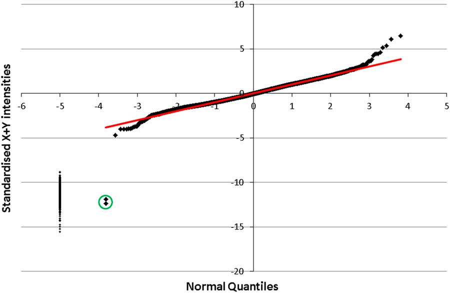
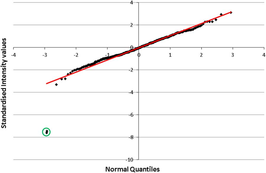
In a case study of a non-mosaic 59,X bovine, Berry et al. (Reference Berry, Wolfe, O’Donovan, Byrne, Sayers, Dodds, McEwan, O’Connor, McClure and Purfield2017) using just Illumina genotypes, reported good diagnostic ability of the mean R-values of SNP genotypes on the X chromosome to differentiate between monosomic and normal females. Berry et al. (Reference Berry, Wolfe, O’Donovan, Byrne, Sayers, Dodds, McEwan, O’Connor, McClure and Purfield2017) represented the mean R-value of the X-chromosome genotypes of each individual in standard deviation units relative to the mean R-value of the entire female population (excluding their monosomy female). The mean (standard deviation) R-value of the X-chromosome genotypes for all females (excluding the two X-monosomy females) in the present study was 1.19 (0.031) which is relatively similar to the mean (standard deviation) of 1.17 (0.022) reported by Berry et al. (Reference Berry, Wolfe, O’Donovan, Byrne, Sayers, Dodds, McEwan, O’Connor, McClure and Purfield2017) from 103 326 female cattle also genotyped on an Illumina platform. The mean R-value of SNPs on the X chromosome for the two 53,X females in the present study was 11.9 and 12.4 standard deviation units lower than the mean of the remaining female population (Figure 1); the next lowest R-value for a female genotyped on the Illumina platform was 4.71 standard deviation units below the female population mean (Figure 1) while the mean R-value of the two other females homozygous for the X chromosome but had given birth to lambs was 2.89 and 4.01. Berry et al. (Reference Berry, Wolfe, O’Donovan, Byrne, Sayers, Dodds, McEwan, O’Connor, McClure and Purfield2017) reported that their X-monosomy bovine female had a mean R-value of X-chromosome genotypes of 16.7 standard deviation units less than the female population mean. The mean allele intensity value of X-chromosome SNP genotypes of the 53,X females genotyped on the AxiomTM platform in the present study was 7.45 and 7.59 standard deviations less than the mean allele intensity value of X-chromosome SNP genotypes of all other females genotyped on the AxiomTM platform; the individual with the next lowest mean allele intensity value for SNPs on the X chromosome was 3.30 standard deviation units less than the population mean (Figure 2). Results from the present study therefore clearly corroborate those of Berry et al. (Reference Berry, Wolfe, O’Donovan, Byrne, Sayers, Dodds, McEwan, O’Connor, McClure and Purfield2017) in cattle and Xiong et al. (Reference Xiong, Tan, Tan, Gong, Zhang, Lu, Luo, Lu and Lin2014) in humans on the usefulness of allele intensity values relative to the normal population statistics for identifying aneuploidy; the conclusion was equally true irrespective of whether an Illumina or AxiomTM genotype platform was used. The mean R-value of the Illumina genotypes on the X chromosome for males was 0.85 (standard deviation of 0.22) and 87% of the males had an R-value greater than the 53,X female with an standardised R-value of 11.9 standard deviation units (Figure 1); limiting the analysis to SNPs on the non-pseudoautosomal region did not greatly impact the differentiation between males and female.
(colour online) Q-Q plot of the standardised sum of X and Y intensity values on the X chromosome for 8930 female sheep genotyped using the Illumina genotype panel with both 53,X females represented as the circled point on the bottom left. Also presented on the vertical line of points on the far left is the mean R-values of the X chromosome of 1815 males.

(colour online) Q-Q plot of the standardised sum of X and Y intensity values on the X chromosome for 383 female sheep genotyped using the AxiomTM genotype panel including the two 53,X females represented as the circled point on the bottom left.

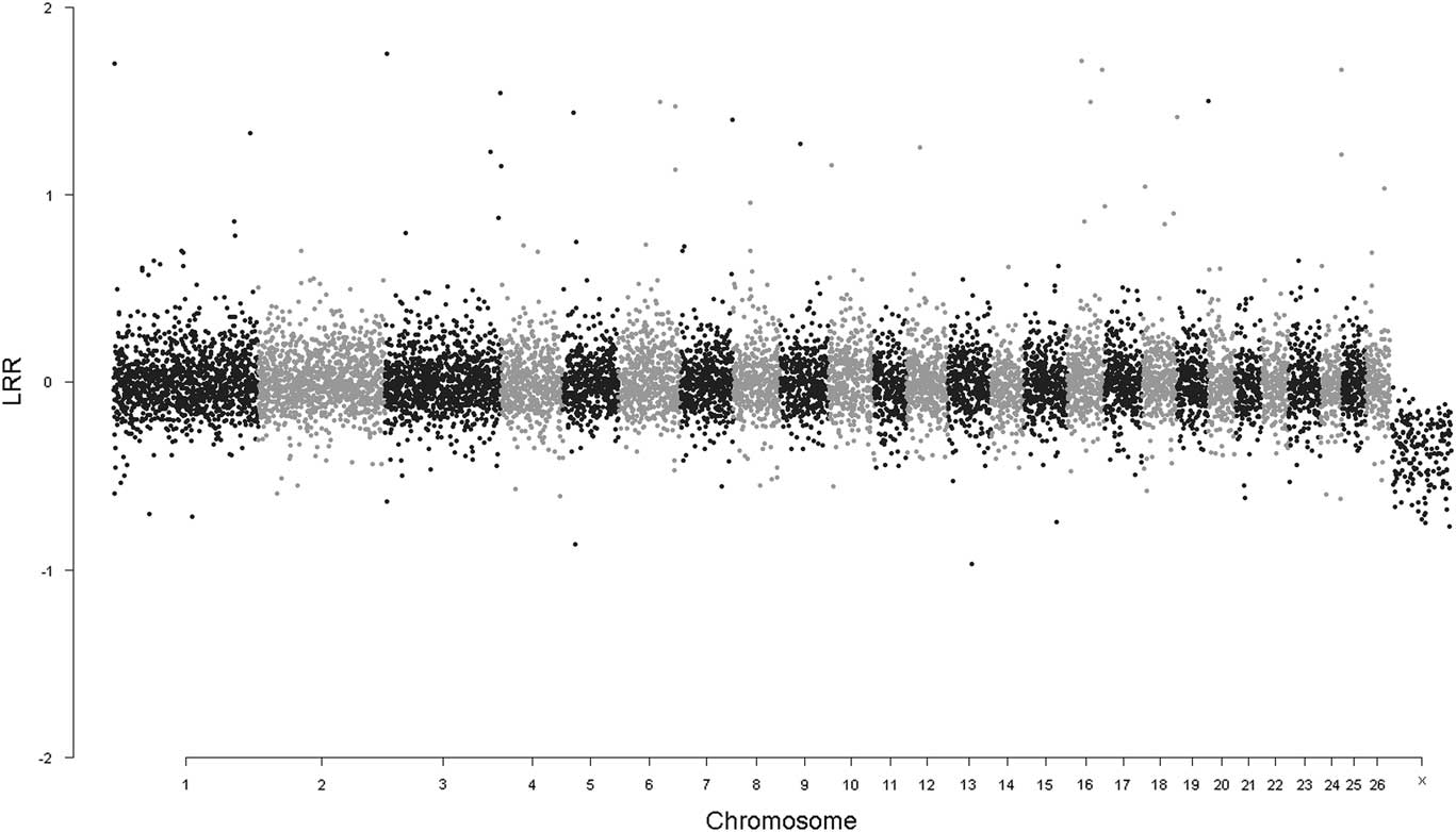
Although the LRR of SNP genotype data can be used to detect copy number variants (Lin et al., Reference Lin, Naj and Wang2014), Berry et al. (Reference Berry, Wolfe, O’Donovan, Byrne, Sayers, Dodds, McEwan, O’Connor, McClure and Purfield2017) concluded that, in cattle at least, the mean LRR of individual female animals’ X-chromosome genotypes, relative to other females, was not particularly good at detecting aneuploidy of the sex chromosomes. Although the 53,X females in the present study had the lowest mean X-chromosome LRR value (−0.54 and −0.36 from the Illumina platform), it was not very different to the mean X-chromosome LRR value of −0.32 for the female with the next lowest LRR value on the Illumina platform; the standard deviation of LRR in the female population (excluding the individuals with X-monosomy) was 0.036. Berry et al. (Reference Berry, Wolfe, O’Donovan, Byrne, Sayers, Dodds, McEwan, O’Connor, McClure and Purfield2017) recommended using the within-individual difference, in standard deviation units, of the mean LRR of X-chromosome genotypes from the mean LRR of the autosomal genotype as an approach to more easily identify individuals with X-monosomy. The mean LRR of the Illumina genotypes for SNPs on the X chromosome of the 53,X individuals relative to the LRR of their respective autosomal genotypes was −23.5 and −11.8 standard deviation units while the next lowest deviating female was −3.35 standard deviation units. Individual SNP LRR values, collated by chromosome, for one of the 53,X females generated on the Illumina platform is in Figure 3; the lower LRR of the SNPs on the X chromosome are clearly visible. The mean LRR of the AxiomTM genotypes for SNPs on the X chromosome of the 53,X females relative to the LRR of their respective autosomal genotypes was −1.57 and −1.68 standard deviation units while the next lowest deviating female was −0.37 standard deviation units. In their analysis of a heifer with X-monosomy, Berry et al. (Reference Berry, Wolfe, O’Donovan, Byrne, Sayers, Dodds, McEwan, O’Connor, McClure and Purfield2017) documented that the within-animal deviation in LRR values of SNPs on the X chromosome v. the autosomes for the X-monosomy female was −3.4 standard deviation units while the female with the next greatest deviation was −0.6 standard deviation units. Therefore, results from the present study again corroborate those of Berry et al. (Reference Berry, Wolfe, O’Donovan, Byrne, Sayers, Dodds, McEwan, O’Connor, McClure and Purfield2017) in cattle on how to use the within-animal relative LRR of genotypes on the X chromosome v. the autosomes to identify aneuploidy from SNP data.
Individual single nucleotide polymorphism logarithm intensity ratio (LRR) per chromsome for one of the 53,X case study female sheep generated using the Illumina platform.

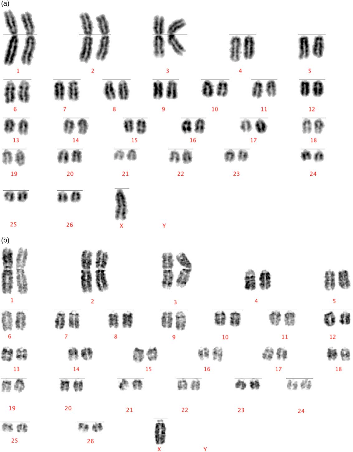
The karyotype of the two sheep with X-chromosome monosomy is in Figure 4. Based on the fact that 20 metaphases in the present study were used for karyotyping each female, only mosaicism of >14% can be excluded with 95% confidence (Hook, Reference Hook1977) in the study females. Therefore, although the likelihood of mosaicism in each case-study sheep is low, it cannot be ruled out. Of the 50 cells analysed by Zartman et al. (Reference Zartman, Hinesley and Gnatkowski1981) from their sheep with X-chromosome monosomy, all but two of the cells had 53 chromosomes with the remaining two cells having 52 chromosomes. Baylis et al. (Reference Baylis, Wayte and Owen1984) documented that 16 of the 20 cells of an infertile sheep they karyotyped had the normal complement of chromosomes with one copy of the X chromosome missing from three of the remaining cells suggesting mosaicism X0/XX.
(colour online) (a, b) Karyotype of the two 53,X case study female sheep.

Analysis of the X-chromosome genotypes of both 53,X sheep in the present study and their two parents revealed that the single X chromosome originated from the sire in both instances. Furthermore, the correlation between the Illumina autosomal allele counts of both females with X-monosomy was 0.53 indicating that they were, in fact, fraternal twins. While most previous studies, albeit limited to human populations, suggest a greater propensity for the single X chromosome of Turner females to originate from the dam (Mathur et al., Reference Mathur, Stekol, Schatz, MacLaren, Scott and Lippe1991; Uematsu et al., Reference Uematsu, Yorifuji, Muroi, Kawai, Mamada, Kaji, Yamanaka, Momoi and Nakahata2002), Berry et al. (Reference Berry, Wolfe, O’Donovan, Byrne, Sayers, Dodds, McEwan, O’Connor, McClure and Purfield2017) reported that the single X chromosome of their 59,X bovine originated from the sire.
To our knowledge this is the first example, in any species, of X-monosomy in dizygotic female twins. Based on a single case study in humans, Rovet and Netley (Reference Rovet and Netley1981) reported on a single case of female dizygotic twins, but only one of the twins was monosomic for the X chromosome. Pescia et al. (Reference Pescia, Ferrier, Wyss-Hutin and Klein1975) reported on a case study of monozygotic human twin sisters both of which had Turner’s syndrome. Although the conclusion differed by what age the sample was taken (i.e. prenatal, at birth, at 10 months of age), and what tissue was analysed (i.e. blood lymphocytes v. fibroblasts), in their analysis of monozygotic human twins, Gilbert et al. (Reference Gilbert, Yardin, Briault, Belin, Lienhardt, Aubard, Battin, Servaud, Philippe and Lacombe2002) reported varying levels of 46,XX/45,X mosacism in their two case study females. The incidence of Klinefelter’s syndrome (i.e. XXY) has, however, been associated with a greater incidence of twinning in humans (Nielsen, Reference Nielsen1966; Rehder et al., Reference Rehder, Schoner, Kluge, Louwen, Schwinger and Neesen2012).
Animal external and reproductive tract characteristics
The two case study 53,X Charollais in the present study were born as twins on the 28 January 2015 to a 3-year old dam and reared together as twins; the sire of the lambs was 787 days old when the twins were born. The birth weight of each twin was 5.2 and 6.7 kg. The 53,X individuals weighed 40 kg (heaviest born twin) and 33 kg (lightest born twin) at 110 days of age. When compared with other twin female lambs born on the farm in the year 2015 to the same sire, the mean adjusted 100 day weight of the monosomy females was 32.00 kg which was not different to the mean of 32.44 kg (SE=2.82 kg) of the eight paternal half-sib contemporaries. Both sheep with X-chromosome monosomy in the present study were exposed to a ram during one breeding season but were pregnancy diagnosed not in lamb before culling. Zartman et al. (Reference Zartman, Hinesley and Gnatkowski1981) also reported sterility in their detected sheep with X-chromosome monosomy in New Mexico.
The females in the present study were slaughtered at 742 (heaviest born twin) and 806 (lightest born twin) days of age. No live weight of the heavier born 53,X sheep was available at slaughter but the lightest born 53,X sheep was 38 kg before slaughter and appeared smaller and lighter than her contemporaries. Both females were assessed to be in poor body condition at the time of slaughter. It therefore appears that the sheep monsomic for the X chromosome grew as normal and were not noticeably distinguishable (in size) from their contemporaries up to ~110 days of age after which the 53,X sheep did not grow much. This further proves the utility of being able to (indirectly) karyotype individuals from routinely available SNP data as the size of these growing females as lambs relative to contemporaries does not appear to be a useful diagnostic tool.
The smaller than expectation size of mature 53,X sheep have been described elsewhere in humans (Rongen-Westerlaken et al., Reference Rongen-Westerlaken, Corel, van den Broeck, Massa, Karlberg, Albertsson-Wikland, Naeraa and Wit1997; Gravholt, Reference Gravholt2005) and cattle (Romano et al., Reference Romano, Raussdepp, Mulon and Villadóniga2015; Berry et al., Reference Berry, Wolfe, O’Donovan, Byrne, Sayers, Dodds, McEwan, O’Connor, McClure and Purfield2017). Other than both 53,X females being considerably smaller in size than expected and of poor body condition, there were no other obvious external abnormal phenotypic characteristics of either female. Zartman et al. (Reference Zartman, Hinesley and Gnatkowski1981) failed to identify any ‘exceptional’ difference in external appearance of their sheep with X-monosomy, despite being 5 years old, citing her to be ‘physically normal phenotype in very good flesh’.
Vaginal glandular tissue development was absent in one of the females in the present study while the vaginal glandular tissue development was limited in the other female. The ovaries of both sheep with X-monosomy in the present study were small for their age and evidence of prior ovulation was not appreciated at gross inspection of the tissues (i.e. no corpora lutea or luteal scars were observed). Compared with the ovarian dimensions of 12 to 24-month-old sheep surveyed at slaughter (Mohammadpour, Reference Mohammadpour2007), the ovaries of the two females with X-monosomy in the present study were hypoplastic. There was microscopic evidence of corpus luteum formation in one ovary in one of the sheep. In both 53,X sheep in the present study, the density of primordial follicles in the ovarian cortex was lower than would normally be found in ovine ovaries and primary follicle development was not observed in any of the sections of ovary examined. In the uterine tubes, uterus and vagina, glandular tissue was either inactive and/or hypoplastic.
Mammary gland development in the 53,X sheep in the present study was very limited. While teats were present in the 53,X sheep in the present study, no associated gland development could be detected visually or by palpation. On dissection, mammary gland tissue could not be appreciated grossly and while mammary gland ducts and acini could be identified on histology, the tissue was, in our opinion, hypoplastic. More developed mammary tissue would be expected in sexually immature XX chromosome female sheep. Although several studies on domesticated animals suffering from X-monosomy have cited no obvious external phenotypic abnormalities (Zartman et al., Reference Zartman, Hinesley and Gnatkowski1981; Baylis et al., Reference Baylis, Wayte and Owen1984), it is not always clear what exactly was examined and therefore not possible to conclude on an absence of abnormality in the mammary tissue of these case studies. In their case study of a 3-year-old Longhorn heifer monosomic for the X chromosome, Romano et al. (Reference Romano, Raussdepp, Mulon and Villadóniga2015) reported that ‘the udder was underdeveloped for the age of the animal’. Visscher et al. (Reference Visscher, Metcalfe and Semple2015) reported on a case study of a human female non-mosaic for Turner Syndrome who had not received any recombinant growth hormone therapy; as well as being smaller in size, at the age of 14 she had symmetrical mammary hypoplasia. The poor mammary gland development may be associated with the absence or delay in pubertal development that is known to exist in human females monosomic for the X chromosome (Gravholt, Reference Gravholt2005).
Conclusion
To our knowledge, this is the first ever report, in any species, of dizygotic female twins monosomic for the X chromosome; both females are likely non-mosaic 53,X and inherited their single X chromosome from their sire. Both 53,X females appear incapable of producing an offspring, consistent with a generally assumed sterility of (untreated) females with X-monosomy (Raudsepp and Chowdhary, Reference Raudseep and Chowdhary2016). Of particular interest in the present study was the ability of routinely available SNP allele intensity values to detect such aneuploidy and the fact that SNP genotypes now routinely being generated using either an Illumina or an AxiomTM platform could be used. This is particularly important given the cost of most commercial female sheep relative to the cost of cytogenetic analysis.
Acknowledgements
Financial support from the Irish Department of Agriculture, Stimulus Research Fund project OVIGEN is gratefully acknowledged. Gratitude is also extended to Ram Verma from Thermo Fisher Scientific.
Declaration of interest
Patil and Ho are employees of ThermoFisher Scientific whose technology was used to generate some of the genotype data used in the present study. All analyses were undertaken by the Berry and ThermoFisher Scientific staff were blind to all samples.
Ethics statement
The study involved the analysis of pre-existing data with the histology analyses performed post-slaughter. Blood samples were collected as part of a veterinary diagnostic examination.
Software and data repository resources
Data are available on request from the corresponding author.