Introduction
Fascioliasis, or liver fluke disease, is one of the most economically important helminth diseases of livestock. Globally, it is estimated to cost the agricultural sector approximately US$3.2 billion per annum (Spithill et al., Reference Spithill, Smooker, Sexton, Bozas, Morrison, Creaney, Parsons and Dalton1999; Bennett & IJpelaar, Reference Bennett and IJpelaar2005; McGonigle et al., Reference McGonigle, Mousley, Marks, Brennan, Dalton, Spithill, Day and Maule2008), with an estimated 600 million animals infected worldwide (Hillyer & Apt, Reference Hillyer and Apt1997). In some areas, there has been a significant rise in the incidence of fascioliasis in both cattle and sheep, over the past few years. For example, in Scotland, the Veterinary Investigation Diagnosis Analysis (VIDA) service reported that the incidence of fascioliasis had increased from 3 to 19% and from 0.5 to 15% of diagnosable submissions in cattle and sheep, respectively, between the early 1990s and 2008 (Kenyon et al., Reference Kenyon, Sargison, Skuce and Jackson2009; van Dijk et al., Reference van Dijk, Sargison, Kenyon and Skuce2010).
Fascioliasis in domestic livestock is traditionally controlled using fasciolicidal drugs. Triclabendazole has been the predominant drug of choice for treating liver fluke infections for over 20 years because of its efficacy against both juvenile and adult flukes (Brennan et al., Reference Brennan, Fairweather, Trudgett, Hoey, McCoy, McConville, Meaney, Robinson, McFerran, Ryan, Lanusse, Mottier, Alvarez, Solana, Virkel and Brophy2007). However, the emergence of triclabendazole-resistant parasites (Overend & Bowen, Reference Overend and Bowen1995) has spurred an interest into developing vaccines as an alternative means of control. Several promising Fasciola vaccine candidates have been isolated to date, including glutathione-S-transferase (GST; Sexton et al., Reference Sexton, Milner, Panaccio, Waddington, Wijfels, Chandler, Thompson, Wilson, Spithill, Mitchell and Campbell1990), cathepsin L-like proteases (CatL; Dalton et al., Reference Dalton, Neill, Stack, Collins, Walshe, Sekiya, Doyle, Mulcahy, Hoyle, Khaznadji, Moiré, Brennan, Mousley, Kreshchenko, Maule and Donnelly2003) and leucine aminopeptidase (LAP; Acosta et al., Reference Acosta, Cancela, Piacenza, Roche, Carmona and Tort2008). GST was selected because of parallels with the human blood flukes (Brophy & Pritchard, Reference Brophy and Pritchard1994), CatLs due to their prominence in the excretory/secretory (E/S) products of adult and juvenile flukes (Dalton et al., Reference Dalton, Neill, Stack, Collins, Walshe, Sekiya, Doyle, Mulcahy, Hoyle, Khaznadji, Moiré, Brennan, Mousley, Kreshchenko, Maule and Donnelly2003) and LAP on the basis that it is associated with the epithelial cells that line the parasite's digestive tract (Acosta et al., Reference Acosta, Cancela, Piacenza, Roche, Carmona and Tort2008). LAP is the most effective antigen isolated to date, with reductions in fluke burdens of 89% in vaccinated sheep compared to controls (Acosta et al., Reference Acosta, Cancela, Piacenza, Roche, Carmona and Tort2008). Also, H11, an aminopeptidase, is the most effective antigen isolated to date from Haemonchus contortus (Knox & Smith, Reference Knox and Smith2001). The LAP study, the recent progress in vaccination against Haemonchus as well as positive outcomes in vaccination against the blood-feeding tropical cattle tick, Rhipicephalus (Boophilus) microplus, which has led to the development of a commercially available vaccine, TickGARD™ (Willadsen et al., Reference Willadsen, Bird, Cobon and Hungerford1995), all indicate that targeting proteins expressed on the luminal surface of the intestine and required for digestion of the blood meal in haematophagous parasites could be a rich source of efficacious vaccine candidates. Fasciola hepatica lives in a highly vascularized environment within the liver system, feeding on surrounding tissue, and although blood may not be the primary part of its diet, the adult fluke's gut is known to be filled with partially digested blood. Furthermore, blood is ingested, whether deliberately or not, by the juvenile fluke migrating through the liver parenchyma (Olsen, Reference Olsen and Olsen1986). Thus F. hepatica may be vulnerable to the gut antigen approach to vaccination.
Most, if not all, of the candidate hidden or gut antigens identified to date have been membrane-bound glycoproteins. Lectins have been valuable tools for identifying and separating these glycosylated antigens from protein-rich extracts (Smith et al., Reference Smith, Smith and Murray1994). Lectins are carbohydrate-binding proteins derived from plants, animals and micro-organisms, with specificity for terminal or subterminal carbohydrate residues (Leathem & Brooks, Reference Leathem, Brooks, Rhodes and Milton1998). In Haemonchus, the major vaccine candidates isolated from the intestine have all been localized to the microvillar surface of the intestinal cells (Knox & Smith, Reference Knox and Smith2001), the equivalent structure to the gut lamellae in Fasciola. Here, our ambition was to identify lectin(s) with specificity for the F. hepatica gut in an effort to develop a procedure to isolate intestinal integral membrane proteins, which may be the equivalent of those purified from the other parasites noted above and, hence, may be strong vaccine candidates.
Materials and methods
Parasitological procedures
Adult flukes were recovered from naturally infected ovine hosts and were kindly supplied by Professor N. Sargison (Royal (Dick) School of Veterinary Studies, Edinburgh), Professor I. Fairweather (Queen's University, Belfast) and Mr S. Stevenson (Duncan Stevenson (Meats) Ltd, Dunblane). The parasites were washed in phosphate-buffered saline (PBS) then either frozen at − 80°C for somatic antigen extraction or fixed in 4% paraformaldehyde and processed to paraffin wax for histological sectioning. The latter procedure was performed on an automated processor (80% ethanol, 45 min; 95% ethanol, 75 min; 99% ethanol, 3 × 75 min; isopropanol, 2 × 90 min; isopropanol/xylene (1:1 v:v), 90 min; xylene, 2 × 90 min; paraffin wax, 2 × 10⁵ min). Sections (5 μm thick) were cut using a microtome and mounted on treated glass slides (Superfrost® Plus glass slides, Thermo Scientific, Epsom, Surrey, UK). These were dried overnight at 37°C and stored at 4°C until required.
Conjugated lectins
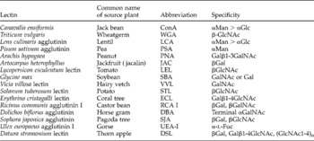
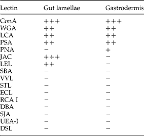
Sixteen fluorescently labelled lectins of differing specificities (Vector Laboratories, Peterborough, Cambs, UK) were used to screen mounted histological sections, as listed in table 1. Lectins were diluted to working concentrations of 5 μg/ml in lectin wash buffer (LWB; 50 mm Tris, 150 mm NaCl, 2 mm MgCl2, 1 mm CaCl2). Biotinylated lectins ConA, WGA, LCA, PSA, PNA, JAC, LEL and SBA (Vector Laboratories) were diluted 1:1000 (ConA 1:2000) in TNTT (10 mm Tris, 0.5 m NaCl, 0.05% Tween-20, 0.01% Thiomersal, pH 7.4).
Conjugated lectins used for screening histological sections of adult Fasciola hepatica.

Lectin probing of histological sections
Mounted sections of adult flukes were de-waxed by an automated procedure (xylene, 90 s; xylene, 165 s; methylated spirit industrial 74 O.P. (74OP) 2 × 90 s; 74OP+distilled water (dH2O), 60 s; dH2O, 90 s) and then probed with one of the panel of 16 lectins listed in table 1. Slides were placed horizontally in a dark, moist chamber, 500 μl of lectin solution was pipetted directly on to the section and incubated for 2 h. Slides were then washed in darkness three times for 5 min in LWB. A negative control was prepared in the same way, replacing lectin with an equal volume of LWB alone. Slides were mounted with a coverslip using Prolong® antifade with diamidino-2-phenylindole (DAPI) (Invitrogen, Paisley, Renfrewshire, UK) and stored at 4°C until viewed on an Axiovert 200 fluorescent microscope (Carl Zeiss Microimaging, Thornwood, New York, USA).
Preparation of somatic extracts from F. hepatica
Frozen flukes were thawed on ice and homogenized using a Ultra-turrax® T25 (IKA® – Labortechnik, Staufen, Germany) with 5 ml of homogenizing buffer (HB; PBS, 2 mm EDTA, 1 mm phenylmethylsulphonyl fluoride pH 7.4) and centrifuged at 20,000 g for 20 min at 0°C. The supernatant (S1) was retained. The pellet was resuspended and hand homogenized in HB with 0.1% Tween-20 then centrifuged at 16,500 g for 20 min at 2°C. The supernatant (S2) was retained. This Tween-20 extraction was repeated, the supernatant discarded and the pellet was then resuspended and hand homogenized in 2% w/v reduced Triton X-100 in PBS pH 7.4 and mixed for 30 min at 4°C. This extract was then centrifuged at 50,500 g for 30 min at 2°C and the supernatant (S3) retained. This resulted in the production of three crude somatic fractions representing water-soluble proteins (S1), membrane-associated proteins (S2) and detergent-soluble proteins (S3), respectively. Protein concentrations of each extract were estimated using a BCA™ Protein Assay Kit (Pierce, Rockford, UK).
Lectin probing of somatic extracts
The F. hepatica S3 fraction was separated by electrophoresis using a 4–12% Bis-Tris NUPAGE Novex SDS–PAGE (Invitrogen) gel, then electroblotted on to nitrocellulose membrane (NC) at 150 mA for 50 min. The NC membranes were incubated for 2 h at room temperature in TNTT, then divided into lanes to enable them to be incubated separately in one of the biotinylated lectins, WGA, LCA, PSA, PNA, JAC and LEL, at a 1:1000 dilution (or ConA at 1:2000) in TNTT for 1 h. Strips were then washed three times in TNTT before incubation in streptavidin–horseradish peroxidase (HRP) (Thermo Scientific) at 1:1000 in TNTT for 1 h and washing as before. Strips were developed with Sigmafast™ 3,3′-diaminobenzidine (DAB) with metal enhancer (Sigma-Aldrich, Poole, Dorset, UK).
Lectin affinity purification
One milligram of protein was diluted fourfold in Wash buffer (WB; 10 mm Tris-HCl, 0.5 m NaCl, 0.1 m CaCl2, 1 μm MgCl2 pH 7.4). The diluted extract was fractionated on a jacalin lectin–agarose column at 4°C. First, the sample was loaded on to a fresh column at 6 ml/h ensuring any unbound protein was collected. Bound proteins were then eluted by washing the column with 0.8 m galactose in WB and collecting the elutate. Bound and unbound proteins were buffer exchanged into 10 mm Tris, pH 7.4 by passing through a Sephadex G-25 column to remove detergents and sugars which may interfere in later assays. Samples were then concentrated by ultracentrifiltration in Amicon Ultracel 10K concentrators (Millipore (UK) Ltd, Watford, UK). The protein concentration for each sample was then estimated using a BCA™ Protein Assay Kit (Pierce) and samples stored at − 80°C. For analytical purposes, collected samples were separated by SDS–PAGE (4–12%) and silver stained using Silverquest™ silver staining kit (Invitrogen).
Results
Lectin probing of histological sections
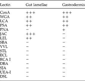
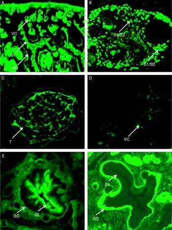
Seven of the lectins showed an affinity for the gut region of F. hepatica, as shown in table 2. However, none of these lectins bound exclusively to the gut. Most of the organ systems within tissue sections of the adult flukes could be labelled by one or more of the panel of lectins screened. There were some examples of tissue-specific localization as shown in fig. 1A–D. The lectins STL, DBA and SJA bound exclusively to carbohydrates located on the testes membrane, the testes follicles and the subtegumental cells, respectively. These were the only lectins to show single-tissue specificity but the intensity of staining in each of these cases was relatively low. Two lectins, UEA-I and DSL, showed no affinity for any organ systems within the parasite. ConA bound intensely to all tissues throughout the parasite, with the exception of the tegumental spines. A similar pattern was seen with the lectins WGA, LCA and PSA, but the staining intensity varied between the organ systems. JAC and LEL bound to molecules on the gut lamellae but not the gastrodermal cells, whereas PNA bound to the gastrodermal cells but not the gut lamellae (fig. 1E and F). Lectins of particular interest, i.e. those binding to the gut of F. hepatica, identified from the screen were, therefore, ConA, WGA, LCA, PSA, PNA, JAC and LEL.
Summary of lectin binding within the gut of adult Fasciola hepatica.
Lectin binding within the tissues of Fasciola hepatica. (A) The non-specific binding of concanavalin A to many tissues throughout the parasite, including the gut lamellae (GL), gastrodermis (GD) and vitelline cells (VC). (B) Binding of Lens culinaris agglutinin to the presumed S1/S2 cells (arrows) of the Mehlis' gland. The selective binding of Lycopersicon esculentum lectin to the male tissues, for example, the testes (T) is shown in (C) and of Erythrina culinaris agglutinin I to the female tissues, for example, the vitelline follicles (V) in (D). Preferential binding is also shown in the case of (E) binding of jacalin lectin to the gut lamellae (GL) and (F) binding of peanut lectin to the gastrodermal cells (GD).

Lectin probing of somatic extracts
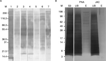
Western blots (fig. 2A) of the S3 protein extract were incubated with the panel of lectins which reacted with the gut material, as identified above; ConA, WGA, LCA, PSA, PNA, JAC and LEL. All the lectins tested reacted with glycan moieties in the S3 extract and there was a suggestion that the staining patterns differed depending on the lectin used. The possibility that differential lectin binding could be used to enrich for different subsets of membrane proteins was then tested by comparing the profiles of the glycoproteins which specifically bound JAC and PNA lectins.
Using conjugated lectins to probe and subfractionate a somatic extract of Fasciola hepatica. (A) Lectin binding to the membrane-bound (S3) extract with the respective lectins. Lanes: M, markers; 1, ConA; 2, WGA; 3, LCA; 4, PSA; 5, PNA; 6, JAC; 7, LEL. (B) Lectin affinity chromatography of the crude detergent soluble fraction (as shown in lane S3). The unbound fractions for peanut (PNA) and jacalin (JAC) are shown in lanes UB under the respective lectin name. Eluted protein fractions from PNA and JAC are shown in lane E under the respective lectin name.

Lectin-affinity purification
The S3 extract of parasite proteins was applied to JAC or PNA affinity columns and the unbound and bound fractions separated using SDS–PAGE and visualized by silver staining (fig. 2B). Both lectins appeared to exhibit selectivity and bound relatively few proteins compared to the original extract. The eluant from the PNA affinity column contained a number of peptides over a broad size range but, nonetheless, clearly represented a subset of the original crude fraction. By contrast, JAC appeared to be more selective, the eluant containing only two faint bands around 62–65 kDa.
Discussion
Here we have used lectin binding in an attempt to facilitate the isolation of gut-specific glycoproteins from adult F. hepatica. Seven of the lectins tested, namely ConA, WGA, LCA, PSA, PNA, JAC and LEL bound to the gut lamellae or to the gastrodermal cells. In addition, they also bound to elements within other tissues and organ systems but the intensity of staining differed depending on the lectin in question. For example, ConA lectin stained almost every anatomical region with equally bright intensity while others (LEL, ECL, JAC, PNA) bound selectively to male (LEL) and female (ECL) tissues, the gut lamellae (JAC) but not the gastrodermal cells, and the gastrodermal cells (PNA) but not the lamellae (fig. 1A–F). When membrane-bound protein extracts were further fractionated using PNA or JAC affinity chromatography (fig. 2B), it became clear that lectins did offer a means to isolate distinct, but relatively simple, protein subsets from detergent-soluble extracts of adult fluke.
Lectins have been used with considerable success for isolating vaccine antigens by affinity chromatography, as shown in the preparation of protective antigens against the parasitic nematode, H. contortus, including H11 and H-gal-GP (Smith et al., Reference Smith, Smith and Murray1994, Reference Smith, Smith and Pettit2000a, Reference Smith, Smith, Pettit, Newlands and Skuceb; Knox & Smith, Reference Knox and Smith2001). The gastrodermal cells of Fasciola contain several known vaccine candidates for fluke, including cathepsin L1 and GST (Collins et al., Reference Collins, Stack, O'Neill, Doyle, Ryan, Brennan, Mousley, Stewart, Maule, Dalton and Donnelly2004; Creaney et al., Reference Creaney, Wijffels, Sexton, Sandeman, Spithill and Parsons1995). Lectin screening described here indicated that JAC had some specificity for the gut lamellae and PNA showed some specificity for glycoproteins within the gastrodermis. While the Western blots described here confirmed that the selected lectins bound to glycosylated molecules within the S3 somatic fraction (fig. 2A), these blots did not indicate that binding was particularly selective with, for example, the PNA and JAC binding profiles being almost indistinguishable. However, when used as affinity ligands, these lectins selected quite distinct subsets of glycoproteins from the original protein fraction (fig. 2B). The eluant from PNA contained a series of bands undoubtedly less complex than the original sample and, surprisingly, the JAC eluant contained two predominant protein bands around 62–65 kDa in size. The gut lamellae of F. hepatica are structurally equivalent to the brush border in H. contortus, which has proven to be a rich source of antigens and both JAC and PNA have been used to purify protective antigens from somatic fractions of H. contortus (Smith et al., Reference Smith, Smith, Pettit, Newlands and Skuce2000b; Knox & Smith, Reference Knox and Smith2001; Knox et al., Reference Knox, Redmond, Newlands, Skuce, Pettit and Smith2003).
This work also provides valuable information regarding the distribution of carbohydrates throughout the parasite's complex organ systems. It is evident that some carbohydrates are shared amongst tissues, whereas others have a more specific tissue distribution. The lectins ConA, WGA, LCA, PSA, PNA, JAC and LEL all bound to molecules in gastrodermal tissues and have specificity for differing sugars such as α-d-mannose, α–d-glucose, N-acetylglucosamine, N-acetylgalactosamine, glucose and galactose. The selective binding of JAC to molecules on the gut lamellae and PNA to those on the gastrodermal cells indicate that the sugars α-galactosidase and galactosyl (β-1,3) N-acetylgalactosamine are present and may be specific to these tissues within the gut region. Previous studies have indicated that α-linked acetylgalactosamine sugars, derivatives of galactose, are on proteins in the gut of F. hepatica (Wuhrer et al., Reference Wuhrer, Grimm, Dennis, Idris and Geyer2004), and this is consistent with the present findings. In addition, the sugars N-acetylglucosamine and N-acetylgalactosamine have also been identified on the gut of the blood fluke Schistosoma japonicum, a homologous trematode species (Gobert et al., Reference Gobert, Jones and Stenzel1998). H-gal-GP, the highly protective antigen identified from the gut of H. contortus, is a galactose-containing glycoprotein complex (W.D. Smith et al., Reference Smith, Smith and Murray1994; S.K. Smith et al., Reference Smith, Pettit, Newlands, Redmond, Skuce, Knox and Smith1999) and is purified using lectins which have specificity for N-acetylgalactosamine (Smith et al., Reference Smith, Smith and Murray1994). This screen indicates that glycoproteins are also present in the gut tissues of F. hepatica. Follow-up proteomic analyses of the JAC- and PNA-binding proteins will establish the nature of these proteins and their relationship to existing fluke vaccine candidates and those identified in other blood-feeding helminths. The next step would be to test the vaccine efficacy of these protein subsets.
This lectin screen highlights the diversity and complexity of glycoconjugates present throughout the organ systems of F. hepatica. The significance of the presence or absence of certain carbohydrates is not fully understood, but the patterns seen here are consistent with previous studies in F. hepatica and other homologous trematode species. Our aim here was to identify any lectins that could selectively isolate glycoproteins from tissues of specific interest, in particular the gut. Unfortunately, many carbohydrate molecules which are present on these tissues are also shared with other organ systems and no single lectin bound exclusively to the gut. We exploited the selective binding of JAC to molecules on the gut lamellae and not those on the gastrodermal cells in order to purify a relatively simple subset of proteins from a detergent extract of adult fluke. Future work will focus on defining these proteins using proteomics and possibly testing the vaccine efficacy of this subset of antigens.
Acknowledgements
The authors would like to thank Mrs Jeanie Findlayson for her help with embedding and sectioning of the adult parasites and also Dr Susan Ekoja, Dr Stuart Smith and Dr George Newlands for all their advice on protein extraction and affinity chromatography.