The mammalian retina has long been a model system for study of development of neural circuits in the central nervous system. The development of the retina requires several steps. First, the seven cell types that comprise the retina need to be generated in the right proportion. Second, postmitotic cells leave the ventricular zone and migrate to one of three cell layers. Third, they start to form synaptic connections with other retinal neurons. Finally, these groups of synaptically coupled neurons evolve into the circuits contained in the adult retina.
Acetylcholine (ACh) signaling plays a key role throughout these developmental stages. Prior to synapse formation, paracrine action of ACh is essential for regulating early developmental events, such as regulation of the cell cycle and in the growth of neurites. In addition, cholinergic synapses are among the earliest to mature and thereby constitute the earliest functional circuits in the retina. Here, we review the maturation of the retinal cholinergic circuit, describing its role in mediating early spontaneous activity and in the development of noncholinergic circuits.
ACh in the retina is produced by a subtype of amacrine cell, termed the starburst amacrine cell (SAC). Of the roughly 30 morphologically distinct amacrine cell types (MacNeil et al., Reference MacNeil, Heussy, Dacheux, Raviola and Masland1999), the SAC is perhaps the best characterized (Famiglietti, Reference Famiglietti1983; Tauchi & Masland, Reference Tauchi and Masland1984; Vaney, Reference Vaney1984). So named for its radially symmetric processes, SACs are the only neurons to produce ACh in the adult retina (Hayden et al., Reference Hayden, Mills and Masland1980). The population of SACs is divided into two subpopulations with somas located in the proximal inner nuclear layer (regular) or in the ganglion cell layer (displaced) (Famiglietti, Reference Famiglietti1983). In addition to releasing ACh, SACs also release GABA (O’Malley & Masland, Reference O’Malley and Masland1989) and adenosine (Blazynski, Reference Blazynski1989). In the mature retina, ACh release acts on both muscarinic and nicotinic receptors to modulate response properties of many different types of ganglion cells (Masland & Ames, Reference Masland and Ames1976; Masland et al., Reference Masland, Mills and Cassidy1984; Schmidt et al., Reference Schmidt, Humphrey and Wässle1987; Baldridge, Reference Baldridge1996; Strang et al., Reference Strang, Andison, Amthor and Keyser2005). The SAC is of particular importance in a retinal circuit that detects motion (Fried et al., Reference Fried, Münch and Werblin2002; reviewed in Taylor & Vaney, Reference Taylor and Vaney2003), the topic of another review in this series.
In mature retina, ACh released from SACs acts exclusively on other cell types (Baldridge, Reference Baldridge1996). However, during development, cholinergic signaling occurs between SACs. Long before eye opening, ACh is produced by SACs and functional cholinergic receptors exist on SACs as well as other cells of the developing retina. SACs are spontaneously active and release ACh onto neighboring SACs and ganglion cells. We refer to this early transient signaling between SACs as the cholinergic network.
A key developmental function for the cholinergic network is the generation of retinal waves. During an extended period of time prior to visual input, the developing retina exhibits propagating spontaneous activity, termed retinal waves (Meister et al., Reference Meister, Wong, Baylor and Shatz1991; Wong et al., Reference Wong, Meister and Shatz1993; Feller et al., Reference Feller, Wellis, Stellwagen, Werblin and Shatz1996). The cholinergic network appears at birth in mice and mediates the initiation and propagation of waves until the second postnatal week when waves are mediated by glutamatergic circuits (Bansal et al., Reference Bansal, Singer, Hwang, Xu, Beaudet and Feller2000; Wong et al., Reference Wong, Myhr, Miller and Wong2000; Zhou & Zhao, Reference Zhou and Zhao2000).
In this review, we first describe the assembly of the transient cholinergic network, which involves early expression of proteins associated with release and action of ACh. Second, we review the process by which SACs orchestrate the wave generating circuit. Third, we describe the transition from cholinergic waves to glutamatergic waves, which requires the disassembly of the cholinergic connections that mediate waves. Last, we review the evidence that SACs and waves play a role in the development of retinal circuits, including dendritic refinement and stratification. [The role of retinal waves in the targeting and refinement of retinofugal synapses has been reviewed elsewhere (Torborg & Feller, Reference Torborg and Feller2005; Huberman et al., Reference Huberman, Feller and Chapman2008a; Feldheim & O’Leary, Reference Feldheim and O’Leary2010)].
Assembly of the cholinergic network
The proteins associated with cholinergic synapses appear early in development (Fig. 1). SACs first begin to express choline acetyltransferase (ChAT), the enzyme responsible for synthesis of ACh, at embryonic day 17 (E17) in rats (Kim et al., Reference Kim, Lee, Kim, Park and Chun2000). SAC cell bodies organize into two layers on opposite sides of the nascent inner plexiform layer (IPL) shortly before birth. By postnatal day 3 (P3), two distinct bands are apparent within the IPL, representing regular and displaced populations of SACs (Bansal et al., Reference Bansal, Singer, Hwang, Xu, Beaudet and Feller2000; Kim et al., Reference Kim, Lee, Kim, Park and Chun2000). Expression of the vesicular transporter for ACh follows a similar developmental profile, with punctate expression found at birth (Stacy & Wong, Reference Stacy and Wong2003). Evidence for presynaptic release machinery, including synaptic vesicle protein 2C (Wang et al., Reference Wang, Janz, Belizaire, Frishman and Sherry2003), synaptophysin (Dhingra et al., Reference Dhingra, Ramamohan and Raju1997), and synaptosomal-associated protein 25 (SNAP-25) (West Greenlee et al., Reference West Greenlee, Finley, Wilson, Jacobson and Sakaguchi1998) are present at birth as well.
Timeline showing major developmental events in mouse retina. Starburst cells become postmitotic between E11 and E17. ChAT expression is first seen at E17 and increases during the first week after birth. The IPL first appears at E17 and SAC processes form two discrete bands by P3. Conventional synapses first appear between amacrine and ganglion cells in the IPL at P3. Retinal waves transition through three stages during development: gap junction, cholinergic, and glutamatergic. Bipolar cells first innervate the IPL at P7. Eyes open around P12. Ganglion cell dendrites stratify into lamina for an extended time beginning around P9 and continuing through the first month. The action of GABA switches from depolarizing to hyperpolarizing around P8. SACs are initially responsive to ACh action on nAChRs but lose this responsiveness with the corresponding transition to glutamatergic waves. References: 1Voinescu et al. (Reference Voinescu, Emanuela, Kay and Sanes2009), 2Kim et al. (Reference Kim, Lee, Kim, Park and Chun2000), 3Hinds and Hinds (Reference Hinds and Hinds1983), 4Fisher (Reference Fisher1979), 5Bansal et al. (Reference Bansal, Singer, Hwang, Xu, Beaudet and Feller2000), 6Johnson et al. (Reference Johnson, Tian, Caywood, Reimer, Edwards and Copenhagen2003), 7Xu and Tian (Reference Xu and Tian2004), 8Barkis et al. (Reference Barkis, Ford and Feller2010), and 9Zheng et al. (Reference Zheng, Lee and Zhou2004) (approximate age from rabbit data).

The broad distribution of nicotinic and muscarinic receptors at birth indicates that ACh released from SACs is poised to act on several cell types. Messenger RNA expression of nicotinic acetylcholine receptors (nACHRs) subunits alpha 3–4 and beta 2–4 is present in the embryonic and early postnatal retina (Hoover & Goldman, Reference Hoover and Goldman1992). Dividing cells in the ventricular zone have M1 muscarinic receptor-dependent calcium increases in response to carbachol application, whereas differentiated amacrine and ganglion cells have voltage-gated channel-dependent calcium increases in response to nicotine (Wong, Reference Wong1995). Among the responsive amacrine cells are SACs themselves, which are depolarized at this early age by ACh acting on nicotinic receptors (Zheng et al., Reference Zheng, Lee and Zhou2004). The presence of nAChRs on SACs indicates that they have the potential to form a recurrent excitatory network at this early stage of development.
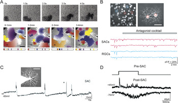
Despite the abundance of pre- and postsynaptic components for cholinergic signaling, there is little ultrastructural evidence of conventional synapses at this early stage of development. Conventional synapses between amacrine and ganglion cells begin to form after P3 in mice (Fisher, Reference Fisher1979) and ChAT expression precedes synapse formation by at least 2 days in chick retina (Spira et al., Reference Spira, Millar, Ishimoto, Epstein, Johnson, Dahl and Morgan1987). However, despite the lack of structural evidence for synapses, there is physiological evidence for cholinergic synaptic transmission between pairs of SACs at this early age. Paired recordings between SACs from embryonic rabbit showed that SACs can indeed release both ACh and GABA to excite neighboring SACs in a reciprocal fashion (Zheng et al., Reference Zheng, Lee and Zhou2004) (Fig. 2D). Postsynaptic nAChR-mediated currents are slow, appearing to reflect an asynchronous and/or diffuse release of ACh from the presynaptic SAC. Paired recordings of SACs in early postnatal mice reveal similar slow currents (Ford et al., Reference Ford, Felix and Feller2010).
Properties of cholinergic waves are generated by the SAC network. (A) Spatial and temporal properties of waves assessed with calcium imaging. Top: Low power calcium imaging of Fura-2AM-labeled neonatal ferret retinas show decreases in fluorescence following calcium increases associated with waves. Bottom: Spatial propagation of 30 waves during four consecutive minutes. Each panel shows the propagation of waves that occurred during a 1-min period. Cholinergic retinal waves initiate at random locations and propagate over finite regions. Waves occur at intervals between waves of around 1 min for a given region (e.g., see asterisk). Adapted from Feller et al. (Reference Feller, Wellis, Stellwagen, Werblin and Shatz1996). (B) Cell-autonomous spontaneous depolarization of SACs may initiate retinal waves. SACs (red regions and traces) from perinatal rabbit retinas loaded with Fura-2AM show spontaneous depolarizations in the presence of antagonists to ionotropic and metabotropic glutamate, GABA, glycine, and ACh receptors, whereas retinal ganglion cells do not (blue regions and traces). Adapted from Zheng et al. (Reference Zheng, Lee and Zhou2006). (C) The interwave refractory period may be due to a sAHP in SACs. Current-clamp recording from a SAC in rabbit retina showing wave evoked and spontaneous (asterisk) depolarizations followed by sAHPs. Adapted from Zheng et al. (Reference Zheng, Lee and Zhou2006). (D) Waves may propagate via reciprocal connections between SACs. Voltage-clamp recordings from pairs of neighboring SACs show both fast GABAergic postsynaptic currents and slow cholinergic postsynaptic currents. Top: Schematic of voltage command of presynaptic SAC (−70 to 0 mV). Bottom: Evoked PSCs at positive and negative holding potentials to isolate GABAergic and cholinergic currents, respectively. Adapted from Zheng et al. (Reference Zheng, Lee and Zhou2004).

SACs also form GABAergic synapses with other SACs and with subpopulations of ganglion cells. Unlike most GABAergic neurons in the retina, SACs express the GABA-synthesizing enzyme glutamate decarboxylase (GAD) 67 rather than the GAD-65 isoform (Brandon & Criswell, Reference Brandon and Criswell1995). GAD-67 expression is present early in development (Famiglietti & Sundquist, Reference Famiglietti and Sundquist2010). SACs can release GABA onto neighboring SACs during the first postnatal week in rabbit (Zheng et al., Reference Zheng, Lee and Zhou2004). Direction-selective ganglion cells also form GABAergic synapses with SACs well before eye opening (Lee et al., Reference Lee, Kim and Zhou2010; Wei et al., Reference Wei, Hamby, Zhou and Feller2011).
A distinguishing feature of the SAC network is that the somas of SACs form a mosaic—a term used to describe the regular arrangement of somas with a minimal spacing between them (reviewed in Cook & Chalupa, Reference Cook and Chalupa2000; Vaney, Reference Vaney1990; Novelli et al., Reference Novelli, Resta and Galli-Resta2005). SAC mosaics arise very early in development. SAC somas are initially positioned randomly with respect to each other as they migrate from the ventricular zone toward the developing ganglion cell layer. By birth in mice, SAC somas have distributed in a mosaic fashion so that there is a minimum distance observed between somas (Galli-Resta et al., Reference Galli-Resta, Resta, Tan and Reese1997, Reference Galli-Resta, Novelli, Volpini and Strettoi2000). The formation of the soma mosaic correlates with the complete coverage of the IPL by SAC processes.
Homotypic interactions between SAC processes are thought to instruct the initial formation of the mosaic (Reese & Galli-Resta, Reference Reese and Galli-Resta2002). The developmental window during which these interactions establish the mosaic is finite, as disruption of microtubules (Galli-Resta et al., Reference Galli-Resta, Novelli and Viegi2002) or ablation of subsets of SACs by excitotoxicity (Farajian et al., Reference Farajian, Raven, Cusato and Reese2004) a few days after birth fails to disrupt the established mosaic. At this point, the mechanisms by which interactions between SAC processes mediate the development of mosaics are unknown.
The role of SACs in generation of retinal waves
The most well-studied function of the early cholinergic network in the retina is the generation of retinal waves (Fig. 2). Retinal waves consist of bursts of action potentials in retinal ganglion cells that propagate in a wave front across the plane of the retina. Retinal waves occur in many species including turtle (Sernagor et al., Reference Sernagor, Young and Eglen2003), chick (Catsicas et al., Reference Catsicas, Bonness, Becker and Mobbs1998; Wong et al., Reference Wong, Sanes and Wong1998; Sernagor et al., Reference Sernagor, Eglen and O’Donovan2000), mice (Mooney et al., Reference Mooney, Penn, Gallego and Shatz1996; Bansal et al., Reference Bansal, Singer, Hwang, Xu, Beaudet and Feller2000), rabbit (Zhou, Reference Zhou1998), ferret (Meister et al., Reference Meister, Wong, Baylor and Shatz1991; Wong et al., Reference Wong, Meister and Shatz1993; Feller et al., Reference Feller, Wellis, Stellwagen, Werblin and Shatz1996), and primate (Warland et al., Reference Warland, Huberman and Chalupa2006). Across all species, waves share similar characteristic features. Waves initiate at random positions and propagate at speeds ranging from ~100 μm/s (mouse, Singer et al., Reference Singer, Mirotznik and Feller2001) up to 400 μm/s (chick, Sernagor et al., Reference Sernagor, Eglen and O’Donovan2000). Individual waves stop at discrete but shifting borders that reflect recently active regions of retina (Feller et al., Reference Feller, Wellis, Stellwagen, Werblin and Shatz1996). This refractory period following a wave lasts for approximately 1 min (Feller et al., Reference Feller, Wellis, Stellwagen, Werblin and Shatz1996, Reference Feller, Butts, Aaron, Rokhsar and Shatz1997), shortly after which an additional wave can be initiated and spread over the same area (Fig. 2A). In addition, this refractory period dictates the frequency with which a local region of the retina experiences a wave.
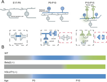
The circuitry underlying the generation of retinal waves progresses through three stages that reflect the maturation of retinal circuits (Blankenship & Feller, Reference Blankenship and Feller2010) (Fig. 3A). Before formation of the IPL [E17 in mice (Kim et al., Reference Kim, Lee, Kim, Park and Chun2000), E25 in rabbit (Sharma & Ehinger, Reference Sharma and Ehinger1997)], retinal waves propagate through the developing ganglion cell layer. These early-stage waves are blocked by gap junction antagonists (Syed et al., Reference Syed, Lee, Zheng and Zhou2004b). With the initial extension of SAC processes to form the IPL, waves transition from propagation via gap junctions to propagation via activation of nAChRs (Feller et al., Reference Feller, Wellis, Stellwagen, Werblin and Shatz1996). Finally, a third stage of waves develop several days before the eyes open and coexist with light responses for a short period of time (Demas et al., Reference Demas, Eglen and Wong2003). These waves are blocked by ionotropic glutamate receptors (Wong et al., Reference Wong, Myhr, Miller and Wong2000; Zhou & Zhao, Reference Zhou and Zhao2000; Blankenship et al., Reference Blankenship, Ford, Johnson, Seal, Edwards, Copenhagen and Feller2009).
Wave circuits transition through checkpoints. (A) Schematics of changing circuits that mediate waves. Left: Prior to birth waves are thought to propagate via gap junctions between ganglion cells. Middle: Postnatal day 0–10, waves are propagated via SAC release of ACh onto other SACs (black box). ACh also depolarizes ganglion cells. During this period of development, the gap junction signaling between ganglion cells is reduced (red box). Right: P10–P15 bipolar cells release glutamate to propagate waves in a mechanism that is thought to involve spillover of glutamate to excite neighboring bipolar cells (black box). Cholinergic signaling between SACs is reduced (red box). Adapted from Blankenship and Feller (Reference Blankenship and Feller2010). (B) Summary timeline of how genetic disruption of cholinergic or glutamatergic waves result in an extended action of the previous wave generating circuit. In wild-type mice, gap junction-mediated waves (gray) are followed by cholinergic waves (blue) starting at P0, then glutamatergic waves (green) at P10. In mice lacking the beta2 subunit of the nicotinic ACh receptor, gap junction-mediated waves persist until ~P8. In mice lacking vesicular glutamate transporter VGLUT1, cholinergic waves persist through the second postnatal week.

The cholinergic network plays an exclusive role in generating waves during an extended period of development. Neighboring ganglion cells and SACs receive simultaneous slow nAChR- and GABA-A-mediated currents that give rise to correlated action potential firing (Feller et al., Reference Feller, Wellis, Stellwagen, Werblin and Shatz1996; Zhou, Reference Zhou1998). In perinatal mice, ferrets, and rabbits, waves monitored by calcium imaging are blocked by nAChR antagonists (Feller et al., Reference Feller, Wellis, Stellwagen, Werblin and Shatz1996; Penn et al., Reference Penn, Riquelme, Feller and Shatz1998; Bansal et al., Reference Bansal, Singer, Hwang, Xu, Beaudet and Feller2000; Zhou & Zhao, Reference Zhou and Zhao2000) but not by ionotropic or metabotropic GABA or glutamate receptor antagonists (Zhou & Zhao, Reference Zhou and Zhao2000). Gap junction signaling seems to play a role in the generation of these waves in chick (Catsicas et al., Reference Catsicas, Bonness, Becker and Mobbs1998) but is involved to a lesser extent in mammalian retina (Singer et al., Reference Singer, Mirotznik and Feller2001; Syed et al., Reference Syed, Lee, Zheng and Zhou2004b).
The features of the developing cholinergic network that give rise to waves are now beginning to be understood. The generation of waves requires a source of depolarization for wave initiation, a network of excitatory interactions for propagation, and a source of inhibition that limits the spatial extent of waves and dictates the minimum interval between waves. We discuss the properties of the cholinergic network that give rise to these features below.
Initiation
SACs themselves are the likely source of wave initiation. In order for a wave to start, a cell or group of cells must spontaneously excite neighboring cells to initiate the spread of depolarization. Waves initiate at random locations (Feller et al., Reference Feller, Wellis, Stellwagen, Werblin and Shatz1996), suggesting that the cells responsible for the spontaneous excitation that drives wave initiation are present uniformly across the retina. In the presence of neurotransmitter antagonists, SACs from rabbit retinas exhibit spontaneous depolarizations that could be measured with cell attached, whole-cell recordings, and calcium imaging (Zheng et al., Reference Zheng, Lee and Zhou2006) (Fig. 2B). Spontaneous depolarizations occur fairly regularly with a frequency of about once per 20 s in cell attached and current-clamp recordings, though spontaneous calcium transients were less regular. These spontaneous depolarizations presumably lead to release of ACh onto neighboring SACs, which may trigger the start of a wave, though this has not yet been demonstrated directly.
The ion channels that underlie spontaneous depolarizations in SACs are unknown. Activation of TTX-sensitive sodium channels, which are present in developing SACs (Zhou & Fain, Reference Zhou and Fain1996), are unlikely to be the cause of spontaneous depolarization, as TTX does not block retinal waves (Stellwagen et al., Reference Stellwagen, Shatz and Feller1999). Spontaneous opening of calcium channels may provide a depolarizing drive. Adult SACs have N-, P/Q-, and R-type calcium channels (Cohen, Reference Cohen2001; Kaneda et al., Reference Kaneda, Ito, Morishima, Shigematsu and Shimoda2007), but these channels are activated at depolarized potentials. Some L-type voltage-gated calcium channels are activated at hyperpolarized potentials (Koschak et al., Reference Koschak, Reimer, Huber, Grabner, Glossmann, Engel and Striessnig2001) and have been implicated in pacemaking neurons (Putzier et al., Reference Putzier, Kullmann, Horn and Levitan2009). Retinal waves are blocked by the L-type channel antagonist nifedipine (Singer et al., Reference Singer, Mirotznik and Feller2001), suggesting that these channels may play a role in either the spontaneous depolarization or propagation of waves.
Propagation
Waves propagate through a cholinergic circuit that depends on activation of a specific subtype of nAChRs. Studies of adult rabbit retina have demonstrated strong expression of alpha7 homomeric channels (Dmitrieva et al., Reference Dmitrieva, Strang and Keyser2007) as well as alpha3-/beta2-containing heteromeric channels (Keyser et al., Reference Keyser, MacNeil, Dmitrieva, Wang, Masland and Lindstrom2000) throughout the IPL. Expression of alpha3- and alpha8-containing receptors during the period of cholinergic waves has been shown in chick retina (Hamassaki-Britto et al., Reference Hamassaki-Britto, Gardino, Hokoc, Keyser, Karten, Lindstrom and Britto1994). Waves are blocked by α-conotoxin MII, a toxin that is most specific for alpha3-/beta2-containing nAChRs but not α-bungarotoxin (targeted to homomeric alpha7 nAChRs) or α-conotoxin-AU1B (targeted to alpha3-/beta4-containing receptors) (Penn et al., Reference Penn, Riquelme, Feller and Shatz1998; Bansal et al., Reference Bansal, Singer, Hwang, Xu, Beaudet and Feller2000). Further evidence for a unique set of receptors mediating waves came from studies using knockout mice—mice lacking either alpha3 or beta2 subunits lacked cholinergic retinal waves (Bansal et al., Reference Bansal, Singer, Hwang, Xu, Beaudet and Feller2000; Sun et al., Reference Sun, Warland, Ballesteros, van der List and Chalupa2008; Stafford et al., Reference Stafford, Sher, Litke and Feldheim2009). Hence, wave propagation depends upon activation of specific heteromeric alpha3/beta2 nAChRs even though other nAChRs exist in the retina.
In addition to ACh, SACs and ganglion cells receive GABAergic input through GABA-A receptors during waves (Feller et al., Reference Feller, Wellis, Stellwagen, Werblin and Shatz1996; Zhou, Reference Zhou1998). At this developmental age, GABA-A-mediated currents are depolarizing due to the accumulation of intracellular chloride (Zhang et al., Reference Zhang, Pathak, Coulter, Freed and Vardi2006b; Barkis et al., Reference Barkis, Ford and Feller2010). GABA-A receptor activation is not required for the propagation of waves in rabbit or mice (Syed et al., Reference Syed, Lee, Zheng and Zhou2004b; Wang et al., Reference Wang, Blankenship, Anishchenko, Elstrott, Fikhman, Nakanishi and Feller2007) but does influence the structure of the firing patterns (Wang et al., Reference Wang, Blankenship, Anishchenko, Elstrott, Fikhman, Nakanishi and Feller2007). Not all studies have reached this conclusion—early waves in both turtle (Sernagor et al., Reference Sernagor, Young and Eglen2003) and ferret (Fischer et al., Reference Fischer, Lukasiewicz and Wong1998; but see Stellwagen et al., Reference Stellwagen, Shatz and Feller1999) depend on GABA-A receptor activation.
A possible explanation for the widespread effects of ACh during waves is that ACh is released by volume transmission—the diffuse release of neurotransmitter in the absence of pre- and postsynaptic specializations (for a comparison of direct vs. volume ACh transmission see Sarter et al., Reference Sarter, Parikh and Howe2009). Several lines of evidence support this. First, retinal ganglion cells with dendritic arbors at opposite ends of the IPL are synchronized during waves, despite the fine stratification of SAC processes (Wong & Oakley, Reference Wong and Oakley1996). Second, slow synaptic currents recorded between pairs of SACs (Zheng et al., Reference Zheng, Lee and Zhou2004) (Fig. 2D) indicate that ACh acts diffusely. Third, cholinergic retinal waves are present prior to the formation of conventional synapses, as identified in electron microscopy studies (Fisher, Reference Fisher1979; Greiner & Weidman, Reference Greiner and Weidman1981). Finally, in rabbit retina, calcium waves propagate through the ventricular zone, which are correlated with waves in the ganglion cells (Syed et al., Reference Syed, Lee, He and Zhou2004a). The ventricular zone waves are blocked by muscarinic receptor antagonists as well as nAChR antagonists, implying that ACh released during waves in the ganglion cell layer diffuses tens of microns to the ventricular zone where it activates muscarinic receptors.
Refractory period
The frequency of retinal waves is limited by a refractory period following waves. Over a period of about 1 min, waves initiated at random points appear to tile the retina, avoiding regions active recently (Feller et al., Reference Feller, Wellis, Stellwagen, Werblin and Shatz1996) (Fig. 2A). Depolarization by focal application of high potassium solution could also cause a refractory period within the affected region. This refractory period is thought to be imparted by a slow after-hyperpolarization (sAHP) following depolarization in SACs (Zheng et al., Reference Zheng, Lee and Zhou2006) (Fig. 2C). The sAHP is blocked by cadmium, suggesting that it is activated by calcium entry through voltage-gated calcium channels. Interestingly, the channel underlying the sAHP is inhibited by elevating levels of cAMP, similar to the calcium-activated potassium channel which is thought to underlie sAHPs in various other regions of the brain (Alger & Nicoll, Reference Alger and Nicoll1980; Lancaster & Adams, Reference Lancaster and Adams1986; Sah & Isaacson, Reference Sah and Isaacson1995; Vogalis et al., Reference Vogalis, Furness and Kunze2001), the molecular identity of which is unknown. Elevating cAMP increases the frequency of retinal waves (Stellwagen et al., Reference Stellwagen, Shatz and Feller1999; Hanganu et al., Reference Hanganu, Ben-Ari and Khazipov2006), consistent with the notion that the sAHP limits the frequency of waves.
Recent studies have incorporated the measured features of the cholinergic network into computational models. Modeling studies are of use in identifying the relevant features of a circuit that give rise to the physiological output. For instance, two modeling studies have shown that in order to achieve propagating waves that have finite borders, that is, do not encompass the entire retina, there must be local variability in the refractory state of the retina (Godfrey & Swindale, Reference Godfrey and Swindale2007; Hennig et al., Reference Hennig, Adams, Willshaw and Sernagor2009). This variability is generated by random spontaneous depolarizations of SACs followed by sAHPs that occur in between waves. While spontaneous depolarization of SACs between waves has been observed in rabbit (asterisk Fig. 2C) (Zheng et al., Reference Zheng, Lee and Zhou2006), we have not observed these in mouse (Ford et al., Reference Ford, Felix and Feller2010). As the details of the SAC network are worked out for different species, there will likely be different, and perhaps disparate, solutions that generate very similar waves.
Role of cholinergic signaling in establishing noncholinergic retinal circuits
Influence on later progenitor cells:
Cholinergic retinal waves occur during a period of rapid retinal development. Progenitor cells in the ventricular zone are dividing and differentiating into each of the seven types of retinal cells (Fig. 1). These newly differentiated cells must then migrate to the proper position within the retina and extend processes to form the neuropil layers. The onset of cholinergic waves correlates with the formation of the IPL, where amacrine and ganglion cells processes stratify into 10 discrete layers (in mice) that process visual information in parallel (Masland, Reference Masland2001; Roska & Werblin, Reference Roska and Werblin2001; Wassle, Reference Wassle2004). Once stratified in these layers, bipolar, amacrine, and ganglion cells must form synapses with their proper partners to form functional circuits that process visual information. Before the onset of vision, these discrete synaptic layers are visible (Kim et al., Reference Kim, Lee, Kim, Park and Chun2000; Drenhaus et al., Reference Drenhaus, Morino and Veh2003; Famiglietti & Sundquist, Reference Famiglietti and Sundquist2010) and some functional circuits are already established (Elstrott et al., Reference Elstrott, Anishchenko, Greschner, Sher, Litke, Chichilnisky and Feller2008; Wei et al., Reference Wei, Hamby, Zhou and Feller2011). To what extent does the cholinergic network shape the outcome of these developmental events?
Rhythmic increases in intracellular calcium play an important role in cell proliferation and differentiation (reviewed in Martins & Pearson, Reference Martins and Pearson2008) for retinal neurons born during cholinergic waves. ACh drives increases in intracellular calcium of progenitor cells in the ventricular zone through activation of M1 muscarinic receptors (Wong, Reference Wong1995), which can alter their progression in the cell cycle (Pearson et al., Reference Pearson, Catsicas, Becker and Mobbs2002). Blocking muscarinic receptors in chick eyes leads to an increase in the size of the eye (Pearson et al., Reference Pearson, Catsicas, Becker and Mobbs2002). A probable source for ACh acting on muscarinic receptors is from cholinergic waves, which drive correlated calcium waves in the ventricular zone (Syed et al., Reference Syed, Lee, He and Zhou2004a). In other parts of the nervous system, elevation of intracellular calcium can also influence neuronal migration (reviewed in Komuro & Kumada, Reference Komuro and Kumada2005) and neurotransmitter selection (reviewed in Spitzer et al., Reference Spitzer, Root and Borodinsky2004). It remains to be seen whether calcium increases caused by retinal waves have similar effects.
IPL stratification
The retina is a highly laminar structure. Cell bodies reside in three vertically oriented layers composed of the distinct cell classes. The IPL comprises axons from bipolar cells, processes from amacrine cells, and dendrites of ganglion cells. These processes are separated into the ON and OFF layers corresponding to the functional responses to light of the corresponding cells. Within ON and OFF layers, processes are further segregated into at least 10 different strata where distinct neural computations are performed. The structural layering and corresponding functional separation has made the retina an ideal model system for studying synapse specificity—that is, the process by which pre- and postsynaptic neurons wire to their appropriate partners.
The development of lamina in the IPL occurs in several steps. First, amacrine cells extend processes into the IPL. The processes of different amacrine cells identified by cell-specific antibody staining form discrete layers as soon as they first appear (Famiglietti & Sundquist, Reference Famiglietti and Sundquist2010). Second, the dendrites of ganglion cells extend into the IPL. While some ganglion cells initially stratify within their appropriate mature lamina, a large portion of ganglion cells stratifies diffusely throughout future ON and OFF layers. Next, ON and OFF bipolar cell axons extend into the IPL where they stratify into the appropriate layers (Johnson et al., Reference Johnson, Tian, Caywood, Reimer, Edwards and Copenhagen2003). Finally, ganglion cells restrict their dendrites to their mature lamina (reviewed in Xu & Tian, Reference Xu and Tian2004). Below we discuss how SACs could play a role in these developmental steps.
A growing body of literature from zebrafish studies points to a critical role for amacrine cells in patterning the IPL. The processes of amacrine cells have directed growth within their target layers without exuberant growth in other layers (Godinho et al., Reference Godinho, Mumm, Williams, Schroeter, Koerber, Park, Leach and Wong2005). While ganglion cells are the first-born cells in the retina, they play a lesser role in formation of layers in the IPL. A genetic model in which ganglion cells are never born have delayed IPL formation, although this is partially corrected and normal lamination of the IPL occurs throughout most of the retina (Kay et al., Reference Kay, Roeser, Mumm, Godinho, Mrejeru, Wong and Baier2004). Hence, interactions between amacrine cells are sufficient to give rise to the layering within the IPL. Interestingly, bipolar cell axons target correctly to the normal IPL in these mutants but fail to innervate regions of abnormal IPL (Kay et al., Reference Kay, Roeser, Mumm, Godinho, Mrejeru, Wong and Baier2004), suggesting that amacrine cells provide the laminar cues for bipolar cell axon targeting. Finally, ganglion cells exhibit directed outgrowth of their processes within established layers of amacrine cell processes (Mumm et al., Reference Mumm, Williams, Godinho, Koerber, Pittman, Roeser, Chien, Baier and Wong2006), implying that amacrine cells can provide lamination cues for ganglion cells. A zebrafish homolog of the SAC has not been conclusively identified (though see Kay et al., Reference Kay, Roeser, Mumm, Godinho, Mrejeru, Wong and Baier2004) so it is unclear what role SACs in particular play in these developmental events.
In mammals, SACs are among the first cells to extend processes within the IPL (Kim et al., Reference Kim, Lee, Kim, Park and Chun2000) and could potentially serve as a foundation for the layering of subsequent strata within the IPL. Lamination signals, such as members of the immunogblobulin superfamily (Yamagata & Sanes, Reference Yamagata and Sanes2008) or semaphorins (Matsuoka et al., Reference Matsuoka, Nguyen-Ba-Charvet, Parray, Badea, Chédotal and Kolodkin2011), expressed on SAC processes could instruct the processes of other amacrine, ganglion, and bipolar cells to stratify above, below, or between the two cholinergic strata. Despite this early genesis, it is unlikely that SACs provide lamination cues for other amacrine and bipolar cells. Retinal deletion of the transcription factor islet-1 results in a near complete loss of ChAT-positive amacrine cells (Elshatory et al., Reference Elshatory, Everhart, Deng, Xie, Barlow and Gan2007). Despite this loss of differentiated SACs, several markers for other amacrine cell types still form layers within the IPL. Similarly, a genetic model of retinal blastoma lacks several markers of mature SACs, but stratification of other amacrine and bipolar cell types are not altered (Chen et al., Reference Chen, Opavsky, Pacal, Tanimoto, Wenzel, Seeliger, Leone and Bremner2007). Furthermore, SACs ablated by immunotoxin (Gunhan et al., Reference Gunhan, Choudary, Landerholm and Chalupa2002) or by excitotoxicity with l-glutamate (Reese et al., Reference Reese, Raven, Giannotti and Johnson2001) at 2–3 days after birth show no defects in the stratification of bipolar cells, suggesting that SAC processes do not provide an instructive role for stratification of these cell types.
While SACs are unlikely to guide the lamination of other amacrine and bipolar cells, they may instruct the localization of some types of ganglion cell dendrites. Using random dye labeling, a certain class of bistratified ganglion cell, mostly likely corresponding to direction-selective ganglion cells, was found to costratify with the cholinergic processes shortly after birth (Stacy & Wong, Reference Stacy and Wong2003), although other monostratified ganglion cells never stratified with the cholinergic bands. Recently, genetically labeled mouse lines of functionally identical ganglion cell classes have been developed that allow for consistent characterization across development (Huberman et al., Reference Huberman, Manu, Koch, Susman, Lutz, Ullian, Baccus and Barres2008b, Reference Huberman, Wei, Elstrott, Stafford, Feller and Barres2009; Kim et al., Reference Kim, Zhang, Yamagata, Meister and Sanes2008). The dendrites of genetically identified direction-selective ganglion cells costratify with SAC processes shortly after birth (Kim et al., Reference Kim, Zhang, Meister and Sanes2010; Wei et al., Reference Wei, Hamby, Zhou and Feller2011), supporting the hypothesis that SACs provide an instructive role for lamination of certain ganglion cell types.
The dendrites of most retinal ganglion cells initially arborize diffusely within the IPL before restricting their dendrites to distinct lamina (Bodnarenko et al., Reference Bodnarenko, Yeung, Thomas and McCarthy1999; Bansal et al., Reference Bansal, Singer, Hwang, Xu, Beaudet and Feller2000; Sernagor et al., Reference Sernagor, Eglen and Wong2001; Xu & Tian, Reference Xu and Tian2004; Coombs et al., Reference Coombs, Van Der List and Chalupa2007; Kim et al., Reference Kim, Zhang, Meister and Sanes2010). Some studies have implicated a role for activity in bipolar cells in the segregation of ganglion cell dendrites. Hyperpolarizing ON bipolar cells during the period of glutamatergic retinal waves using the drug 2-amino-4-phosphonobutyrate prevents the segregation into ON and OFF layers (Bodnarenko & Chalupa, Reference Bodnarenko and Chalupa1993; Bodnarenko et al., Reference Bodnarenko, Jeyarasasingam and Chalupa1995). Additionally, mice that lack the Major Histocompatibility Complex I receptor CD3zeta have altered glutamatergic retinal waves. The ganglion cells of these mice have reduced dendritic motility and have more diffuse dendrites within the IPL (Xu et al., Reference Xu, Chen, Ding, Xie, Chen, Diao, Wang, Gan, Crair and Tian2010). However, not all manipulations of activity during development alter dendritic stratification. Preventing synaptic release of glutamate from ON bipolar cells by expression of tetanus toxin fails to prevent the stratification of ganglion cell dendrites, although synapse formation onto ON bipolar cells is reduced (Kerschensteiner et al., Reference Kerschensteiner, Morgan, Parker, Lewis and Wong2009).
Is there a role for cholinergic waves in the stratification process? Blocking nAChRs during the period of cholinergic waves reduces the motility of filipodia on the dendrites of ganglion cells (Wong & Wong, Reference Wong and Wong2001), demonstrating that waves can drive structural changes in dendrites. Studies in turtle have demonstrated that blocking cholinergic waves with nAChR antagonists inhibits dendritic growth (Mehta & Sernagor, Reference Mehta and Sernagor2006) and reduces receptive field sizes (Sernagor & Grzywacz, Reference Sernagor and Grzywacz1996). Additionally, mice lacking the beta2 subunit of the nAChR had a delay in, though did not prevent, the fine stratification of ganglion cell dendrites (Bansal et al., Reference Bansal, Singer, Hwang, Xu, Beaudet and Feller2000). These findings indicate that cholinergic waves do influence the outgrowth of ganglion cell dendrites but are not the primary factor dictating their final organization.
One intriguing hypothesis for a role of retinal waves is in the development of direction-selective circuits. Retinal waves have directional information both in their propagation and in the observation that there is a propagation bias, with more waves traveling toward the nasal direction than the other cardinal axes (Stafford et al., Reference Stafford, Sher, Litke and Feldheim2009; Elstrott & Feller, Reference Elstrott and Feller2010). However, direction-selective ganglion cells participate equally in all waves irrespective of propagation direction (Elstrott & Feller, Reference Elstrott and Feller2010), and complete blockade of retinal activity using intraocular muscimol injections did not prevent the development of direction selectivity (Wei et al., Reference Wei, Hamby, Zhou and Feller2011). These findings indicate that waves are not likely to provide the instructive signal for the establishment of asymmetric direction-selective circuits. However, whether retinal waves play a role in the refinement of direction selective circuits remains to be determined.
Is there a role for cholinergic waves in the transitions between wave generating circuits?
Another role of cholinergic waves in development of the retina circuitry may be to terminate early-stage gap junction-mediated waves (Fig. 3). Knockout animals that lack nAChR receptor subunits still exhibit wave-like activity under certain recording conditions, such as elevated temperature [alpha3(−/−) (Bansal et al., Reference Bansal, Singer, Hwang, Xu, Beaudet and Feller2000), beta2(−/−) (Sun et al., Reference Sun, Warland, Ballesteros, van der List and Chalupa2008; Stafford et al., Reference Stafford, Sher, Litke and Feldheim2009)]. This activity is likely mediated by gap junctions (Sun et al., Reference Sun, Warland, Ballesteros, van der List and Chalupa2008), as in earlier stage waves. Similarly, a study using a genetic model that eliminates ChAT in a large portion of the retina found normal cholinergic waves in the spared region but compensatory waves in the region lacking ChAT (Stacy et al., Reference Stacy, Demas, Burgess, Sanes and Wong2005). These studies point to a sequential maturation of the retinal circuitry that relies on checkpoints to make transitions from one stage (gap junction-mediated waves) to the next (cholinergic transmission-mediated waves). This sort of checkpoint model of neuronal development (Ben-Ari & Spitzer, Reference Ben-Ari and Spitzer2010) is further evidenced in the disassembly of the cholinergic network to make way for glutamatergic signaling (Blankenship & Feller, Reference Blankenship and Feller2010).
Disassembly of the cholinergic network
Shortly before eye opening, retinal waves transition from being mediated by ACh to glutamate. Bipolar cell axons invade the IPL and begin to release glutamate at the end of the first week after birth in rodents (Johnson et al., Reference Johnson, Tian, Caywood, Reimer, Edwards and Copenhagen2003). Shortly thereafter, waves change their pharmacological profile and spatiotemporal properties. Waves are no longer blocked by nAChR antagonists but rather are sensitive to a combination of AMPA and NMDA antagonists (Wong et al., Reference Wong, Sanes and Wong1998; Zhou & Zhao, Reference Zhou and Zhao2000; Blankenship et al., Reference Blankenship, Ford, Johnson, Seal, Edwards, Copenhagen and Feller2009). These glutamatergic waves occur as periodic clusters of activity followed by periods of silence (Kerschensteiner & Wong, Reference Kerschensteiner and Wong2008; Blankenship et al., Reference Blankenship, Ford, Johnson, Seal, Edwards, Copenhagen and Feller2009). Waves exist simultaneously with light responses and disappear a few days after eye opening in a manner that is independent of visual experience (Demas et al., Reference Demas, Eglen and Wong2003).
With the onset of glutamatergic waves, the SAC network loses the features necessary for propagation of cholinergic waves. First, mature SACs are no longer depolarized by application of nicotine (Baldridge, Reference Baldridge1996). This is the result of a rapid decline in nAChR activation that coincides with the start of glutamatergic waves (Zheng et al., Reference Zheng, Lee and Zhou2004). Second, acetylcholinesterase, the enzyme that degrades ACh, is increased during this time window (Hutchins et al., Reference Hutchins, Bernanke and Jefferson1995), which would decrease the spread of ACh. Third, the action of GABA becomes hyperpolarizing a few days before the onset of glutamatergic waves (Zhang et al., Reference Zhang, Pathak, Coulter, Freed and Vardi2006b; Barkis et al., Reference Barkis, Ford and Feller2010). Intracellular chloride is reduced as the expression of the potassium–chloride transporter KCC2 is increased in ganglion cells (Zhang et al., Reference Zhang, Fina and Vardi2006a). Mature SAC processes have a graded expression of KCC2 that increases along the length of the process (Gavrikov et al., Reference Gavrikov, Nilson, Dmitriev, Zucker and Mangel2006). When this graded distribution of KCC2 occurs is unknown. Fourth, SACs become less excitable because maturation of processes increases the electronic space constant. The long and skinny processes of SACs likely prevent the spread of depolarization between processes, which would serve to electrically isolate the soma from the processes. A further decrease in the excitability of SACs is imparted by the loss of voltage-gated sodium channels (Zhou & Fain, Reference Zhou and Fain1996) and an increase in expression of the voltage-gated potassium channel Kv3.1 (Ozaita et al., Reference Ozaita, Petit-Jacques, Völgyi, Ho, Joho, Bloomfield and Rudy2004; Zheng et al., Reference Zheng, Lee and Zhou2006).
At this same time, other circuits involving SACs are rapidly maturing. GABAergic connections between SACs are maintained during this period (Zheng et al., Reference Zheng, Lee and Zhou2004) and cholinergic and GABAergic synapses onto direction-selective ganglion cells develop (Lee et al., Reference Lee, Kim and Zhou2010; Wei et al., Reference Wei, Hamby, Zhou and Feller2011). SACs greatly extend their processes as much as sevenfold and develop varicosities at the distal portions that are presumed to be the sites of GABA release (Wong & Collin, Reference Wong and Collin1989).
The nature of cholinergic transmission in the adult retina is not well understood. Many ganglion cells with dendrites distal to the cholinergic strata within the IPL still respond to ACh released from SACs (Schmidt et al., Reference Schmidt, Humphrey and Wässle1987). Paired recordings between SACs and direction-selective ganglion cells reveal symmetrical sites of ACh release (Lee et al., Reference Lee, Kim and Zhou2010); however, ultrastructural reconstruction of synaptic contacts onto direction-selective ganglion cells failed to find evidence for these cholinergic synapses (Briggman et al., Reference Briggman, Helmstaedter and Denk2011). Furthermore, expression of SNAP-25, a SNARE protein involved in evoked release of neurotransmitter, is transiently highly expressed in SAC processes during the period of cholinergic waves (West Greenlee et al., Reference West Greenlee, Finley, Wilson, Jacobson and Sakaguchi1998), suggesting that the presynaptic machinery for neurotransmitter release might change during development.
The onset of glutamatergic waves may play an active role in the disassembly of cholinergic circuits, similar to the transition from gap junction to cholinergic waves (Blankenship & Feller, Reference Blankenship and Feller2010) (Fig. 3). Mice lacking the vesicular glutamate transporter VGLUT1 lack glutamate release from bipolar cells. These mice still have retinal waves at the period of time when littermate control animals have glutamatergic waves. Interestingly, these later waves in the VGLUT1 knockout mice are unaffected by glutamate receptor antagonists but are blocked by nAChR antagonists (Blankenship et al., Reference Blankenship, Ford, Johnson, Seal, Edwards, Copenhagen and Feller2009). Glutamate signaling from bipolar cells is therefore required to dismantle the cholinergic network.
The mechanisms underlying this transition are not understood. SACs have functional kainate, AMPA, and NMDA receptors shortly after birth (Acosta et al., Reference Acosta, Chua and Kalloniatis2007), suggesting that they can respond to glutamate when it is first released by bipolar cells. In addition to ionotropic receptors, SACs also express the metabotropic glutamate receptor 2 starting shortly after birth in rodents (Koulen et al., Reference Koulen, Malitschek, Kuhn, Wässle and Brandstätter1996). Action of glutamate on either ionotropic or metabotropic receptors on SACs could result in downregulation of functional nAChRs and perhaps the regulation of other proteins that are necessary for cholinergic waves. The mechanisms involved in this transition will likely be of general importance, as several other developing neural circuits such as the spinal cord, hind brain, cochlea, and hippocampus transition through different stages of spontaneous activity before reaching their mature state (Blankenship & Feller, Reference Blankenship and Feller2010).
Conclusions
Here, we have reviewed the assembly, disassembly, and function of cholinergic circuits in retinal development. SACs release ACh and nicotinic and muscarinic ACh receptors appear early in development to provide signaling that influences events prior to synapse formation. The processes of SACs define the early strata of the developing IPL and therefore may play a role in organizing processes of other cells. For a brief period of development, SACs can mutually excite each other. These recurrent excitatory connections provide the substrate for the initiation and propagation of retinal waves. Future work will continue to elucidate how other transient features of developing starburst cells and the nature of their synaptic connections influences the propagation properties of retinal waves.
One outstanding question that remains is how early signaling in the retina influences the formation of retinal circuits. The assembly of this cholinergic circuit is critical for driving the transition from an earlier, gap junction-mediated wave generating circuit. Similarly, maturation of the glutamatergic wave generating circuit drives the disassembly of the cholinergic network. Hence, each circuit is critical for eliminating the circuit before it. In addition, retinal waves drive correlated depolarizations of many synaptic partners (e.g., SACs and direction-selective ganglion cells). Whether these early signaling events are critical for the maturation of these functional circuits remains an open question. Answers to these questions require a deeper understanding of the interplay between activity and other cellular mechanisms that underlie the wiring up of functional circuits.