Introduction
Spontaneous cerebrospinal fluid (CSF) leakage into the middle ear and adjacent spaces is a disease process occurring in the absence of an obvious cause. It is unrelated to traumatic head injuries, tumours, skull base surgery or congenital abnormalities. There appears to be an increased incidence of spontaneous CSF leak being reported in the literature, both locally and internationally.Reference Nelson, Gantz and Hansen1,Reference Hendriks, Bala, Rodrigues and Kuthubutheen2 Spontaneous CSF otorrhoea puts patients at risk of life-threatening intracranial infections such as meningitis.Reference Hendriks, Bala, Rodrigues and Kuthubutheen2 Prompt and accurate diagnosis is essential but difficult given the non-specific presentation of a seemingly benign middle-ear effusion of unknown cause, often thought to be due to Eustachian tube dysfunction. A combination of thorough history and examination, in addition to radiological imaging and biochemical analyses, is generally required in order to make a definitive diagnosis.
The condition is commonly observed in middle-aged obese females; however, the pathophysiology is largely unknown.Reference Vemuri, Karanam, Manchikanti, Dandamudi, Puvvada and Vemuri3,Reference Rao and Redleaf4 Idiopathic intracranial hypertension is a frequently cited contributing factor for developing spontaneous CSF leak and has been explored in several papers.Reference Schlosser, Wilensky, Grady and Bolger5–Reference Allen, Perez, Kutz, Gerecci, Roland and Isaacson9 Skull base thickness and pneumatisation are potentially important anatomical parameters in patients with spontaneous CSF leak, given the increased frequency in those with thin tegmina and well-pneumatised temporal bones. These variations have been demonstrated to be associated with spontaneous CSF leak, and appear to be related to idiopathic intracranial hypertension and obesity.Reference Qahtani7,Reference Stevens, Rizk, McIlwain, Lambert and Meyer10–Reference Gacek, Gacek and Tart17
Imaging modalities vary according to institutional practices, but high-resolution computed tomography (CT) of the temporal bone is an important early investigation in the diagnostic pathway. Isolated opacification of the middle ear or mastoid cavities on temporal bone CT should immediately raise suspicion of a spontaneous CSF leak, and the tegmen must be meticulously inspected for defects. This prompts clinicians to sample middle-ear fluid and send for beta-trace protein to confirm the diagnosis.Reference Meco, Oberascher, Arrer, Moser and Albegger18 Alternatively, if the diagnosis is confirmed biochemically after clinical concern, a temporal bone CT should be performed to help to determine the location(s) and potential cause of the leak, which can guide surgical management.
Despite the proposed theories for the development of spontaneous CSF leak and associated diagnostic features (biochemical and radiological), few studies have specifically assessed the key radiological findings in these patients.Reference Stone, Castillo, Neelon and Mukherji19,Reference Lloyd, DelGaudio and Hudgins20 We sought to describe the temporal bone radiological findings in a cohort of patients with spontaneous CSF leak, to assist clinicians in diagnosing this condition with more accuracy and to further characterise this growing cohort.
Materials and methods
A retrospective cohort study was conducted across tertiary hospitals in Western Australia over a seven-year period (March 2013 to March 2020). Adult patients (aged over 18 years) with biochemically confirmed spontaneous CSF leak (beta-trace protein of more than 1.3 mg/l) who were managed by ENT surgeons were included. Spontaneous CSF leaks were defined as non-iatrogenic and non-traumatic CSF leaks of the temporal bone (with or without otorrhoea). Patients were excluded if a dedicated temporal bone CT scan had not been performed, or if magnetic resonance imaging (MRI) was the sole imaging study undertaken, given the inaccuracy in diagnosing osseous defects with this modality.
Demographic data including age, gender and body mass index (BMI) were recorded in addition to: symptomatology on presentation to the ENT surgeon, examination findings and beta-trace protein analysis of middle-ear fluid. The prevalence of obstructive sleep apnoea (OSA) and idiopathic intracranial hypertension in the cohort was documented because of the association between these conditions and spontaneous CSF leak. Patients diagnosed with OSA were required to undergo a formal polysomnogram and receive a diagnosis by a sleep or respiratory physician. A diagnosis of idiopathic intracranial hypertension was confirmed on the documented presence of raised opening CSF pressures above 20 cm H2O on lumbar puncture.
Specific radiological features identified on temporal bone CT and/or MRI were documented. Patient imaging was reviewed twice by different head and neck subspecialist neuroradiologists (radiology reviews one and two). Several parameters were assessed, including: skull base defect size and location, presence of fluid within the middle ear or mastoid (nil, partial, subtotal or total), presence of a cephalocele, degree of mastoid development (well, moderately or poorly developed), tegmen height (at, above, or below the arcuate eminence, or down-sloping) and presence of superior semicircular canal dehiscence. Skull base defect locations were divided into: tegmen mastoideum anterior, tegmen mastoideum posterior and tegmen tympani. Tegmen mastoideum anterior defects were defined as occurring anterior to the level of the mastoid antrum (including lateral, supra-meatal air cells), whilst tegmen mastoideum posterior defects were defined as occurring at or posterior to the level of the antrum. The MRI findings were limited to the presence or absence of cephaloceles, given the variety of MRI imaging protocols utilised in the cohort. These parameters were assessed bilaterally for each patient.
The simple student's t-test was used to compare continuous variables. Chi-square analysis was used to compare nominal or categorical variables. For all analyses, we considered an alpha value of 0.05 as statistically significant. The study was approved by the South Metropolitan Health Services Ethics Committee (approval number: RGS0000001008). A waiver of consent was granted for the study in line with the Australian Government National Health and Medical Research Council National Statement on Ethical Conduct in Human Research (2007).
Results
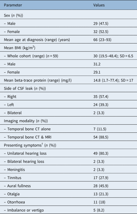
Sixty-one adult patients with biochemically confirmed spontaneous CSF leak were identified during the study period (Table 1) and formed the final cohort of the study. There was a slight female predominance (n = 32) and the mean BMI of the cohort was 30 kg/m2. Right-sided spontaneous CSF leak was most common (n = 35) and two patients presented with bilateral spontaneous CSF leak. Unilateral hearing loss (80 per cent) and aural fullness (46 per cent) were the most frequently presenting symptoms. Two patients (3 per cent) presented emergently to hospital with meningitis. Nine patients had polysomnographic evidence of OSA (15 per cent), whilst only one patient had idiopathic intracranial hypertension. The mean beta-trace protein level for the whole cohort was 14.8 mg/l.
Patient characteristics*

* Total n = 61. †Some patients presented with more than one symptom. BMI = body mass index; SD = standard deviation; CSF = cerebrospinal fluid; CT = computed tomography; MRI = magnetic resonance imaging
All patients underwent radiological investigation in the form of temporal bone CT and/or MRI. Fifty-four patients (88.5 per cent) underwent both imaging modalities, and the remaining underwent temporal bone CT imaging alone.
Despite only two patients presenting with bilateral spontaneous CSF leak, over 75 per cent of patients were identified by radiologists as having bilateral tegmen defects on imaging. There was some variation between radiologists regarding involvement of the non-affected side. Forty-eight patients (78.7 per cent) were identified as having bilateral tegmen defects on radiology review one, whilst 53 patients (86.9 per cent) were identified on radiology review two. Of those with bilateral defects after radiology review one (n = 48), 30 patients (62.5 per cent) had at least three defects on both the affected and the contralateral side. Similarly, after radiology review two, the majority of patients (62.3 per cent) had at least three defects identified bilaterally. Few patients had single-sided defects only (radiology review one n = 8, radiology review two n = 7); the largest of these defects identified was 6 mm, whereas the remainder were less than 3 mm.
There were two patients without an obvious defect noted radiologically (despite beta-trace protein positive middle-ear aspirate). The radiologists noted a transmastoid vein in one of these patients, which appeared to mimic a dehiscence laterally. The other patient had a partially opacified mastoid cavity, but no obvious osseous defect.
Assessment of the affected or presenting side of spontaneous CSF leakage demonstrated anterior tegmen mastoideum dehiscence as the most common defect, representing almost 50 per cent of all defects identified (in both radiology review one and radiology review two). Tegmen tympani dehiscence was the second most common defect. Only a marginal difference was noted between the number of anterior tegmen mastoideum dehiscences present on the contralateral side and the symptomatic side on both radiology reviews (radiology review one – 56 vs 65 defects, and radiology review two – 70 vs 80 defects).
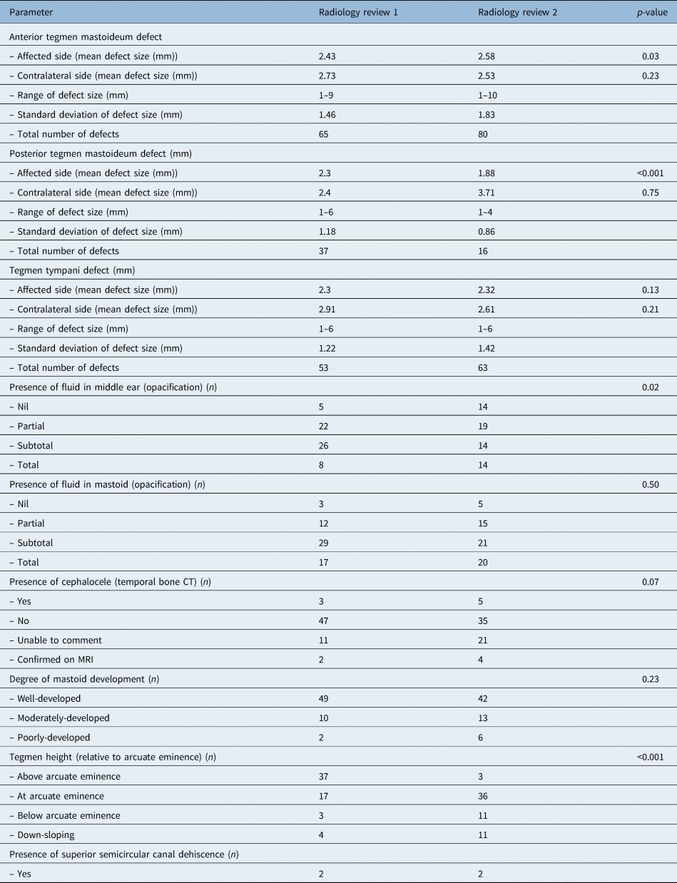
A degree of fluid was present in the middle ear and mastoid in the majority of the cohort. Cephaloceles were suspected on temporal bone CT in a few patients (radiology review one n = 3, radiology review two n = 5). On subsequent MRI, the diagnosis was confirmed in most of these cases (radiology review one n = 2, radiology review two n = 4). Two patients had radiological evidence of semicircular canal dehiscence noted on both radiology reviews. Notably, the imaging reports for index temporal bone CT (conducted prior to independent radiology reviews in our study) in 11 patients (18 per cent) failed to highlight skull base defects. Imaging characteristics for the cohort are presented in Table 2.
Imaging characteristics*

* For affected side unless stated otherwise. CT = computed tomography; MRI = magnetic resonance imaging
Statistical analysis identified several variations between the radiology reviews, in terms of: mean tegmen mastoideum anterior affected size, mean tegmen mastoideum posterior affected size, presence of fluid in the middle ear, and tegmen height. For defects on the affected side, variation between radiology reviews was lower with larger defects. For both radiology reviews, defects over 5 mm were identified in 85 per cent of cases and defects over 3 mm were identified in only 74 per cent of cases.
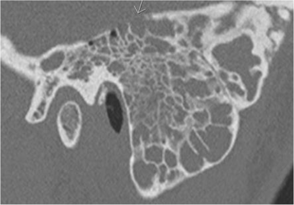
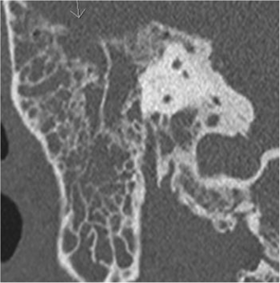
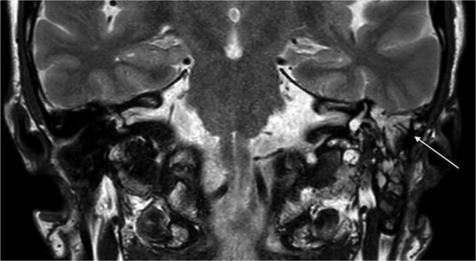
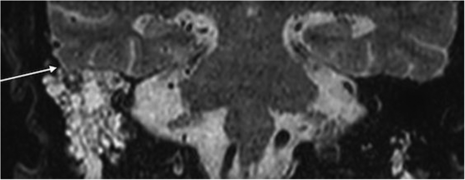
The majority of patients with OSA were male (n = 7), with a mean BMI of 32.3 kg/m2. Five patients had more than three defects of the affected side noted on both radiology reviews. The contralateral side was involved in the majority of cases (radiology review one n = 7, radiology review two n = 8). The average affected and contralateral defect sizes (all defects included) in the OSA subgroup were greater than the average affected and contralateral defect sizes of the whole cohort. Figures 1–4 show examples of tegmen dehiscences seen in the cohort.
Coronal computed tomography scan demonstrating multiple left-sided tegmen mastoideum dehiscences (arrow).

Coronal computed tomography scan demonstrating a large right-sided tegmen mastoideum dehiscence (arrow).

Coronal magnetic resonance imaging scan demonstrating a thin tegmen mastoideum and fluid within the left mastoid cavity (arrow).

Coronal magnetic resonance imaging scan demonstrating a likely dehiscence in the posterolateral aspect of the tegmen mastoideum and fluid within the right otomastoid cavity (arrow).

Discussion
Spontaneous CSF otorrhoea appears to be an emerging clinical entity, as evident by the substantial increase in the number of patients presenting with spontaneous CSF leak. Despite a relatively unclear aetiology, recent studies highlight idiopathic intracranial hypertension, obesity and OSA as factors contributing towards the development of spontaneous CSF otorrhoea.Reference Schlosser, Wilensky, Grady and Bolger5,Reference Allen, Perez, Kutz, Gerecci, Roland and Isaacson9,Reference Rabbani, Saltagi, Ye, Patel, Manchanda and Nelson13,Reference Stucken, Selesnick and Brown21–Reference LeVay and Kveton23 Importantly, the vast majority of patients presenting with spontaneous CSF leak exhibit subtle signs and symptoms that often masquerade as common otological conditions, most commonly a benign unilateral middle-ear effusion thought to be secondary to Eustachian tube dysfunction. Radiological investigation is paramount for establishing an accurate and prompt diagnosis, and is important for assisting with surgical planning, typically a middle cranial fossa repair.
The literature to date is scarce regarding radiological parameters in patients with spontaneous CSF leak as a primary outcome, but several papers have explored temporal bone CT and MRI findings as secondary outcomes.Reference Stone, Castillo, Neelon and Mukherji19,Reference Lloyd, DelGaudio and Hudgins20,Reference Schuknecht, Simmen, Briner and Holzmann24–Reference Degnan and Levy26 Lloyd et al. examined imaging modalities in several skull base CSF leak cohorts, including those with post-traumatic, non-traumatic and spontaneous causes, with a predominant focus on anterior leaks and rhinorrhoea rather than otorrhoea.Reference Lloyd, DelGaudio and Hudgins20 Thin-sliced temporal bone CT imaging was the initial modality of choice in that article, with CT cisternography utilised if more than one osseous defect was noted. This was followed by magnetic resonance cisternography, which was reserved for cases suspected of having soft tissue adjacent to the defect, to exclude cephalocele and encephalocele.Reference Lloyd, DelGaudio and Hudgins20
Stone and colleagues’ retrospective cohort study assessed 52 patients with CSF leaks; 6 of these patients presented with otorrhoea deemed to be secondary to surgical intervention.Reference Stone, Castillo, Neelon and Mukherji19 Initial imaging in this cohort consisted of high-resolution CT, and in five of the six patients with otorrhoea, a tegmen defect was identified. Defects were identified in 71 per cent of patients (whole cohort, n = 52) through the use of high-resolution CT, and subsequent CT or radionuclide cisternography did not reveal any further defects in these patients. Hence, this study concluded that high-resolution CT is appropriate for initial investigation, and cisternography may often be unnecessary.Reference Stone, Castillo, Neelon and Mukherji19 Similarly, in our cohort, temporal bone CT studies were extremely sensitive in detecting skull base defects, and when used in combination with MRI, further information regarding the presence or absence of cephaloceles was obtained. Cisternography (MRI, CT or radionuclide) was not utilised in our cohort, as this is not routine practice at our institutions, and so we were unable to compare the efficacy of this with temporal bone CT in this study.
A recent review of imaging of CSF leaks (both rhinorrhoea and otorrhoea) reiterated the importance and sensitivity of high-resolution CT in the detection of skull base defects. The authors noted that when multiple osseous defects are identified through high-resolution CT, determining the site of active leakage is difficult, so further imaging modalities (such as cisternography) are suggested.Reference Vemuri, Karanam, Manchikanti, Dandamudi, Puvvada and Vemuri3 More than half of our cohort had multiple osseous defects noted radiologically. This is important in planning surgical repair, especially when the culprit site of leakage is not known. However, based on the literature to date, temporal bone skull base defect repairs for spontaneous CSF leak routinely entail a multi-layered repair over a broad area of middle cranial fossa, where the tegmen is often diffusely thin and dehiscent.Reference Hendriks, Bala, Rodrigues and Kuthubutheen2,Reference Nelson, Roche, Gantz and Hansen27–Reference Gonen, Handzel, Shimony, Fliss and Margalit29 All defects (regardless of size or whether actively leaking) are addressed in this procedure, so pre-operative investigation with a somewhat invasive imaging modality like cisternography becomes less relevant.
Increased hydrostatic forces on inherently weak skull base sites such as the cribriform plate and well-pneumatised bone regions (ethmoid, sphenoid and mastoid) have also been proposed as a contributing factor to the development of spontaneous CSF leak.Reference Qahtani7,Reference Shetty, Shroff, Fatterpekar, Sahani and Kirtane12,Reference Gacek, Gacek and Tart17 An early paper by Gacek et al. assessed the demographics, radiological features and surgical management of 21 patients with spontaneous CSF otorrhoea and 2 patients with spontaneous CSF rhinorrhoea.Reference Gacek, Gacek and Tart17 Nineteen patients had arachnoid granulations located in the floor of the middle cranial fossa. The authors hypothesised that as the arachnoid granulations became larger, CSF pulsation resulted in bone erosion, particularly if this bone was thin or well pneumatised.Reference Gacek, Gacek and Tart17,Reference Schuknecht, Simmen, Briner and Holzmann24 Shetty et al. explored radiological findings in sphenoid sinus fistulae demonstrating excessive pneumatisation of the sphenoid sinus with lateral extensions in 91 per cent of the cohort.Reference Shetty, Shroff, Fatterpekar, Sahani and Kirtane12 Arachnoid pits were also a common finding. Pneumatisation is variable, and 23 out of 100 temporal bone CT images of age-matched controls demonstrated excessive pneumatisation.Reference Shetty, Shroff, Fatterpekar, Sahani and Kirtane12 In our cohort of patients, the mastoid air cells were well developed in most patients. The reason for this association is uncertain. When poor pneumatisation of the temporal bones occurs, such as in chronic middle-ear disease or chronic Eustachian tube dysfunction, the mastoids become sclerotic, which perhaps makes them less porous.
Skull base thickness was not assessed in this cohort of patients but appears important, with recent studies highlighting the relationship between OSA, obesity and a thin skull base. Rabanni et al. conducted a large retrospective analysis of over 1000 patients and revealed that OSA was independently associated with intracranial (calvarial and skull base) thinning, leading the authors to hypothesise that OSA is a potential contributing factor to the development of spontaneous CSF otorrhoea.Reference Rabbani, Saltagi, Ye, Patel, Manchanda and Nelson13 A similar conclusion was reached by Nelson et al., who found thinner squamous temporal bones in patients with spontaneous CSF otorrhoea, although extracranial zygoma thickness was not significantly different, indicative of an intracranial process.Reference Nelson, Hansen, Gantz and Hansen15 Comparable findings were highlighted by Stevens et al., with significantly thinner skull bases present in an obese control group and a spontaneous CSF leak group when compared with non-obese controls.Reference Stevens, Lambert, Rizk, McIlwain, Nguyen and Meyer11
With this information, further assessment of patients in our cohort with OSA was undertaken. As observed in our cohort, patients with OSA were unsurprisingly heavier, but also appeared to have larger defects on average on both the affected and the contralateral temporal bones. The number of bilateral defects, however, was largely similar to that of the whole cohort, and, given the small sample size, no power calculation was performed to assess statistical significance between these cohorts.
There were four patients in our cohort with radiological signs of superior semicircular canal dehiscence without symptomatology. The estimated prevalence of superior semicircular canal dehiscence in asymptomatic individuals is usually 3–12 per cent, which is similar to that of our cohort (6.6 per cent). However, findings from our study appear to be in contrast to the published literature, which proposes an association between spontaneous CSF leak and superior semicircular canal dehiscence.Reference Sood, Rana, Chauhan, Shukla and Nandolia30 Stevens et al. highlighted a relationship between spontaneous CSF leak and superior semicircular canal dehiscence in his retrospective review, identifying significantly lower lateral skull base heights in these two groups of patients as compared with other groups in the study (those with acoustic neuromas and otosclerosis).Reference Stevens, Hock, Samy and Pensak31 Allen et al. also demonstrated a higher than average prevalence of superior semicircular canal dehiscence identified intra-operatively in a cohort of patients undergoing surgical repair for spontaneous CSF leaks.Reference Allen, Perez, Isaacson, Roland, Duong and Kutz32
The significance of superior semicircular canal dehiscence and spontaneous CSF leak as seen in our cohort is unclear, with a prevalence within the expected range. Further studies of larger case series are required to investigate this relationship. An important aspect to consider, however, is the benefit of a middle cranial fossa (rather than transmastoid) approach to superior semicircular canal dehiscence, which is the mainstay approach for management of spontaneous CSF leak.Reference Nguyen, Lagman, Sheppard, Romiyo, Duong and Prashant33 Clinicians should have a high index of suspicion for superior semicircular canal dehiscence in patients with spontaneous CSF leak when reviewing imaging, such that surgical repair could address both pathologies if required.
A significant finding in our study was the identification of bilateral defects in the majority of patients, despite most presenting with unilateral otological symptoms. The presence of bilateral defects suggests that spontaneous CSF leak is not only acquired (given the onset in adulthood), but affects the temporal bones bilaterally. The association between OSA and spontaneous CSF leak with consequent thinning of the skull base is therefore consistent with this observation. It remains uncertain why one side tends to leak despite seemingly numerous defects bilaterally, although right-sided leaks appear to be more common, for reasons that are not evident in our cohort.Reference Gubbels, Selden, Delashaw and McMenomey34 Local factors such as inherent mastoid pneumatisation, arachnoid granulations or unequal dural venous sinus drainage are possible explanations.
Furthermore, in the absence of a dural defect (despite obvious osseous defects), CSF leakage into the middle ear or mastoid cavities presumably does not occur. This raises the question of why some patients develop both osseous and dural defects with obvious spontaneous CSF leak, whereas others have only osseous defects without clinically or radiologically apparent spontaneous CSF leak, which suggests that the dura remains intact. Closer examination of the dura in this situation would be informative and relevant to many cases in our series with contralateral defects but no spontaneous CSF leak. A larger sample size, targeted imaging and/or intra-operative assessment of the skull base and dura may help to uncover this.
Middle-ear and/or mastoid opacification of the affected side was present in the vast majority of temporal bone CT scans examined in this cohort. These findings typically indicate an effusion of some description. This can be secondary to an infective process such as acute otitis media, inflammatory such as otitis media with effusion, or related to obstruction due to a nasopharyngeal mass or Eustachian tube dysfunction.Reference Juliano35 In the setting of an isolated finding (particularly unilateral and in the absence of obvious sinonasal inflammatory disease), opacification of the middle ear and/or mastoid must prompt assessment of the skull base for dehiscences. This is especially important given the current increasing incidence of spontaneous CSF leak.Reference Nelson, Gantz and Hansen1
Patient imaging was independently reviewed twice by different radiologists to reduce reporting bias; however, some statistically significant variations were apparent. This is an important point to consider; despite the accuracy of diagnostic temporal bone CT and/or MRI in delineating skull base defects, there will frequently be discrepancy. In the context of this study, skull base defects of between 1 mm and 3 mm were reported most frequently, and, unfortunately, greater variation existed in the reporting of these smaller defects as compared with defects over 5 mm, for example, which were identified on both radiology reviews in 85 per cent of the cohort.
Several factors can lead to a discrepancy in the reporting of these defects, often associated with their typically minute size. Imaging protocols, radiation dose, artefact and patient body habitus are examples of technical factors that can potentially affect the reporting of imaging studies.Reference Brady36 Inadequate clinical information when the scan was requested and variations in radiologist experience are further factors that may result in inter-reader variation. For example, in a substantial proportion of patients in our cohort, skull base defects were not mentioned on the index temporal bone CT report. Following a positive beta-trace protein result and/or further MRI, these scans were reviewed and subsequently amended with skull base defects highlighted. An additional variable is the fluid within the middle ear and mastoid, which makes it more difficult to define skull base defects due to a loss of the air–bone interface. This was noted in our study in the context of cephaloceles, where suspicion existed on temporal bone CT based on a large tegmen defect with adjacent opacification. On subsequent review of MRI, cephaloceles were excluded in two patients despite this initial suspicion. Consequently, we recommend a multidisciplinary team (MDT) approach for assessing these patients, given the complexity of the condition with regard to the inevitable variation in interpretation of imaging findings, and the further investigation and subsequent management required.
• Spontaneous cerebrospinal fluid (CSF) temporal bone leakage is increasingly prevalent and a cause for concern given the morbidity and mortality risk
• Patients may present with chronic and subtle symptoms of aural fullness, unilateral hearing loss and otorrhoea
• Previously, computed tomography (CT) or magnetic resonance cisternography was performed to localise CSF leakage, but this is often unnecessary
• All spontaneous CSF leak patients should undergo temporal bone CT, with skull base examination for potential dehiscence
• Magnetic resonance imaging is useful for further evaluation where a cephalocele is suspected
• Spontaneous CSF leak aetiology and pathophysiology must be further explored given the incidence of numerous, large and bilateral skull base defects and often unilateral symptomatology
The study has several other limitations, including its retrospective nature, which inherently introduces bias, and the variety of imaging protocols used by the external radiological providers, which may have impacted the detail and/or content of the images. In addition, assessment of several radiological parameters, such as mastoid development and tegmen height, was subjective rather than objective.
Conclusion
Spontaneous CSF leaks of the temporal bone are a growing cause for concern, as there has been a substantial increase in the proportion of patients presenting with this disease process in recent years. An accurate diagnosis is difficult, given the subtle symptoms associated with spontaneous CSF leak, so clinicians must have a high index of suspicion when assessing these patients. The authors advocate temporal bone CT imaging at a minimum for patients with spontaneous CSF leak, and close examination of the skull base for dehiscence in the context of middle-ear and/or mastoid opacification. Identifying the number, size and location of defects via temporal bone CT is valuable for ENT surgeons for pre-operative planning, in addition to MRI to confirm or exclude an encephalocele. However, there are limitations of imaging (particularly temporal bone CT) in patients with spontaneous CSF leak, so discussion in an MDT forum with radiologists and surgeons present is advisable.
This observational study adds to the existing literature through its identification of a cohort of patients suffering with multiple large bilateral defects despite predominately unilateral symptomatology. The significance of bilateral defects in terms of the potential risk of complications and management options is yet to be determined. Further studies assessing intra-operative findings with reference to pre-operative imaging are required to determine the utility and precision of imaging in spontaneous CSF leaks.
Competing interests
None declared