Introduction
The anthelmintic effects of plant-derived cysteine proteinases (CPs) from protease Family C1 (http://merops.sanger.ac.uk; Rawlings et al., Reference Rawlings, Barrett and Bateman2012) against nematodes in vivo have been demonstrated successfully in pigs (Satrija et al., Reference Satrija, Nansen, Bjorn, Murtini and He1994; Levecke et al., Reference Levecke, Buttle, Behnke, Duce and Vercruysse2014), mice (Satrija et al., Reference Satrija, Nansen, Murtini and He1995; Stepek et al., Reference Stepek, Lowe, Buttle, Duce and Behnke2006, Reference Stepek, Lowe, Buttle, Duce and Behnke2007a, b), sheep (Buttle et al., Reference Buttle, Behnke, Bartley, Elsheikha, Bartley, Garnett, Donnan, Jackson, Lowe and Duce2011) and even chickens (Mursof & He, Reference Mursof and He1991). They are also highly effective against the three major human intestinal nematodes in naturally infected individuals (Ascaris lumbricoides, Trichuris trichiura and hookworms; Caldwell & Caldwell, Reference Caldwell and Caldwell1929; Hansson et al., Reference Hansson, Veliz, Naquira, Amren, Arroyo and Arevalo1986).
In comparison, little is known about the effects of CPs on cestode infections. Based on in vitro assays, there is evidence that CPs are detrimental to the survival of cestodes (He et al., Reference He, Tiuria and Retnani1992; Stepek et al., Reference Stepek, Lowe, Buttle, Duce and Behnke2007c; Mansur et al., Reference Mansur, Luoga, Buttle, Duce, Lowe and Behnke2014) but data also indicate that this was not reflected in a reduction of parasite burdens when CPs were administered to infected hosts (He et al., Reference He, Tiuria and Retnani1992; de Amorin et al., Reference de Amorin, Borba, Carauta, Lopes and Kaplan1999). To date, few results based on well-designed in vivo trials have been published, and whether CPs have in vivo activity against cestodes is still debatable. Nevertheless, it is clearly important to establish whether they are effective against cestodes in vivo if potential drugs based on CPs are to be taken forward to clinical trials and eventually developed into useful therapeutic agents with wide-spectrum activity against helminths.
Three rodent cestode model systems are maintained widely in laboratories (Hymenolepis microstoma, H. diminuta and H. nana) and have been used extensively to study the biology and host–parasite relationships of cestodes and for screening novel compounds for cestocidal activity (Siles-Lucas & Hemphill, Reference Siles-Lucas and Hemphill2002). Here, we used H. microstoma owing to the ease of maintaining this species in the laboratory and its practical advantages over H. nana which, without an absolute requirement for an insect intermediate host, can pose problems through autoinfection and can spread to other animals under conventional animal house husbandry (Heyneman, Reference Heyneman1961, Reference Heyneman1962; Isaak et al., Reference Isaak, Jacobson and Reed1977). Hymenolepis microstoma is also a more cost-effective cestode model-system than H. diminuta, which requires costlier maintenance since its definitive host is the rat. Moreover, it is one of the four tapeworm species for which the complete genome has been characterized (Cunningham & Olson, Reference Cunningham and Olson2010). Despite all the advantages, it is somewhat challenging and intriguing that with respect to anthelmintic susceptibility, H. microstoma has proven to be a resilient species, and therefore this model system provides a robust test of the efficacy of cestocidal drugs. From juvenile cysticercoids in the flour beetle intermediate host (Evans et al., Reference Evans, Gray and Novak1979) to bile duct dwelling adult worms, all have shown resilience to benzimidazole-based preparations (McCracken et al., Reference McCracken, Lipkowitz and Dronen1992) compared to the relatively more susceptible H. diminuta (Coles, Reference Coles1983). Exact explanations remain elusive, but one possibility may be that a large portion of the worm is protected from direct contact with drugs passing down the lumen of the intestine, because of its location high up in the bile duct (McCracken et al., Reference McCracken, Lipkowitz and Dronen1992).
Having demonstrated that H. microstoma is susceptible to CPs in vitro (Mansur et al., Reference Mansur, Luoga, Buttle, Duce, Lowe and Behnke2014), we now present the results of experiments that aimed to assess whether CPs also have detectable activity against a natural cestode of mice in its normal host in vivo.
Materials and methods
Enzyme preparation and treatment/infection of mice
The enzymes used in this study were cysteine proteinases (CPs) derived from the supernatant of papaya latex (Buttle et al., Reference Buttle, Behnke, Bartley, Elsheikha, Bartley, Garnett, Donnan, Jackson, Lowe and Duce2011; Luoga et al. Reference Luoga, Mansur, Buttle, Duce, Garnett and Behnke2012; Mansur et al., Reference Mansur, Luoga, Buttle, Duce, Lowe and Behnke2014) containing chymopapain, glycyl endopeptidase, caricain and papain (in order of relative abundance (Buttle et al., Reference Buttle, Dando, Coe, Sharp, Shepherd and Barrett1990)). The molar concentration of the active cysteine proteinase was measured by active-site titration adapted from Barrett et al. (Reference Barrett, Kembhavi and Hanada1981) and Zucker et al. (Reference Zucker, Buttle, Nicklin and Barrett1985), with increasing concentrations of the cysteine proteinase specific inhibitor, l-trans-epoxysuccinyl-l-leucylamido-(4-guanidino) butane (E-64) (Apollo Scientific Ltd, Stockport, Cheshire, UK) with 4 mml-cysteine as a reducing agent and N-α-benzoyl-arginine-p-nitroanilide (BAPNA) (Bachem Ltd, St. Helens, Merseyside, UK) as the substrate.
We used the mouse tapeworm H. microstoma, a widely exploited laboratory model of tapeworm infections (Andreassen, Reference Andreassen1991; Siles-Lucas & Hemphill, Reference Siles-Lucas and Hemphill2002). We refer to H. microstoma rather than Rodentolepis microstoma following Cunningham & Olson (Reference Cunningham and Olson2010). Male, 5-week-old C3H mice (weighing 15–20 g) from Harlan (Bicester, Oxfordshire, UK) were allowed 1 week to adjust to local conditions and then were infected by gavage with five H. microstoma cysticercoids each (using a 1-ml syringe with a ball-tipped, stainless-steel, 38.1 mm, 22-gauge feeding needle (Harvard Apparatus Ltd, Edenbridge, Kent, UK)), freshly dissected on the day of administration from infected flour beetles (Tribolium confusum). The infective dose of cysticercoids, and treatment with the appropriate dose of CP and water, by gavage, was given in a volume not exceeding 0.2 ml. The life cycle of H. microstoma has been maintained in Nottingham in a closed colony, using the intermediate beetle host T. confusum, since 1976, but for further details see Behnke (Reference Behnke, Halton, Behnke and Marshall2001) and Mansur et al. (Reference Mansur, Luoga, Buttle, Duce, Lowe and Behnke2014). All the animals were ear-marked with a unique combination to allow individuals to be identified, and were housed in groups, at a maximum stocking density of five/cage, in standard polypropylene cages containing environmental enrichment, and provided with food and water ad libitum.
For assessment of the effectiveness of treatment, faecal samples were collected from each mouse on days 22, 24, 26, 28, 30 and 32 post-infection (pi). Animals were separated into individual cages early in the morning (09.00 h) and observed at regular intervals until sufficient fresh faeces had been deposited, to allow egg counts to be carried out, after which they were returned to their original groups. The number of eggs present (faecal egg counts, FEC) was counted using the McMaster technique modified from Behnke & Parish (Reference Behnke and Parish1979) as described by Luoga et al. (Reference Luoga, Mansur, Buttle, Duce, Garnett, Lowe and Behnke2014) and expressed as eggs per gram of faeces (EPG). For the determination of parasite burdens, mice were culled on day 32 of infection by an overdose of CO2. Because the scoleces of H. microstoma are mainly attached to the bile duct, the liver was removed first from its attachments to the diaphragm and other gut structures in order to expose an intact bile duct. The bile duct was then opened carefully using sharp-ended dissecting scissors, starting proximally from its base at the liver and then moving distally away from the liver towards the duodenum. This incision was then continued in both directions of the duodenum, towards the stomach and towards the ileo-caecal junction. The opened intestine was placed in a glass Petri dish containing pre-warmed Hanks’ saline (37°C) for 30 min to allow the worms (if any) to move away from the tissues. Individual worms were first identified by their scoleces under the light microscope before being counted and transferred into pre-weighed aluminium foil, which had been fashioned into small cups. They were weighed in these cups and the weight of empty cups was subtracted to obtain the value for the wet biomass of the parasite burdens from individual mice. The dry biomass was determined by baking the worms in an oven at 60°C continuously for 5–10 h daily for two consecutive days, as opposed to the usual method of baking continuously for 24 h (Hopkins et al., Reference Hopkins, Subramanian and Stallard1972), followed by reweighing and subtraction of the weight of the empty cups. The two periods of drying were necessary to conform to local safety regulations aimed at avoiding a fire hazard from unattended overnight use of the oven.
Experimental design
Experiment 1. Papaya latex supernatant extract (PLS) on H. microstoma in vivo
Eighteen mice were used in this experiment and were divided into two groups. Each mouse in treatment group 1 received 240 nmol of PLS while group 2 received water. Both groups were treated for six consecutive days, starting from day 27 until day 32 pi. The effectiveness of treatment was determined through FEC, which were carried out every other day from day 22 until day 32 pi, as well as by autopsy for recovery of worms and assessment of their biomass.
Experiment 2. PLS on the viability of shed eggs
Six 22-day-old H. microstoma worms were used in this experiment, all from the mice in Experiment 1. Three worms were from the mice that had been treated for 6 days with 240 nmol of PLS and three from infected mice treated only with water. From each worm, the most distal proglottid segments of approximately 1 cm length were cut and fed to a group of three previously starved (for 5 days) T. confusum beetles, hence nine beetles were exposed to cestode eggs from each mouse treatment group. One detached proglottid segment/mouse from the colon of the same mice was fed to a group of T. confusum beetles that had been starved for 5 days. After indications that the fed segments had been consumed, the usual beetle diet, consisting of a mixture of white and wholemeal flour, was added for conventional maintenance until after day 30, when all the beetles were dissected in Hanks’ saline to recover and count any developed cysticercoids.
Experiment 3. Are cysteine proteinases in PLS involved?
Twenty mice were used in this experiment and these were divided into four groups. Each mouse in group 1 was treated with 240 nmol of PLS, group 2 with 240 nmol of PLS pre-treated with 280 nmol of E-64 to ensure that the CPs were inactivated prior to delivery to mice, group 3 with 280 nmol of E-64 only and group 4 received water. All doses were given daily and each treatment was sustained for six consecutive days starting from day 17 until day 22 pi. Treatment effects were only assessed at autopsy through recovery of worms and assessment of their biomass.
Experiment 4. Scanning electron microscopical observations of surface changes to worms in PLS-treated animals
This experiment was carried out exactly as above, with the same four treatment groups, to observe the effects of PLS inflicted on H. microstoma in vivo. The efficacy of treatment was assessed through the recovery of detached proglottids found in the large intestine and these were immediately fixed in glutaraldehyde and later dehydrated in ethanol, critical point dried and gold coated, before finally being viewed under scanning electron microscopy (SEM) for signs of any damage.
Experiment 5. Dose-dependent effect of PLS on H. microstoma
An experiment was carried out to assess whether the reduction in worm burdens and biomass in PLS-treated mice, as observed in Experiment 1, was dose-dependent. Twenty-five mice were used; these were arranged into seven groups and treated as follows: group 1 (n =5) were treated with water, group 2 (n= 3) received 30 nmol of PLS, group 3 (n =3) received 120 nmol of PLS, group 4 (n =3) received 180 nmol of PLS, group 5 (n =5) received 240 nmol of PLS, group 6 (n= 3) received 360 nmol and group 7 (n =3) received 480 nmol of PLS. All groups received treatment daily for 6 days, starting from day 17 until day 22 pi. The outcome of treatment was assessed through worm counts and assessment of biomass at autopsy.
Experiment 6. PLS on H. microstoma 1 week after treatment
This experiment was designed to assess whether or not H. microstoma has the ability to recover in vivo post PLS treatment, since tapeworms have been shown to recover quickly and repair damage sustained by chemotherapy and the host immune response (Befus & Threadgold, Reference Befus and Threadgold1975). Twenty mice were used for this experiment and these were divided into four groups with five mice per group. Groups 1 and 3 received 240 nmol of PLS, while groups 2 and 4 received only water. All groups were given treatment daily for six consecutive days from day 17 until day 22 pi. Groups 1 and 2 were culled immediately at the end of the treatment week (day 22 pi) while groups 3 and 4 were left to continue, without any further treatment, for another week, until day 28 pi. The effectiveness of treatment was determined through autopsy for recovery of worms and assessment of their biomass on days 22 and 28 pi.
Experiment 7. PLS on pre-patent stages of H. microstoma
This experiment was designed to assess whether young, and still migrating, H. microstoma, which are present in the lumen of the intestine for 3–4 days before entering the bile duct, are more susceptible to CPs compared with adult worms that have established anchorage in the bile duct. Freshly excysted and migrating juvenile worms should be fully exposed to lumenal CP after treatment. Eighteen mice were used in this experiment and were divided into two groups of nine mice each. Each mouse in treatment group 1 received 240 nmol of PLS daily for 5 days from day 2 until day 6 pi, while mice in control group 2 received only water. Because 6-day-old worms are still too small to recover reliably from the intestine, the mice were not culled until day 14 pi, by which time the worms had attained a more easily quantifiable size (Moss, Reference Moss1971). The effectiveness of treatment was determined through autopsy for recovery of worms and assessment of their biomass. Any effects on the growth of juvenile worms should still be evident on day 14 before the worms reach full adult size (day 19; Moss, Reference Moss1971).
Data analysis
All data were analysed using SPSS software (version 21.0; SPSS Inc., Chicago, Illinois, USA). For FEC, data were log10 transformed to normalize variance and analysed using repeated measures general linear models (rmGLM), fitting time as the within-subject factor and treatment as the between-subject factor. The Huynh–Feldt adjustment to the degrees of freedom was used to interpret significance on the side of caution when the data did not meet the requirements of Mauchley's Test of Sphericity. In experiments assessing the efficacy of treatment on worm burdens, wet and dry biomass of worms, the non-parametric Mann–Whitney U and Kruskal–Wallis tests were used, depending on number of treatment groups being tested. In experiments assessing dose-dependency, data were analysed using Spearman's correlation test.
Results
Experiment 1. Papaya latex supernatant extract (PLS) on H. microstoma in vivo
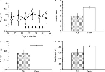
The faecal egg counts (fig. 1A) in the group receiving PLS did not show any evidence of a reduction compared to the control group receiving water. Indeed, arithmetically the mean values were higher than those of the control group following treatment. This difference in FEC between treatment groups was not statistically significant (rmGLM, between-subjects test, F 1,16= 1.578, P= 0.227) and there was no evidence of a divergence between treatment groups with time (within-subjects test, time × treatment interaction, F 3.7,59.2= 0.763, P= 0.544).
Effect of papaya latex supernatant extract (PLS) on H. microstoma in vivo (Experiment 1). (A) The mean log10 of eggs per gram of faeces before (days 22–26) and during (days 28–32) treatment (arrows) of C3H mice infected with H. microstoma with PLS (▲; n= 9) or water (○; n= 9). (B) Mean worm burden of C3H mice infected with H. microstoma and treated with 240 nmol PLS (n= 9, stippled columns) or water (n= 9, open columns). (C) Mean wet biomass and (D) mean dry biomass. Error bars represent the standard error of the mean.

However, there were significant differences in worm burdens (fig. 1B; 35.7% reduction; Mann–Whitney U test, z= –2.789, P= 0.005), wet biomass (fig 1C; 31.9% reduction; Mann–Whitney U test, z= –2.518, P= 0.012) and dry biomass (fig 1D; 29.6% reduction; Mann–Whitney U test, z= –1.987, P= 0.047) between the treated group and the control group.
Experiment 2. PLS on the viability of shed eggs
Tribolium confusum beetles fed with the cut terminal proglottid segments from H. microstoma that had been treated in vivo with PLS were found to harbour intact cysticercoids. The mean number of cysticercoids recovered (11.0 ± 4.8) was not significantly different from that of the control group of beetles fed worms from mice that had been treated with water (13.1 ± 4.7; Mann–Whitney U test, z= 0.134). Beetles fed with detached proglottid segments recovered from the large intestine of treated mice also harboured intact cysticercoids (4.3 ± 2.2) and the mean worm burden was almost identical to that in the control beetle group fed with detached proglottids from mice treated with water (4.0 ± 1.0; Mann–Whitney U test, z= 0.664).
Experiment 3. Are cysteine proteinases in PLS involved?
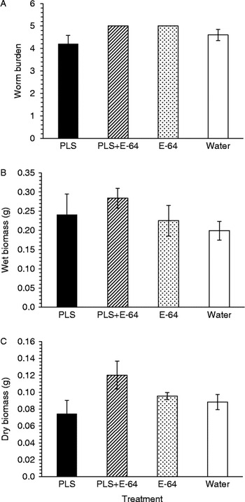
Arithmetically, there was only a 10–15% reduction in worm burdens when comparing mean values between the treated group and control groups (fig. 2A), and the difference between treatment groups was not statistically significant, although close to the cut-off (Kruskal–Wallis test, χ23= 6.97, P= 0.073). Unexpectedly, and surprisingly, in the assessment of the wet biomass, the mean values of the group treated with PLS was marginally higher when compared to control groups, except for the group receiving PLS+E-64 which had the highest mean biomass (fig. 2B), but overall the difference between treatment groups was not significant (Kruskal–Wallis test, χ23= 3.263, P= 0.353). There was a 15–40% reduction in the dry biomass of the PLS-treated group compared with the control groups (fig. 2C) but, as above, this was not statistically significant (Kruskal–Wallis test,  $$\chi _{3}^{2} $$= 4.077, P= 0.253). Despite the lack of statistical significance, the tapeworms from the mice treated with PLS+E-64 had numerically higher mean values for all three indices compared to those given PLS alone.
Involvement of cysteine proteinases in PLS (Experiment 3). C3H mice were infected with H. microstoma and treated either with PLS 240 nmol, PLS 240 nmol pre-treated with 280 nmol E-64 before administration to mice, E-64 280 nmol or water (n= 5 mice for each treatment). (A) Mean worm burden; (B) mean wet biomass; (C) mean dry biomass. Error bars represent the standard error of the mean.

Experiment 4. SEM observations of surface changes to worms in PLS-treated animals
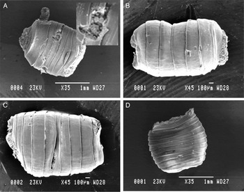
The detached segment recovered from the PLS-treated mouse appears to have sustained treatment-related damage (fig. 3A) with superficial erosions revealing underlying structures which were not present in all the other segments from the other treatment combinations (figs 3B–D), all of which maintained their rather velvety appearance due to the intact brush border layer of the tegument.
Scanning electron microscopical observations of surface changes to worms in PLS-treated animals (Experiment 4). SEM images of detached proglottid segments of H. microstoma recovered from the colons of infected mice treated with (A) PLS 240 nmol; (B) PLS 240 nmol pre-treated with 280 nmol E-64; (C) E-64 280 nmol; (D) water. Note the erosions in (A) revealing internal structure of the worm, shown at increased magnification ( × 200) in the insert, and the otherwise smooth appearance of other segments.

Experiment 5. Dose-dependent effects of PLS on H. microstoma
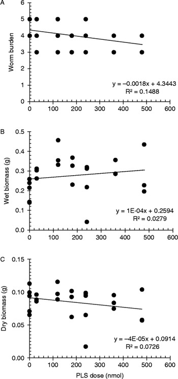
Autopsy revealed a significant negative correlation between PLS dose and worm burden (fig. 4A; r= –0.397, n= 25, P= 0.049). There was also a weak negative trend between PLS dose and dry biomass, which was not significant (fig. 4C; r = –0.265, n= 25, P =0.201), and a weak positive trend of wet biomass with increasing doses of PLS, which was also not significant (fig. 4B; r= 0.206, n= 25, P =0.323).
Dose dependency of the effect of PLS on H. microstoma (Experiment 4). The relationship between increasing PLS dose and worm burden (A), wet biomass (B); and dry biomass (C). All groups were of the same sample size (n= 3) except groups 1 and 5 (n= 5). For statistical analyses and further details of experimental design see text. The line of best fit is provided.

Experiment 6. Permanency of the effect of PLS on H. microstoma
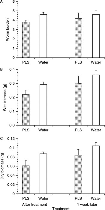
In all three indices studied (number of worms, wet and dry biomass) there were obvious reductions of worms in treated mice compared with those in the controls, irrespective of autopsy time. However, as expected, there were appreciable increases in worm burden and biomass when animals were autopsied later during the course of infection on day 28 pi. Data were explored using two-way GLM to assess the main effects of time and treatment and also the interaction between these two factors (fig. 5).
The effect of PLS on H. microstoma 1 week after treatment (Experiment 6). C3H mice were infected with H. microstoma and culled immediately after 6 days of treatment with either 240 nmol of PLS or water (n= 5 mice for both treatments) and a further 1 week after withholding treatment (n= 5 for both treatments). (A) Mean worm burden; (B) mean wet biomass; (C) mean dry biomass. Error bars represent the standard error of the mean.

The effect of treatment on worm burden was not significant (F 1,16= 2.4, P= 0.141; although treated mice had 17.4% fewer worms than controls after treatment and 8.7% fewer worms than controls when autopsied 1 week later), and neither was the effect of time on worm burden (F 1,16= 0.267, P= 0.613). There was no significant interaction between time and treatment on worm burdens (F 1,16= 0.267, P= 0.613).
The effect of treatment on wet biomass, however, was close to significance (F 1,16= 3.514, P= 0.079) and the percentage reduction in wet biomass was 24.2% post treatment and 16.6% when autopsied 1 week later. The effect of time was significant (F 1,16= 4.596, P= 0.048), wet biomass values being higher in both treated and control mice on day 28 compared with day 22 pi. However, the interaction between treatment and time was not significant (F 1,16= 0.023, P= 0.881).
The effect of treatment on dry biomass was significant (F 1,16= 6.268, P= 0.024), as was that of time (F 1,16= 4.681, P= 0.046). Again, perhaps surprisingly, there was no significant interaction between time and treatment (F 1,16= 0.086, P= 0.773).
Experiment 7. PLS on pre-patent stages of H. microstoma
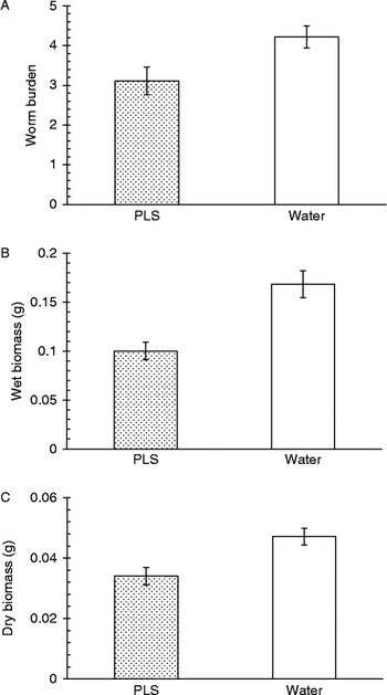
Autopsy revealed significant effects of treatment on worm burden (fig. 6A; Mann–Whitney U test, z= –2.147, P= 0.032) compared with the control group. The wet biomass (fig. 6B; Mann–Whitney U test, z= –3.311, P= 0.001) as well as the dry biomass (fig. 6C; Mann–Whitney U test, z= –2.474, P= 0.013) were also significantly lower in treated mice.
Effects of PLS on pre-patent, migratory stages of H. microstoma (Experiment 7). C3H mice were infected with H. microstoma and treated with PLS 240 nmol (n= 9) or water (n= 9). (A) Mean worm burden; (B) mean wet biomass; (C) mean dry biomass. Error bars represent the standard error of the mean.

Discussion
The experiments described in this paper are the first to test the effects of papaya latex on H. microstoma infections in mice. Although we found generally modest effects on the number of worms surviving treatment and on their size, these experiments set a precedent for further work and are highly instructive with regard to the biological basis of the resilience of H. microstoma in the face of the hostile local environment created by oral therapy of infected mice with CPs. There are some notable parallels between the observations reported here and in previous studies with other anthelmintics, to which H. microstoma was also found to be resilient. For example, this species is far more resistant to benzimidazoles than the considerably more susceptible H. diminuta (McCracken et al., Reference McCracken, Lipkowitz and Dronen1992). Even at the larval stage, H. microstoma was found to be resistant to benzimidazole-based preparations (Evans et al., Reference Evans, Gray and Novak1979, Reference Evans, Hardy and Novak1980).
In Experiment 1, in which FEC were employed as a measure for assessment of treatment efficacy, there was no reduction in the mean EPG in treated groups as compared to the controls, despite the concurrent reductions in worm burden and biomass. This was unexpected because up to 90% reduction has been reported in FEC of mice infected with nematodes such as Heligmosomoides bakeri after treatment with papaya latex (Stepek et al., Reference Stepek, Lowe, Buttle, Duce and Behnke2007b). There are several possible explanations. First, it has to be emphasized that H. microstoma is a bile duct dwelling worm, with only parts of its strobila exposed in the intestinal lumen (de Rycke, Reference de Rycke1966). Since the orally administered CPs pass down the intestinal lumen, the scolex and neck regions are not likely to be exposed to high concentrations of the CPs. Hence, the areas of major contact between CPs and the cestode are the terminal proglottids. In a short-term administration protocol such as this, damage to these terminal gravid proglottids, may be reflected in loss of weight, by their premature loss from the strobila, but also in the liberation of the eggs that had formed in the mature proglottids higher up the strobila. The actual generation and development of eggs in the developing proglottids, however, may be protected from the effects of CPs by their location in the upper sections of the bile duct.
Direct exposure of the surface of parasites to CPs appears to be a critical requirement of this potential novel drug, because CPs are poorly absorbed systemically (Behnke et al., Reference Behnke, Buttle, Stepek, Lowe and Duce2008). This was evident in the case of the sheep nematode Trichostrongylus colubriformis, which tunnels inside the intestinal mucosa and which was not affected by papaya latex treatment, in contrast to the potent effect observed on Haemonchus contortus, which is entirely luminal, in concurrent infections (Buttle et al., Reference Buttle, Behnke, Bartley, Elsheikha, Bartley, Garnett, Donnan, Jackson, Lowe and Duce2011). Even if it appears that treatment with CPs does not affect FEC, this does not establish conclusively that fecundity is not affected. FEC is probably a poor indicator of the worm burdens in tapeworm infections, as evidenced also by the wide error bars of the mean EPG values before treatment, indicating high variability in the number of eggs and their concentration in faeces (fig. 1A). This may be explained by the sporadic discharge of gravid segments as well as their unequal distribution in the faecal mass (Moss, Reference Moss1971; Nilsson et al., Reference Nilsson, Ljungstrom, Hoglund, Lundquist and Uggla1995), as exemplified by infections with the horse tapeworm Anoplocephala perfoliata, where coprological detection of infection is unreliable due to the low sensitivity of FEC (Abbott & Barrett, Reference Abbott and Barrett2008). In this system there was no significant correlation between tapeworm burden and egg detection (Proudman & Edwards, Reference Proudman and Edwards1992; Meana et al., Reference Meana, Luzon, Corchero and Gomez-Bautista1998).
Nevertheless, even though there were many eggs in the faeces of infected mice post treatment, it was possible that in PLS-treated mice the eggs had experienced more subtle harmful effects, limiting their capacity to develop normally. It was therefore imperative to ascertain whether their viability was affected, through an egg viability test. Disappointingly, intact H. microstoma cysticercoids were recovered from Tribolium beetles fed with either cut terminal proglottid segments or detached proglottid segments recovered from the large intestines of mice treated with PLS, in similar numbers to those from beetles given eggs from infections in control mice. Structurally, cestode eggs have a thicker outer shell compared to trichostrongyloid nematode eggs such as those of H. bakeri, but not those of Trichuris and Ascaris species (Smyth & Clegg, Reference Smyth and Clegg1959; Wharton, Reference Wharton1980). This robust eggshell is required because of the need for the eggs to survive under harsh external environmental conditions before ingestion by the intermediate host, and it is likely that the shells of H. microstoma eggs provided adequate protection from exposure to CPs for the hexacanth embryos within. It is also possible that cestode eggs do not have externally exposed amino acid sequences that are sensitive to digestion by CPs and, if they do, that the eggs were exposed to insufficient concentrations of CPs to experience overt damage.
While it is apparent that the arithmetic mean worm counts were numerically lower in groups of treated mice in all the experiments reported here, there was nevertheless considerable variation in the degree of reduction between experiments, and worm counts in treated mice were not all significantly lower than in control mice. An experiment with E-64, a specific inhibitor of CPs, found only a modest inhibition of the effect of PLS, which also lacked statistical significance (fig. 2). Interestingly, the dose of PLS was found to correlate significantly but negatively with worm burden (Experiment 5), suggesting that fewer worms survived treatment when the dose was high. This dose-dependent reduction in worm numbers may suggest that, despite the location of the scoleces in the bile duct, they were, in fact, not all well protected from the effects of treatment with PLS. However, the recovery of tiny scoleces is never 100% accurate and there remains the possibility that, as a result of the stress on the strobila from the CPs, the worms had destrobilated and their scoleces still persisted somewhere in the intestine without having been detected at autopsy and post-mortem examination. Indeed, mice autopsied a week after the completion of treatment, harboured more and bigger worms compared to those autopsied immediately after treatment. Hence, the reductions seen in earlier experiments based on autopsy immediately after the last dose of PLS, were temporary, and surviving H. microstoma did re-grow post-treatment (fig. 5).
When recovered worms were subjected to assessment of their biomass, there were again notable inconsistencies between experiments, especially with regard to the wet biomass, and instead of the expected reductions, in some cases the arithmetic mean worm biomasses were higher than those of the control, untreated groups of mice (fig. 2). In one instance, the PLS dose was found to be positively correlated with wet biomass (fig. 4B). The process of removing excess fluid that surrounds and coats the worms is never entirely reliable and, for this reason, a second biomass assessment was always made based on completely dried worms after removal of all fluid content by incubation in an oven. Cestodes lacking a tough cuticle, such as that of nematodes, might be susceptible to fluid influx due to dysfunctional membrane transport systems from damaged or dead proglottids, thereby resulting in unexpected biomass values. In fact, in contrast to wet biomass, all the mean dry biomass assessments showed an arithmetic reduction compared to the control groups but, nevertheless, despite this consistent trend of lower dried weight, the accompanying statistical analyses were inconsistent and the specific effect of the CPs in PLS was not confirmed even following E-64 treatment (fig. 2). Similarly, the relationship between dry biomass and dose of PLS showed a weak negative trend, but this was not statistically significant. Therefore, despite the consistently lower arithmetic means for values derived for worms from treated groups, the wide variation in data distribution and the lack of a consistently significant effect of PLS suggest that papaya latex, at the doses utilized in the present work, is at best minimally efficacious against H. microstoma in vivo. This conclusion is also supported visually by the scanning electron microscopy of the teguments (fig. 3), where the detached proglottid segments recovered from the mouse treated with PLS showed only minimal surface changes.
Since the relatively weak effect of PLS on H. microstoma may be attributable to its location, residing high in the bile duct where it is largely protected from contact with intestinal lumenal contents (de Rycke, Reference de Rycke1966; McCracken et al., Reference McCracken, Lipkowitz and Dronen1992), an experiment was carried out in which young and still migrating worms, fully exposed in the intestinal lumen, were subjected to CP treatment. Here we might have expected a marked reduction in all autopsy indices but, in actual fact, while significant, the actual percentage reductions compared with controls were again fairly minimal (fig. 6).
It is also relevant that the IC50 values, derived from in vitro experiments in earlier work (Mansur et al., Reference Mansur, Luoga, Buttle, Duce, Lowe and Behnke2014), were higher than for nematodes under similar exposure to CPs (Stepek et al., Reference Stepek, Buttle, Duce, Lowe and Behnke2005). While none of the experiments described in this paper saw treatment-related animal casualties nor poor weight gain (results not shown), nevertheless toxicity to much higher PLS doses than the ones utilized here was experienced in pilot trials, and hence we adopted an upper limit of 240 nmol/day in the current work (except in the dose response, Experiment 5). It is also worth pointing out that oral dosing of sheep and pigs with PLS proved equally sound and safe (Buttle et al., Reference Buttle, Behnke, Bartley, Elsheikha, Bartley, Garnett, Donnan, Jackson, Lowe and Duce2011; Levecke et al., Reference Levecke, Buttle, Behnke, Duce and Vercruysse2014). Hence, despite these possible side-effects, we feel that they should not hinder progress in further developing CPs into useful novel anthelmintics.
In summary, CPs do indeed affect H. microstoma in vivo, albeit minimally and temporarily. Given the high efficacy of PLS observed against nematode models, H. microstoma is not a sensitive-enough model system for screening novel compounds such as naturally occurring plant-derived CPs for potential anthelmintic activity. Hence, there is a need to explore other model systems and also methods for better formulation and improvement in the delivery of CPs. Novel approaches to drug delivery may overcome some of the obstacles to efficacy against H. microstoma identified in the current work, to enable these natural plant products to be developed into useful wide-spectrum anthelmintics for humans and livestock, especially as they have such powerful anthelmintic effects against intestinal lumen dwelling nematodes.
Acknowledgements
We thank Tim Smith for technical support with electron microscopy. We dedicate this paper to Dr Wenceslaus Luoga who sadly was killed in a road accident in July 2013 shortly after returning to Tanzania on the successful completion of his postgraduate studies at the University of Nottingham.
Financial support
F.M. was supported by a postgraduate studentship from the Government of Malaysia, and W.L. from the Government of Tanzania.
Conflict of interest
None.
Ethical standards
The authors assert that all procedures contributing to this work comply with the ethical standards of the national guides on the care and use of laboratory animals in the UK (The Animals (Scientific Procedures) Act 1986) and were locally approved by the Animal Welfare and Ethical Review Body of the University of Nottingham. Importantly, all work was conducted within a recognized culture of care and compliance to meet the expectations of both the University and the UK Home Office Inspectorate.