Introduction
Schizophrenia is a severe and disabling psychiatric illness, affecting approximately 1% of the population. It is a chronic condition in a high proportion of cases. Although the underlying pathophysiology remains unknown, it has been hypothesised that abnormalities in brain white matter play a key role in the pathophysiology of schizophreniaReference Davis, Stewart and Friedman(1). Postmortem studies have identified abnormalities in the myelin sheathReference Uranova, Orlovskaya and Vikhreva(2), oligodendrogliaReference Hof, Haroutunian, Copland, Davis and Buxbaum(3) and interstitial neuronsReference Akbarian, Kim, Potkin, Hetrick, Bunney and Jones(4) in schizophrenic patients compared with those of healthy volunteers. KraepelinReference Kraepelin(5) postulates frontal and temporal lobe abnormalities to be central to the pathology of the disease. Recent postmortemReference Hulshoff Pol, Schnack and Bertens(6) and neuroimaging studies(Reference Andreasen, O'Leary and Flaum7–Reference van Haren, Hulshoff Pol and Schnack15) have provided a considerable amount of evidence for brain abnormalities in the frontal and temporal regions of the brain in schizophrenia. If the pathophysiology of schizophrenia reflects a disturbance in the white matter, then such abnormalities might be observed by using diffusion tensor imaging (DTI) because DTI metrics are sensitive to nerve fibre changes. To date, the uncinate fasciculus and superior longitudinal fasciculus, i.e. the pathways through the frontotemporal lobes, have been well investigated(Reference Jones, Catani and Pierpaoli10,Reference Burns, Job, Bastin, Whalley, MacGillivray and Honstone16,Reference Kubicki, Westin, Maier, Frumin, Nestor and Salisbury17). Several previous studies have reported differences in DTI metrics between schizophrenic patients and healthy volunteers, but results have been inconsistent and differences between patients and volunteers have been observed not only in the frontotemporal lobes but also in the corpus callosumReference Agartz, Andersson and Skare(18,Reference Foong, Maier, Clark, Barker, Miller and Ron19) and cingulate cortexReference Kubicki, Westin, Nestor, Wible, Frumin and Maier(20,Reference Sun, Wang, Cui, Breeze, Du and Wang21), and more widespread abnormalities in the brain have also been reported(Reference Ardekani, Nierenberg, Hoptman, Javitt and Lim22–Reference Lim, Hedehus, Moseley, de Crespigny, Sullivan and Pfefferbaum25). These differences may be attributed, in part, to differences between individual patients. Schizophrenia is characterised as a progressive illness, and it is known that there are structural changes in white matter between patients with first-episode schizophrenia and those with chronic schizophreniaReference Ho, Andreasen, Nopoulos, Arndt, Magnotta and Flaum(9). Differences in illness duration, age at scan, full-scale intellectual quotient (IQ) and image processing and analysis, including the use of region-of-interest (ROI) versus voxelwise approaches, may also account for discrepancies in these various findingsReference O'Sullivan, Jones, Summers, Morris, Williams and Markus(26,Reference O'Sullivan, Morris, Huckstep, Jones, Williams and Markus27). In addition, a study by Andreasen et al.Reference Andreasen, O'Leary and Flaum(7) suggests that impairments of the ‘eocortical and limbic network’ after frontal dysfunction may occur secondarily.
Changes in the DTI metrics of patients with first-episode schizophrenia might be less pronounced than those in chronic patientsReference Szeszko, Ardekani and Ashtari(14,Reference Price, Bagary, Cercignani, Altmann and Ron28), suggesting that the changes in DTI metrics observed in schizophrenics might be attributed, at least in part, to progressive and exaggerated age-dependent rather than to neurodevelopmental abnormalities in the white matter. There have been a few cross-sectional studies showing a decrease in age-related fractional anisotropy (FA) in schizophrenicsReference Mori, Ohnishi and Hashimoto(29) but, to the best of our knowledge, there has been no longitudinal study on neurodegenerative change in schizophrenia to date. On the other hand, Jones et al. mentioned that the previous studies including older schizophrenia patients are less likely to find differences between schizophrenic patients and control than studies including very young subjects, and results may differ depending on the age range of the subjectsReference Jones, Catani and Pierpaoli(10). So, this is in need to examine the neurodegenerative change in older schizophrenics as well as the neurodevelopmental abnormalities in younger patients.
The purpose of the present longitudinal study was to examine whether the older patients with chronic schizophrenia show changed DTI metrics as well as morphometric changes during the course of the disorder, and whether the progress of these changes is affected by other factors such as age at scan, illness duration or the daily dose of antipsychotic drugs, and so on.
Materials and methods
Subjects
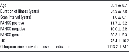
Sixteen males with schizophrenia, defined according to the criteria described in the fourth edition of the Diagnostic and Statistical Manual of Mental Disorders (DSM-IV), were recruited from inpatients at Hospital Bando (Ibaraki, Japan). Their mean age was 58.1 ± 6.7 years (ranging from 47 to 69 years) and their clinical characteristics are presented in Table 1. The psychopathology was assessed using the positive and negative syndrome scale (PANSS)Reference Kay, Fiszbein and Opler(30), and clinical evaluation included full-scale IQ of the Wechsler adult intelligence scaleReference Wechsler(31). All patients were treated with antipsychotic medication. Exclusion criteria included history of head injury, neurological symptoms, speech or hearing difficulties, significant cerebrovascular diseases (cortical infarctions, multiple lacunar lesions or leukoaraiosis) and fulfillment of DSM-IV criteria for abuse of illicit drugs or alcohol at any point during their lifetime. All participants provided their written informed consent and the local ethics committee approved the study protocol. The interval between the initial and follow-up magnetic resonance imaging (MRI) studies was approximately 1 year.
Clinical characteristics of our 16 patients
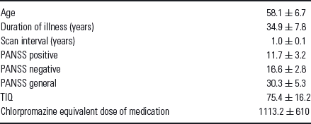
TIQ, total intelligence quotient.
Data acquisition and processing
MRI was performed on a 1.5 Tesla Siemens Magnetom Harmony (Erlangen Germany). DTI was carried out on the axial plane [echo time (TE)/repetition time (TR) = 100/7000 ms; field of view (FOV), 262 × 262 mm; matrix 128 × 128; 40 continuous transverse slices; slice thickness, 4 mm with no slice gap]. To enhance the signal-to-noise ratio, acquisition was repeated four times. Diffusion was measured along 12 non-collinear directions with the use of a diffusion-weighted factor b in each direction of 1000 s/mm2, and one image was acquired without the use of a diffusion gradient. High spatial resolution three-dimensional (3D) T1-weighted images of the brain were obtained for morphometric study. 3D T1-weighted images were scanned on the sagittal plane [TE/TR, 3.93/1460 ms; flip angle, 15°; effective section thickness, 1.5 mm; matrix, 512 × 416; FOV, 250 × 203; one number of excitations (NEX)], yielding 112 contiguous slices through the head. In addition to DTI and 3D T1-weighted images, we also acquired axial T2-weighted turbo spin echo images (TE/TR, 95/3800 ms; slice thickness, 6 mm; intersection gap, 1.2 mm; matrix, 384 × 288; FOV, 220 × 175 mm; acquisition, 1) and fluid attenuation inversion recovery (FLAIR) images on the axial plane (TE/TR, 104/9000 ms; flip angle, 170°; slice thickness, 6 mm; intersection gap, 1.2 mm; matrix, 256 × 192; FOV, 220 × 175 mm; acquisition, 1) to rule out cerebral vascular disease.
The raw diffusion tensor and 3D T1-weighted volume data were transferred to the workstation and the DTI data sets were analysed using DtiStudioReference Jiang, Van Zijl, Kim, Pearlson and Mori(32). The diffusion tensor parameters were calculated on a pixel-by-pixel basis, and FA, mean diffusivity (MD) and b = 0 images were then calculated.
Before analysis, we made a customised anatomical T1 template from the 3D-T1 images of 32 participantsReference Good, Johnsrude, Ashburner, Henson, Friston and Frackowiak(33). We then processed the images using Statistical Parametric Mapping 5 software (SPM5; Wellcome Department of Imaging Neuroscience, London, UK) running on MATLAB 7.0 (Math Works, Natick, MA, USA). Optimised voxel-based morphometry (VBM) was processed using the appropriate SPM5 tool. Details of this process were previously described by Good et al.Reference Good, Johnsrude, Ashburner, Henson, Friston and Frackowiak(33). Briefly, we first normalised the 3D-T1 images of 32 participants to default SPM5 template using optimised VBM. Then, we calculated the original 3D-T1 template image by averaging 3D-T1 images of 32 participants. Second, we normalised the 3D-T1 images of 32 participants to original 3D-T1 template using optimised VBM once again. Normalised segmented 3D-T1 images were modulated by multiplication with Jacobian determinants of the spatial normalisation function to encode the deformation field for each subject as tissue density changes in normal space. Images were smoothed using a 12-mm full-width half maximum of an isotropic Gaussian kernel. We also normalised the DTI metrics. First, each individual 3D-T1 image was coregistered and resliced to its own b = 0 image. Next, the coregistered 3D-T1 image was normalised to the customised anatomical T1 template mentioned above using optimised VBM method. And finally, the transformation matrix was applied to the FA and MD maps. Furthermore, to eliminate the effect of the diffusivity of cerebrospinal fluid (CSF), the FA and MD images were masked with the CSF image derived from the segmented customised anatomical T1 template. Each map was then spatially smoothed by an 8-mm full-width half-maximum Gaussian kernel in order to decrease spatial noise and compensate for the inexact nature of normalisation following the ‘rule of thumb’ developed for functional MRI and positron emission tomography studiesReference Snook, Plewes and Beaulieu(34).
Images of change rate were obtained in all three factors (volume, FA and MD values) to examine the relationship with variable clinical data. They were processed as follow-up normalised scan image minus baseline normalised scan image, divided by baseline normalised scan imageReference Fotenos, Snyder, Girton, Morris and Buckner(35).
Statistical analysis
To clarify the longitudinal changes in schizophrenia, volume data and normalised DTI metrics were compared between the initial and follow-up magnetic resonance (MR) studies using a paired t-test with Statistical Parametric Mapping 2 software (SPM2; Wellcome Department of Imaging Neuroscience) running on MATLAB 6.5 (Math Works). The relationships among the rate of regional brain change per year and disease duration, full-scale IQ, daily dose of antipsychotic drugs and age at scan date were evaluated using a single-regression model.
To identify significant voxel clusters and to minimise type II error, only correlations that met these criteria were deemed statistically significant. In this case, we selected a seed and cluster level of p < 0.001 (uncorrected).
Results
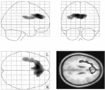
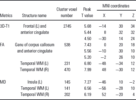
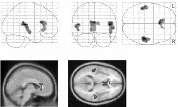
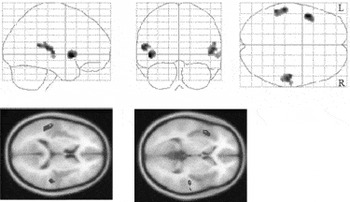
There were significant changes in cortical volume, FA and MD values in the space of a year, respectively. Specifically, a loss of regional cortical volume was observed in the anterior cingulate cortex (ACC) and left prefrontal lobe (Fig. 1; Table 2), a decrease in FA values was seen in the bilateral posterior superior temporal lobe, the genu of the corpus callosum and the ACC (Fig. 2; Table 2), and an increase in MD values was observed in the bilateral posterior superior temporal lobe and left insula (Fig. 3; Table 2).
Regions of grey matter atrophy in schizophrenic patients upon 1-year follow-up (paired t-test, SPM2). Significant volume losses were detected in the left prefrontal cortex and bilateral ACC.

The voxel-based analysis showing significant difference in schizophrenic patients upon 1-year follow-up
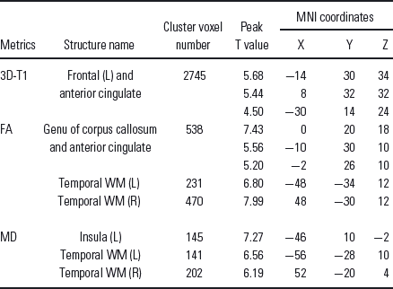
L, left side; MNI, Montreal Neurological Institute; R, right side; WM, white matter.
Regions of decreased FA values in schizophrenic patients upon 1-year follow-up (paired t-test, SPM2). Significant decreases in FA values were detected in the genu of the corpus callosum, the ACC and the bilateral temporal area.

Regions of increased MD values in schizophrenic patients upon 1-year follow-up (paired t-test, SPM2). Significant increases in MD values were detected in the left insula and bilateral temporal area.

No correlations were found between the change rate of the scan images of these three factors and disease duration, age at scan, full-scale IQ or daily dose of antipsychotic drugs (data not shown).
Discussion
To the best of our knowledge, this is the first study that provides a longitudinal evaluation of follow-up white matter changes in chronic schizophrenia. Using MRI and DTI, we found that there were ongoing changes in the brains of schizophrenic patients during the course of the illness. We also detected discrepancies between the results of volume study and DTI analysis.
We observed a volume loss in the left prefrontal cortex. Volumetric, DTI and postmortem studies(Reference Akbarian, Kim, Potkin, Hetrick, Bunney and Jones4,Reference Szeszko, Ardekani and Ashtari14,Reference van Haren, Hulshoff Pol and Schnack15,Reference Kirkpatrick, Messias, Conley and Roberts36) have shown that left prefrontal impairment occurs in schizophrenia; our results from volumetric data were consistent with these observations. With respect to DTI metrics, however, we did not detect any changes in this region. A previous study showed that difference in FA between schizophrenic patients and comparison controls was detected in left superior longitudinal fasciculus, but the difference was pronounced in the younger subjects only and tended to disappear with increasing ageReference Jones, Catani and Pierpaoli(10). In other words, the age-related change of FA in controls caught up with the FA change caused by the illness at the older groups. Some DTI studies have shown that FA and MD changes in white matter can be detected earlier than changes in the cortex in the same region in neurodegenerative illnessReference Müller, Greverus and Weibrich(37,Reference Ota, Sato, Saitoh, Endo, Murata and Asada38). The inconsistencies in the present DTI metrics and volumetric data in the left prefrontal cortex may result from the cessation of the DTI metrics change, which precedes a volume loss of the cortex, and from the continuous robust shrinkage of the left prefrontal cortex after the completion of the white matter change. The relatively low mean full-scale IQ of the participants (75.4 ± 16.2) may be a reflection of this frontal dysfunction.
The present DTI study also showed white matter abnormalities in the bilateral temporal area. Some schizophrenia studies have shown left temporal impairment(Reference Kasai, Shenton and Salisbury11,Reference Szeszko, Ardekani and Ashtari14,Reference van Haren, Hulshoff Pol and Schnack15), whereas the others indicate disruptions of the bilateral temporal area(Reference Andreasen, O'Leary and Flaum7,Reference Mathalon, Sullivan, Lim and Pfefferbaum12,Reference Ardekani, Nierenberg, Hoptman, Javitt and Lim22,Reference Mori, Ohnishi and Hashimoto29). The present study found changes in MD value in the bilateral temporal areas and left insula, and volume changes in the left prefrontal cortex. In a previous study, Jones et al.Reference Jones, Catani and Pierpaoli(10) identified a disturbance of the superior longitudinal fasciculus, which forms a connection from the frontal to the temporal lobe in schizophrenia, and also projects to the insula. Taking these results into consideration, the present results indicate a progressive disruption in the left dominant bilateral temporal areas in schizophrenia. In the present study, FA change of the left insula was unclear, possibly because of predominant subcortical connections and only weak or diffuse cortical connections in the insula. Directionality of diffusion was obscured by certain connections, and we were unable to detect any changes in the FA value.
Previous DTI studies have shown abnormalities in the ACC(Reference Sun, Wang, Cui, Breeze, Du and Wang21,Reference Ardekani, Nierenberg, Hoptman, Javitt and Lim22,Reference Wang, Dunnavant, Jarman and Deutch39). The cingulate cortex has profound reciprocal connections with many other regions, and functional, anatomical and histopathological studies provide considerable evidence that the connections between subregions of the cingulate cortex and other brain regions are disturbed in schizophreniaReference Benes(40,Reference Benes41). Consistent with the findings of previous studies that used not only DTI but also other methodologies, the present study supplies direct in vivo evidence of structural disconnections between the ACC and other brain regions in schizophrenia. Nevertheless, our study did not show any increase in MD in the ACC, possibly because of the partial volume effect derived from the volume loss of the ACC.
In the present study, we confirmed the abnormality of the genu of the corpus callosum; this is consistent with the results of previous studies(Reference Buchsbaum, Friedman and Buchsbaum42–Reference Rotarska-Jagiela, Schönmeyer, Oertel, Haenschel, Vogeley and Linden44). As the largest commissure of the human brain, the corpus callosum plays a central role in the hypoconnectivityReference Innocenti, Ansermet and Parnas(45,Reference Schlosser, Wagner, Kohler and Sauer46) and misconnectionReference Crow(47) models of schizophrenia. Some studies, however, have found decreased FA values only in the splenium(Reference Agartz, Andersson and Skare18,Reference Foong, Maier, Clark, Barker, Miller and Ron19,Reference Ardekani, Nierenberg, Hoptman, Javitt and Lim22). It is difficult to compare these studies as they differ not only in terms of the sample (first-episode vs. chronic schizophrenia patients) but also in their methods of analysis, which range from a whole-brain voxelwise approach to the extraction of diffusion values from small ROIs, which again vary across studies with respect to position and size. It is known that the FA and MD values of corpus callosum subregions can serve as indices of disturbance across their respective projection areasReference Ota, Obata and Akine(48), and that changes in the genu where the projection fibre to the prefrontal cortices runs through it may reflect changes in the prefrontal cortex where there is cortical volume loss was seen in schizophrenic patients(Reference Ho, Andreasen, Nopoulos, Arndt, Magnotta and Flaum9,Reference Mathalon, Sullivan, Lim and Pfefferbaum12,Reference van Haren, Hulshoff Pol and Schnack15). In the present study, only the left prefrontal cortex, and not the other cortices, decreased in volume during the 1-year test period, suggesting that the genu of the corpus callosum may show only secondary change. Our study found no increase in MD values in either the genu or the ACC; however, this may result from the partial volume effect because of the volume loss of the ACC, which lies next to the corpus callosum.
There were several limitations to the present study. First, we analysed only for patients. Volume loss of the brain is known to accelerate with age and the aging effect on white matter occurs relatively slowly compared to cortical changesReference Snook, Plewes and Beaulieu(34). It is also known that 1-year follow-up studies show no age-related change in DTI metrics in healthy older volunteersReference Weaver, Richards and Liang(49,Reference Chang, Wong and Nakama50). The present results indicate no correlation between the rate of change of the brain and age at scan, and may therefore be regarded as showing a progressive disruption during the course of the disorder that is almost free of the effects of aging. Furthermore, it is known that there are no age-related changes in FA in the temporal area in healthy malesReference Hsu, Leemans and Bai(51,Reference Salat, Tuch and Greve52).
Second, it is possible that there may be an effect of long-term medication with antipsychotics. Although daily dose of antipsychotics was not correlated with DTI values in our study, we were unable to estimate accurately the cumulative doses of antipsychotics during each patient's illness. Several morphological MRI studies and animal studies suggest that the administration of antipsychotics could affect brain morphologyReference Wang, Dunnavant, Jarman and Deutch(39,Reference Lieberman, Tollefson and Charles53). It is possible that long-term medication with antipsychotics also affects the microstructure of white matter in schizophrenics; longitudinal animal studies may clarify this issue.
Third, in the present study, we found no correlation between the rate of regional brain change per year and disease duration, full-scale IQ, daily dose of antipsychotic drugs or age at scan date; however, it must be kept in mind that our scan interval was short. Further studies with longer scan intervals may elucidate these points.
In summary, the present study confirms that there are ongoing changes in the brains of schizophrenic patients during the course of illness. Cortical volume loss was observed in the left prefrontal lobe, but changes in the DTI metrics of the left frontal white matter area were not, while there were changes in the DTI metrics of the bilateral temporal areas, but no shrinkage in these areas. These discrepancies between the volume data and DTI metrics in schizophrenic patients around age 60 suggest that the pattern of progressive brain changes in schizophrenia varies by brain region. The present observations remain to be replicated with a larger study population and with healthy controls.