An aberrant right subclavian artery arising from a left aortic arch is the most frequently described congenital aortic arch anomaly, occurring in 0.5–2.3% of the general population.Reference Molz and Burri 1 – Reference Ramos-Duran 3 Despite the retro-oesophageal course of the aberrant subclavian artery, an arterio-oesophageal fistula is an uncommon finding, only previously reported as a very rare complication in critically ill patients with oesophageal instrumentation or foreign-body ingestion.Reference Miller 4 , Reference Epstein and DeBord 5 We describe a unique case of a spontaneous aberrant right subclavian arterio-oesophageal fistula without an inciting event in a 17-month-old child.
Case presentation
A previously healthy 17-month-old child was brought to the emergency department following two episodes of haematemesis, which occurred the previous evening. There was no history of foreign-body ingestion. At presentation, the child was tachycardic and anaemic. She was admitted to the paediatric intensive care unit for observation, and received aggressive fluid resuscitation, first with saline and then blood products. A haematology evaluation was performed, the results of which were normal. Abdominal and chest x-rays were unremarkable, and a chest computed tomography scan displayed minimal right upper lobe disease, a left aortic arch with an aberrant right subclavian artery coursing posterior to the oesophagus. An oesophagogastroduodenoscopy was performed on day 1 at the hospital, which displayed a small mucosal abnormality with an unremarkable white plaque 10 cm from the mouth. There was no active bleeding along the entirety of the oesophagus or extensive blood clots in the stomach, and the patient otherwise presented normal anatomy. A repeat oesophagogastroduodenoscopy was performed on day 2 at the hospital, again displaying the stable white plaque. Later that day, a barium oesophagram with upper gastrointestinal follow-through displayed mucosal irregularity with collection of contrast at the level of the right subclavian artery.
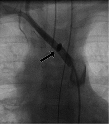
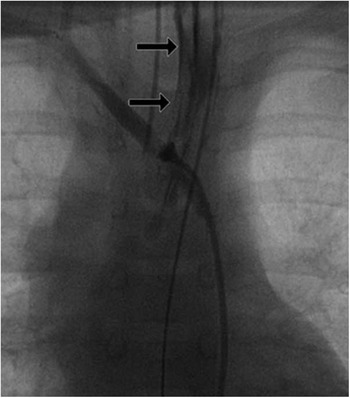
The patient continued to have sporadic episodes of gross haematemesis. On day 4 at the hospital, following an episode of large haematemesis, the paediatric cardiology department was consulted because of concerns for an arterio-oesophageal fistula. The patient was taken promptly to the cardiac catheterisation lab. An aortogram was performed with a pigtail catheter in the ascending aorta, which revealed the previously described left aortic arch, with the first branch giving rise to a common origin of the right and left carotid arteries. The second branch was the left subclavian artery, and the third and last branch was the right subclavian artery (Fig 1). There was no contrast enhancement of the oesophagus. A hyperdense spot of contrast, however, was noted in the aberrant right subclavian as it coursed anterior to the spine. A repeat aortogram was performed in the descending aorta, again displaying the hyperdense area of contrast in the mid aberrant right subclavian artery. The pigtail catheter was exchanged for a right coronary artery catheter, which was advanced into the aberrant right subclavian artery. Several selective hand-injection angiograms were performed in the mid right subclavian artery, with the final angiogram displaying contrast entering the oesophagus due to an arterio-oesophageal fistula (Fig 2).
Angiogram of the anomalous right subclavian artery in the posteroanterior (PA) projection, with a 4-Fr Judkins right coronary artery catheter in the mid portion of the anomalous right subclavian artery. A stagnant area of contrast enhancement is seen overlying the mid spine.

Angiogram of the anomalous right subclavian artery in the posteroanterior (PA) projection, with a 4-Fr Judkins right coronary artery catheter in the anomalous right subclavian artery rotated leftward. Contrast extravastion is seen along the fistula and into the oesophagus.

After displaying the arterio-oesophageal fistula, to prevent additional bleeding, a 5-mm angioplasty balloon was placed in the right subclavian artery and then inflated to temporarily occlude the artery. The patient was taken emergently from the cardiac catheterisation laboratory directly to the operating room for immediate surgical repair. Owing to haemodynamic instability, a median sternotomy was performed, and cardiopulmonary bypass was initiated. The previously placed angioplasty balloon was deflated and removed. The subclavian arterio-oesophageal fistula was identified and it was actively bleeding. The retro-oesophageal portion of the subclavian artery was clamped, with subsequent return of adequate blood pressure. The right subclavian artery was ligated at the origin from the aorta, and then re-implanted onto the proximal right carotid artery via an end-to-side anastomosis. The oesophagus was repaired primarily. Following the surgery, the patient returned to the intensive care unit and had no recurrent episodes of haematemesis. The patient made a complete recovery following prolonged hospitalisation.
Discussion
During embryological development, the left-sided aortic arch remains following dissolution of the right sixth aortic arch and the right dorsal aorta distal to the origin of the seventh intersegmental artery, transforming the right fourth arch into the right subclavian. With the dissolution of the right fourth aortic arch, the distal right dorsal aorta becomes the proximal right subclavian artery, giving the subclavian an aberrant, retro-oesophageal segment with an otherwise normal left aortic arch.Reference Weinburg 6 A left aortic arch with aberrant subclavian artery is the most common congenital aortic arch anomaly, and nearly all patients have an uneventful course with no clinical sequelae of the anatomical abnormality. Previous reports of complications related to this lesion are usually associated with foreign-body ingestion and perforation, direct instrumentation with trauma, or prolonged intubation.Reference Miller 4 , Reference Belkin 7 , Reference Fuentes 8 This case, however, represents a unique presentation of an arterio-oesophageal fistula without any such inciting events. The high mortality of arterio-oesophageal erosions necessitates aggressive treatment in any patient with a symptomatic aberrant right subclavian artery.Reference Millar 9 , Reference Hollander and Quick 10 In our patient, acute bleeding was stopped by placing an angioplasty balloon in the right subclavian artery to temporarily occlude flow, which allowed her to be taken emergently, but safely, to the operating room for re-implantation of the aberrant artery. The patient made a complete recovery.
Conclusion
Aggressive treatments are warranted in any patient with a symptomatic aberrant right subclavian artery, given the potentially disastrous consequences of arterio-oesophageal fistula formation. In our case, the key diagnostic intervention was performing the aortogram followed by selective right subclavian artery angiogram, as the chest tomography scan did not display evidence of a fistula.
Acknowledgements
None.
Financial Support
This research received no specific grant from any funding agency, commercial, or not-for-profit sectors.
Conflicts of Interest
None.