


Introduction
Clostridioides difficile causes more than 225,000 infections in hospitalized patients and 12,000 to 30,000 deaths in the U.S. each year. Reference Kociolek, Gerding and Carrico1 The National HealthCare Safety Network (NHSN) of the Centers for Disease Control and Prevention Division of Healthcare Quality Promotion monitors C. difficile infections (CDI) as a safety measure, 2 but evolving strategies for diagnosis are prompting adjustments to its measure.
Currently, most laboratories diagnose CDI using assays for the presence of toxin A and/or B. This can be done by detecting the genes encoding these enzymes using a nucleic acid amplification test (NAAT) or by using an enzyme immunoassay (EIA) for directly detecting the toxins in stool. The former has greater sensitivity whereas the latter has a greater clinical predictive value for CDI. Reference Kociolek, Gerding and Carrico1 For this reason, some laboratories employ a two-step method for diagnosis. Historically, the NAAT was sometimes performed when the initial EIA was negative to reduce false negatives. More recently, the more sensitive NAAT is done first followed by the more specific toxin EIA if the former is positive. The NHSN recommends that the finding of the last test performed using this two-step testing algorithm should determine if a positive laboratory assay definition is met. 2
However, adoption of a two-step testing method where a positive NAAT is followed by negative toxin EIA may lead to underreporting of patients with C. difficile healthcare-associated infections (HAIs) since the sensitivity of toxin assays is approximately 57%, potentially missing up to 40% of cases. Reference Prosty, Hanula and Khaled3 In VA, use of two-step testing led to a 62% decrease in reportable C. difficile HAIs. Reference McCauley, Evans, Simbartl, Gamage, Kralovic and Roselle4 In 13 Atlanta hospitals, the CDI Standardized Infection Ratio decreased 46% on average once two-step testing was adopted. Reference Goodenough, Sefton and Overton5
The possibility that patients with an active infection may be underreported is also suggested by data showing that NAAT+/toxin– patients are sometimes treated. In VA, more than 29% of NAAT+/toxin– patients were treated with oral vancomycin, metronidazole, or fidaxomicin. Reference Evans, McCauley and Simbartl6 In some non-VA hospitals, 70% to 78% of such patients may be treated. Reference Hecker, Son, Zuccaro, Conti and Donskey7 Guh et al, evaluating data from 36 laboratories at 5 NHSN Emerging Infections Program sites, showed that more than twice as many potentially unreported NAAT+/toxin– cases were treated than the number of reported NAAT+/toxin+ cases. Importantly, NAAT+/toxin– cases were as likely to have CDI-related complications as NAAT+/toxin+ cases. Reference Guh, Fridkin and Goodenough8
Because of these findings, a new surveillance definition, healthcare facility-onset antibiotic-treated C. difficile infection (HT-CDI), is under development by NHSN with the goal of approximating active CDI cases more closely by incorporating clinical decision-making into the metric. In this case, the prescribing of anti-C. difficile antibiotics serves as a proxy for the clinical decision.
To date, however, the impact of adopting this new metric on the number of CDI cases that would qualify for HAI surveillance reporting remains unknown. We chose to explore this by retrospectively evaluating CDI cases using the current HO-HCFA CDI definition and the proposed HT-CDI surveillance definition over a two-year period in a sample of VA medical centers.
Methods
HT-CDI definition
For the purposes of this study, an HT-CDI was defined according to NHSN guidelines as any qualifying C. difficile-positive assay collected in an inpatient location on day 4 or later of a healthcare facility admission, along with the administration of new qualifying antimicrobial therapy for C. difficile started within a window of ≤2 calendar days before or after the specimen collection date and continued for at least 5 calendar days or until the calendar day of/or day prior to discharge, death, transition to hospice care, or transfer to another facility.
Qualifying C. difficile assays included a NAAT specific for C. difficile toxin genes or a C. difficile toxin EIA. NAAT and toxin EIA could be done alone or in combination, or with the toxin EIA combined with a glutamate dehydrogenase (GDH) EIA as a two-step test.
Qualifying therapeutic agents for CDI were defined as enteral vancomycin, metronidazole, or fidaxomicin or intravenous metronidazole. The therapeutic agent was considered new for the purposes of this definition if it had not been given to the patient on either of the 2 calendar days preceding the current start date. The therapeutic agent or route of administration could be changed as long as the start date of at least one agent fell within the window period.
Comparing the number of C. difficile HAIs using two-step algorithms and the HT-CDI definition
The above HT-CDI definition was applied to all acute care inpatients tested for CDI from July 1, 2022, through June 30, 2024, in 25 volunteer acute care VA medical centers. These centers included both the most and least complex facilities. 9 Centers were chosen based on the use of NAAT alone, NAAT followed by toxin EIA, GDH followed by toxin EIA with discrepant results arbitrated by NAAT, GDH plus toxin EIA, toxin EIA alone, or GDH plus NAAT. In all cases, either a positive NAAT, GDH, or toxin EIA allowed identification of a patient. It was assumed that NAAT-positive, toxin EIA-negative patients or GDH-positive, toxin EIA-negative patients would not be reported to a national surveillance database since these are two-step algorithms where the last test done determines the result of the test. 2
The monthly CDI diagnostic testing algorithms used nationwide by all 124 acute care medical facilities during the two-year study period was determined from data entered into the Inpatient Evaluation Center (IPEC) database by local infection prevention and control personnel. Reporting by a facility was limited by the IPEC program to the six algorithms described above. Facilities in the study sample were recruited to gather data from those with the same distribution of testing algorithms as seen nationally.
The testing algorithm and use of CDI-specific antibiotics during the HT-CDI window period was determined by reviewing patient electronic health records from each of the recruited facilities using VA’s Compensation and Pension Record Interchange system or Joint Longitudinal Viewer. The difference in CDI case rates using the HT-CDI definition was compared to that which would be reported using current NHSN guidelines. 2
Results
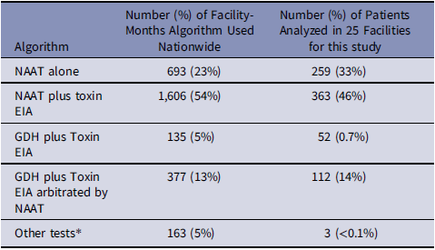
Over the two-year analysis period, the distribution of testing algorithms in the 124 acute care VA facilities nationwide and the distribution of patients in our sample from 25 facilities diagnosed with CDI using these algorithms was similar (Table 1). In every case where metronidazole was used, the drug was prescribed for CDI and not some other indication (e.g., anaerobes in a diabetic foot infection). In terms of treatment for patients identified as HT-CDI cases, 62% were treated with Vancomycin PO, 37% with Fidaxomycin PO, and 1% each with Metronidazole IV and PO respectively.
Distribution of testing algorithms used by 124 acute care VA facilities nationwide compared to distribution of patients analyzed in sample of 789 patients from 25 facilities (July 2022 through June 2024)

Table 1. Long description
A table comparing the distribution of testing algorithms used by 124 acute care VA facilities nationwide and the distribution of patients analyzed in a sample of 789 patients from 25 facilities. The table has four columns and five rows. The columns are labeled Algorithm, Number (%) of Facility-Months Algorithm Used Nationwide, and Number (%) of Patients Analyzed in 25 Facilities for this study. The rows are labeled with different algorithms: NAAT alone, NAAT plus toxin EIA, GDH plus Toxin EIA, GDH plus Toxin EIA arbitrated by NAAT, and Other tests. Row 1: NAAT alone, 693 (23 percent), 259 (33 percent). Row 2: NAAT plus toxin EIA, 1606 (54 percent), 363 (46 percent). Row 3: GDH plus Toxin EIA, 135 (5 percent), 52 (0.7 percent). Row 4: GDH plus Toxin EIA arbitrated by NAAT, 377 (13 percent), 112 (14 percent). Row 5: Other tests, 163 (5 percent), 3 (<0.1 percent).
* Included Toxin EIA alone, GDH plus NAAT, cell culture cytotoxin neutralization assay and toxigenic culture.
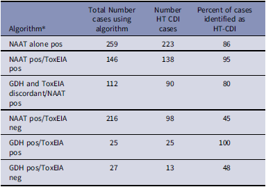
The most common diagnostic approach in our sample of 25 facilities was the use of NAAT either alone or in combination with the toxin EIA (Tables 1 and 2). NAAT was used in 734 (93%) of the 789 CDI cases analyzed (Figure 1), with 451 (61%) of these meeting the NHSN definition for reporting a HT-CDI case. An additional 98 (13%) cases were NAAT positive/toxin EIA negative. These may not have been reported based on the NHSN recommendation that the finding of the last test performed using a two-step testing algorithm should determine if a positive laboratory assay definition is met. 2 GDH was used in 52 (6.5%) of the 789 cases. Twenty-five of these were also toxin EIA positive and would have been reported, whereas 13 were toxin EIA negative and my not have been reported. Overall, adoption of the HT-CDI definition could potentially increase the number of reportable cases by 23.2% (Figure 1). The impact was most pronounced for patients diagnosed with two-step testing (NAAT followed by toxin EIA or GDH plus toxin EIA) (Table 2) where presumably the patient would not be reported to a national database based on the last test done. 2 Most patients diagnosed with NAAT alone or with NAAT as the last test done (GDH plus toxin EIA arbitrated by NAAT) were treated.
Effect of applying healthcare facility-onset antibiotic-treated Clostridioides difficile infection (HT-CDI) definition on reportable healthcare-associated C. difficile healthcare-associated infections. (*No NAAT done, **no NAAT or GDH done).

Relationship between diagnostic testing algorithm and healthcare facility-onset antibiotic-treated Clostridioides difficile infection (HT-CDI) in VA acute care medical facilities (July 2022 through June 2024)

Table 2. Long description
A table with six rows and four columns. The columns are labeled Algorithm, Total Number cases using algorithm, Number HT CDI cases, and Percent of cases identified as HT-CDI. The rows are labeled with different diagnostic algorithms: NAAT alone pos, NAAT pos/ToxEIA pos, GDH and ToxEIA discordant/NAAT pos, NAAT pos/ToxEIA neg, GDH pos/ToxEIA pos, and GDH pos/ToxEIA neg. Each row lists the total number of cases using the algorithm, the number of HT CDI cases, and the percent of cases identified as HT-CDI. For example, the first row shows NAAT alone pos with 259 total cases, 223 HT CDI cases, and 86 percent identified as HT-CDI.
* NAAT, nucleic acid amplification test, EIA, enzyme immunoassay, GDH, glutamate dehydrogenase.
Discussion
We found that application of the HT-CDI surveillance definition to Veterans Affairs patients could lead to an estimated 23% increase in the number of cases identified and eligible for reporting to a national database such as NHSN. This is a modest increase in comparison to studies previously done in private sector hospitals where increases in identifiable cases were as high as 70%. These cases may be a better indicator of the true burden of CDI in acute health care compared to those identified by two-step algorithms which rely on a relatively insensitive second test (toxin EIA) as the final answer. This was demonstrated by the common use of an anti-CDI antibiotic in these cases. Use of HT-CDI emphasizes that the diagnosis of CDI and treatment decisions may be driven by a patient’s clinical presentation and risk factors rather than by the type of test used.
Incorporating clinical decision-making manifested by the decision to treat may maintain (or increase) the ability to detect true-positive and decrease false-negatives or false-positives cases and promote improvement in patient care and safety by driving infection prevention practices, diagnostic stewardship, and antimicrobial stewardship while reducing unintended consequences.
Alternatively, it is unclear whether a clinician’s judgement is a better measure of real disease than laboratory tests since there is no gold standard diagnostic test to test this hypothesis. Clinician decisions to treat may be based on thoughtful considerations of risk factors (e.g., advanced age, receipt of high-risk antibiotics, history of hospitalization, and history of CDI) Reference Patterson, Fajnzylber and Nero10 but ultimately are arbitrary. Additionally, there may be facility incentive for physicians to not treat since treatment increases the number of publicly reportable cases for the facility Reference Guh, Fridkin and Goodenough8 and increases the potential for financial implications. 11 This could have a significant impact on patients since NAAT+/toxin– cases may be as likely to have CDI-related complications as NAAT+/toxin+ cases, and the mortality in untreated NAAT+/toxin– patients may be higher than for NAAT+/toxin– patients who are treated. Reference Prosty, Hanula and Khaled3,Reference Guh, Fridkin and Goodenough8 Currently, it is unknown what proportion of NAAT+/toxin– patients should be treated or how we can identify them. Reference Gerding12 Ultrasensitive quantitative C. difficile toxin assays which are being developed, may improve detection and the need for treatment, but these tests are currently unavailable commercially and there may be overlap between asymptomatic colonized and CDI patients. Reference Gerding12
There are several limitations to our work. It was often impossible for us to determine the order tests were done from the electronic health record since many tests had the same time stamp, which was presumably the time the stool specimen was collected. For our analysis, we assumed that when the NAAT was done along with the toxin EIA, the latter followed the former. We also could not identify the sequence of GDH followed by toxin with discrepant results arbitrated by NAAT for the same reasons. This may be important since patients diagnosed using this algorithm may be treated more often. Reference Gerding12 If this is the case, we may have identified more reportable CDI cases. The interpretative wording supplied by the laboratory when reporting the results may also be important for influencing whether clinicians treat the patient, especially if the wording suggests the results indicate colonization and not infection. Reference Guh, Fridkin and Goodenough8 Unfortunately, we were unable to track this meaningfully since report interpretation often changed over the two-year study period making it difficult to correlate with treatment.
Our data suggest that adoption of the HT-CDI definition will increase the number of cases eligible to be reported to a national surveillance database. In the absence of a gold standard for the diagnosis of CDI, it will remain to be seen what effect adding clinician intuition to current laboratory testing will have on accurately estimating the burden of CDI.
Acknowledgments
None.
Author contribution
The authors wish to thank Ms. Denise Leaptrot, MBA, SM/BSM(ASCP), CIC, Epidemiologist/Infection Preventionist, Contractor for National Healthcare Safety Network (NHSN), Centers for Disease Control and Prevention for providing subject matter expertise pertaining to the NHSN development of the HT-CDI surveillance criteria. The authors wish to thank the following MDRO Prevention Coordinators and Infection Preventionists for providing patient data from their respective facilities: Adebisi Adeyeye, VA New York Harbor Health Care System; Rowena Aseo, VA Palo Alto Health Care System; Lourdes Blanco, James A. Haley VA Medical Center; Jaime Conger, Carl T. Hayden Veterans’ Administration Medical Center; Vanessa Christian, Dayton VA Medical Center; Mang Ensell, Lt. Col. Luke Weathers, Jr. Memphis VA Medical Center; Jennifer Espinosa, VA Long Beach Health Care System; Elizabeth Flannery, Louis Stokes VA Medical Center; Delvina Ford, South Texas Veterans Health Care System, Audie L. Murphy Division; Leslie Furr, Tennessee Valley Healthcare System-Nashville Campus; Diana Greathouse, Richard L. Roudebush VA Medical Center; Anastashia Guillory, Jennifer Moreno Department of Veterans Affairs Medical Center; Meagan Hedin, Minneapolis VA Health Care System; Christine House, West Roxbury VA Medical Center; Angela Jordan, William S. Middleton Memorial Veterans Hospital; Debra Manderville, Malcom Randall Department of Veterans Affairs Medical Center; Avester Mitchell, West Palm Beach VA Health Care System; Paula Montgomery, Clement J. Zablocki VA Medical Center; Cindy Muyna, South Texas Veterans Health Care System; Illa (Marie) Penavs, Boise VA Medical Center; Amit Pulickan, Michael E. DeBakey Veterans Affairs Medical Center; Michael Raczynski, Salem VA Health Care System; Sherry Reid, VA North Texas Health Care System; Christine Southard, Central Texas Veterans Health Care System-Olin E. Teague; Teyobesta Tewabe, Iowa City VA Health Care System; Kelly Villar, VA New York Harbor Health Care System, and Christine Wilson, Montana VA Health Care System.
Financial support
None.
Competing interests
All authors report no conflicts of interest relevant to this article.