INTRODUCTION
The Indonesian archipelago forms a part of the ‘Coral Triangle’ (Veron, Reference Veron1995), a region recognized for the high diversity of its marine species (Briggs, Reference Briggs1999; Hoeksema, Reference Hoeksema and Renema2007; Bellwood & Meyer, Reference Bellwood and Meyer2009). However, the genetic diversity of the marine biota in this region remains incompletely studied and poorly understood (Veron, Reference Veron1995; Hoeksema, Reference Hoeksema and Renema2007). Gaining this knowledge is pivotal to the assessment of genetic diversity within populations and species, and to understand past factors and future triggers of how organisms radiate and disperse in a given region. Such information is important for managing ecosystems and directing conservation efforts towards better resilience in times of global warming and sea-level changes (Sutherland et al., Reference Sutherland, Porter and Torres2004; Jones et al., Reference Jones, Srinivasan and Almany2007; Lopez-Legentil & Pawlik, Reference Lopez-Legentil and Pawlik2009).
Sponges (Phylum Porifera) are an abundant and diverse group of marine organisms in the Indo-Pacific region, for which the genetic connectivity is also not fully understood (Wörheide et al., Reference Wörheide, Sole-Cava and Hooper2005; Bentlage & Wörheide, Reference Bentlage and Wörheide2007; Blanquer & Uriz, Reference Blanquer and Uriz2007). Sponges play an important role in marine ecosystems due to their impact on substrate (e.g. as bioeroders, sediment aggregators, or for reef restoration), chemical cycling, symbiotic associations (see review in Bell, Reference Bell2008) and might be a key component for the sustainability of reef ecosystems in oligotrophic seas (de Goeij et al., Reference de Goeij, van Oevelen, Vermeij, Osinga, Middelburg, de Goeij and Admiraal2013). Sponge biodiversity studies have been shown to assist in monitoring the influence of environmental factors such as depth, light, tidal amplitude, water flow rate, temperature, velocity, salinity and suspended sediment load (Carballo et al., Reference Carballo, Naranjo and García-Gómez1996; Bell, Reference Bell2007; Cleary & de Voogd, Reference Cleary and de Voogd2007; de Voogd & Cleary, Reference de Voogd and Cleary2007; Becking et al., Reference Becking, Cleary and de Voogd2013). In addition, many sponge species contain various secondary metabolites, which have been a focus of drug discovery for decades (e.g. Newman & Cragg, Reference Newman and Cragg2012).
An iconic sponge species in the Indonesian archipelago is the petrosiid demosponge Xestospongia testudinaria (Lamarck, 1815), also known as the ‘Giant Barrel Sponge’ (van Soest, Reference van Soest1989; van Soest et al., Reference van Soest, Boury-Esnault, Vacelet, Dohrmann, Erpenbeck, de Voogd, Santodomingo, Vanhoorne, Kelly and Hooper2012). Xestospongia testudinaria is known to be widespread in the Indo-Pacific and is one of the more common species on Indonesian coral reefs (van Soest, Reference van Soest1989; de Voogd & Cleary, Reference de Voogd and Cleary2008). Research on X. testudinaria has been focused mainly on new bioactive compounds (Calcul et al., Reference Calcul, Longeon, Mourabit, Guyot and Bourguet-Kondracki2003; Cao et al., Reference Cao, Foster, Brisson, Lazo and Kingston2005), ecology (Powell, Reference Powell2013), aspects of reproduction (Fromont, Reference Fromont and Choat1988; Fromont & Bergquist, Reference Fromont and Bergquist1994) and taxonomy (Fromont, Reference Fromont1991). More recently, research was conducted on the genetic connectivity of X. testudinaria, but so far only in a narrow geographic scale in northern (Swierts et al., Reference Swierts, Peijnenburg, de Leeuw, Cleary, Hörnlein, Setiawan, Wörheide, Erpenbeck and de Voogd2013) and south-east Sulawesi (Bell et al., Reference Bell, Smith, Hannan, Haris, Jompa and Thomas2014a). Swierts et al. (Reference Swierts, Peijnenburg, de Leeuw, Cleary, Hörnlein, Setiawan, Wörheide, Erpenbeck and de Voogd2013) found that in an area of less than 200 km2, X. testudinaria comprises four distinct morphotypes, which are not obviously affected by environmental factors, and could be differentiated using mitochondrial and nuclear markers. One of the most intriguing findings of that work was the overlap of mitochondrial haplotypes between the Indo-Pacific X. testudinaria with the Caribbean barrel sponge Xestospongia muta (Schmidt, 1870) (see also Montalvo & Hill, Reference Montalvo and Hill2011). These intriguing findings highlight the need for a better understanding of barrel sponge species and population structures on a wider, preferably global scale. This study should include a critical re-evaluation of how sponge species have been delineated in the past, and can act as models for the future (see e.g. Solé-Cava & Wörheide, Reference Solé-Cava, Wörheide, Custódio, Lôbo-Hajdu, Hajdu and Muricy2007), using barrel sponges as an example.
Throughout the entire history of sponge systematics (see Boury-Esnault, Reference Boury-Esnault2006) morphological apomorphies and specific character combinations were the major discriminatory factors in taxonomy (cf. Diagnosable Species Concept, see de Queiroz, Reference de Queiroz2007). Even now, the most recent supra-specific classification of sponges, Systema Porifera (Hooper & van Soest, Reference Hooper and van Soest2002), is predominantly based on morphological criteria. This concept, however, apparently fails to distinguish between all barrel sponge species. On the one hand few morphological features differentiate X. testudinaria from a second Indo-Pacific barrel sponge species, X. bergquistia Fromont (Fromont, Reference Fromont1991), as recently supported by molecular (microsatellite) data (Bell et al., Reference Bell, Smith, Hannan, Haris, Jompa and Thomas2014a, b). However, so far there have been no unequivocal morphological apomorphies established to differentiate these two Pacific species from the Caribbean X. muta barrel sponge. Likewise, biochemical compounds, occasionally used as a source for apomorphies in demosponges (Erpenbeck & van Soest, Reference Erpenbeck and van Soest2007), are inconclusive for barrel sponges. One X. muta specimen shared a similar, but not species-specific sterol composition to X. bergquistia specimens, and this was different from those found in specimens of X. testudinaria (Fromont et al., Reference Fromont, Kerr, Kerr, Riddle and Murphy1994).
Ecological parameters can provide apomorphies to differentiate sponge species (see e.g. Wapstra & van Soest, Reference Wapstra, van Soest, Vacelet and Boury-Esnault1987), where taxa are distinguishable by their respective ecological niches (cf. Ecological Species Concept, see de Queiroz, Reference de Queiroz2007). For example, among the oviparous and gonochoric barrel sponges, different spawning periods act as a potential reproductive barrier between the two Indo-Pacific taxa X. testudinaria and X. bergquistia (Fromont, Reference Fromont and Choat1988; Fromont & Bergquist, Reference Fromont and Bergquist1994). Likewise, the geographic distance and continental barrier that separates the Caribbean X. muta from the Indo-Pacific barrel sponges acts as a potential reproductive barrier and is regarded as an important distinguishing feature (Montalvo & Hill, Reference Montalvo and Hill2011).
The advent of molecular systematics in sponges (Kelly-Borges et al., Reference Kelly-Borges, Bergquist and Bergquist1991) facilitated the delimitation of taxa based on the coalescence or common ancestry of lineages (Monophyletic and Genealogical Species Concepts, de Queiroz, Reference de Queiroz2007) (see examples in Cárdenas et al., Reference Cárdenas, Pérez and Boury-Esnault2012). Many genetic analyses of barrel sponge species use the mitochondrial cytochrome oxidase subunit 1 (cox1), particularly its 3′ partition, which is shown to better resolve species-level differences than the standard barcoding fragment (Erpenbeck et al., Reference Erpenbeck, Hooper and Woerheide2006b). The 3′ partition enabled the recognition of four X. muta haplotypes in the Caribbean (Lopez-Legentil & Pawlik, Reference Lopez-Legentil and Pawlik2009), and six among Indo-Pacific barrel sponges (Swierts et al., Reference Swierts, Peijnenburg, de Leeuw, Cleary, Hörnlein, Setiawan, Wörheide, Erpenbeck and de Voogd2013). Swierts et al. (Reference Swierts, Peijnenburg, de Leeuw, Cleary, Hörnlein, Setiawan, Wörheide, Erpenbeck and de Voogd2013) however, also corroborated earlier studies that failed to distinguish the geographically separated X. muta and X. testudinaria based on cox1 sequences alone (Montalvo & Hill, Reference Montalvo and Hill2011). So far, barrel sponge cox1 contradicts earlier studies on the use of this marker for unravelling demosponge species level phylogenies (Erpenbeck et al., Reference Erpenbeck, Breeuwer, Parra-Velandia and van Soest2006a) (see also Lopez-Legentil & Pawlik, Reference Lopez-Legentil and Pawlik2009; Xavier et al., Reference Xavier, Rachello-Dolmen, Parra-Velandia, Schönberg, Breeuwer and van Soest2010; Reveillaud et al., Reference Reveillaud, van Soest, Derycke, Picton, Rigaux and Vanreusel2011).
These findings prompted the present study, which aims to analyse the haplotype diversity of barrel sponges at the largest geographic scale attempted so far, stretching from the western Indian Ocean to the west Pacific, and including the type material of all three barrel sponge species. We explore the suitability of one of the most frequently used mitochondrial markers, cox1, to distinguish the three barrel sponge species. Furthermore, this study aims to further our understanding of the genetic population structure of the barrel sponge species at a much wider geographic scope than previous studies that were limited to populations surrounding Sulawesi (Swierts et al., Reference Swierts, Peijnenburg, de Leeuw, Cleary, Hörnlein, Setiawan, Wörheide, Erpenbeck and de Voogd2013; Bell et al., Reference Bell, Smith, Hannan, Haris, Jompa and Thomas2014a).
MATERIALS AND METHODS
Fragments of 187 specimens of Xestospongia testudinaria were freshly collected from three regions: Java (West, Central, and East), Bali and Sulawesi (Lembeh, Spermonde, Wakatobi). Immediately after collection, samples were cut, rinsed and soaked in 99% ethanol before being kept in fresh 99% ethanol. An additional 24 samples were studied from Saudi Arabia, Tanzania, Taiwan and Thailand, provided by the Naturalis Biodiversity Center, Leiden, the Netherlands, and samples from the Great Barrier Reef (GBR) and Solomon Islands provided by the Queensland Museum (QM) Brisbane, Australia. Furthermore, the Queensland Museum also provided five individuals of Xestospongia bergquistia, including the holotype specimen (see Supplementary Material). In addition, we also obtained the neotype (BMNH 1881.10.21.266) and another, associated specimen of Ridley's (1881) material (BMNH 1881.10.21.267) of X. testudinaria from the British Museum of Natural History, London (Hooper & Wiedenmayer, Reference Hooper, Wiedenmayer and Wells1994). For Xestospongia muta, two syntypes were investigated (MCZ PORa-6449, MCZ PORa-6450, of which we designate the former as lectotype) and additional sequences were taken from GenBank (http://www.ncbi.nlm.nih.gov).
Samples were extracted based on previously published methods established for sponge barcoding (Vargas et al., Reference Vargas, Schuster, Sacher, Büttner, Schätzle, Läuchli, Hall, Hooper, Erpenbeck and Wörheide2012). The I3-M11 partition of cox1 was amplified with the Polymerase Chain Reaction (PCR) using primers CJ-J2165 and C1-Npor 2760 (Erpenbeck et al., Reference Erpenbeck, Breeuwer, van der Velde and Soest2002). The 25 µL PCR mix consisted of 5 µL 5× green GoTaq ® PCR Buffer (Promega Corp, Madison, WI), 4 µL 25 mM MgCl2 (Promega Corp, Madison, WI), 2 µL 10 mM dNTPs, 1 µL each primer (5 µM), 9.8 µL H2O, 0.2 µL GoTaq ® DNA polymerase (5 µ μL−1) and 2 µL DNA template. The PCR regime comprised an initial denaturation at 94°C for 3 min, 35 cycles of 30 s denaturation at 94°C, 20 s annealing at 42°C and 60 s elongation at 72°C each, followed by a final elongation at 72°C for 5 min. Re-amplifications with an internal primer set of the partition I3-M11 (CO1porF1 and CO1porR1, Erpenbeck et al., Reference Erpenbeck, Knowlton, Talbot, Highsmith and van Soest2003) and combinations (CJ-J2165 – CO1porR1 and CO1porF1 – C1-Npor 2760) were required for obtaining PCR products from the type specimen of X. bergquistia. For the 133 year old specimen of X. testudinaria (BMNH 1881.10.21.267), the primer set CJ-J2165 and COX1-R1 (5′-TGTTGRGGGAAAAARGTTAAATT-3′) (Rot et al., Reference Rot, Goldfarb, Ilan and Huchon2006) was amplified under the use of additional 2 µL Bovine Serum Albumin (BSA, 10 mg mL−1) and the PCR regime of 45 s annealing at 42°C, 45 s elongation at 72°C and a final elongation at 72°C for 7.5 min. All of the PCR products were cleaned by ammonium acetate precipitation. Sequencing of forward and reverse strand was performed with the ABI BigDye v3.1 chemistry (Applied Biosystems, CA, USA) and the amplification primers following the manufacturer's protocol on an ABI 3730 Automated Sequencer in the Genomic Sequencing Unit of the LMU Munich. Sequences were assembled, trimmed and analysed by Geneious version 6.1.7 (http://www.geneious.com). Sequences were checked with BLAST against GenBank (http://www.ncbi.nlm.nih.gov) for contaminations. In total 211 sequences were aligned with MUSCLE version 3.5 (Edgar, Reference Edgar2004) as implemented in Geneious under default settings. Sequences of X. testudinaria, which are identical to the haplotype of sequences in the study of Swierts et al. (Reference Swierts, Peijnenburg, de Leeuw, Cleary, Hörnlein, Setiawan, Wörheide, Erpenbeck and de Voogd2013), are numbered according to that study (haplotypes C1-C6, GenBank accession numbers KC424439–KC424444). Conversely, new haplotypes (one haplotype C7, two type specimens (X. testudinaria and X. bergquistia) and one X. bergquistia haplotype) were submitted and deposited to NCBI GenBank under accession numbers KM014752-KM014755.
TCS v 1.21 (Clement et al., Reference Clement, Posada and Crandall2000) was used to create a statistical parsimony haplotype network among the analysed barrel sponges (X. testudinaria, X. bergquistia and X. muta) to map barrel sponge haplotype distributions according to their localities. DnaSP v. 5.10.01 (Librado & Rozas, Reference Librado and Rozas2009) was recruited to obtain genetic diversity indices (π) and to test neutral evolution of I3-M11 alleles from X. testudinaria by calculating Tajima's D (Tajima, Reference Tajima1989). For the analysis of the genetic structure of X. testudinaria, regional samples were pooled as follows: Saudi Arabia (N = 3), Tanzania (N = 13), Taiwan (N = 7), Thailand (N = 12), west Java (N = 14), central Java (N = 9), east Java & Bali & Sumba (N = 10), north Sulawesi (N = 126), south & southeast Sulawesi (N = 10), and the GBR and Solomon Islands (N = 7). A hierarchical analysis of molecular variance (AMOVA) was performed and pairwise F ST values calculated in Arlequin v 3.5.1.2 (Excoffier et al., Reference Excoffier, Laval and Schneider2005) with a permutation test of 10,000 replicates. The significance of F ST values was amended after a Bonferroni correction (Rice, Reference Rice1989). Phylogenetic reconstructions among barrel sponges were performed under Maximum-likelihood (ML) and Bayesian inferences criteria. A Maximum-likelihood phylogram was inferred by RAxML v. 7.0.4 in raxmlGUI v. 1.3 (Silvestro & Michalak, Reference Silvestro and Michalak2012) using 1000 rapid bootstrap replications (Stamatakis et al., Reference Stamatakis, Hoover and Rougemont2008) and the GTR + I + Γ model as suggested by jModeltest v. 2.1.3 (Darriba et al., Reference Darriba, Taboada, Doallo and Posada2012) under the Akaike Information Criterion (Akaike, Reference Akaike1974). Likewise, a Bayesian phylogram was inferred using MrBayes v. 3.2.1 (Ronquist et al., Reference Ronquist, Teslenko, van der Mark, Ayres, Darling, Höhna, Larget, Liu, Suchard and Huelsenbeck2012) under the same model of evolution. Each analysis consisted of two independent runs of four Metropolis-coupled Markov-chains under default temperature with trees sampled at every 1000th generation. Analyses were terminated automatically when the chains converged significantly as indicated by an average standard deviation of split frequencies <0.01.
Spicule re-measurements were made for the three type specimens of barrel sponges X. testudinaria (BMNH 1881.10.21.266), X. bergquistia (QM G25018) and X. muta (MCZ PORa-6449). Spicule preparations were made by dissolving a small piece of the specimen in commercial bleach, after which the residue was rinsed four times with water, and once with 96% ethanol. The spicules were air-dried on microscopic slides and prepared for study with a light microscope by mounting in Ultrabed. Spicule dimensions are given as the mean length times mean width of 25 spicule measurements.
RESULTS
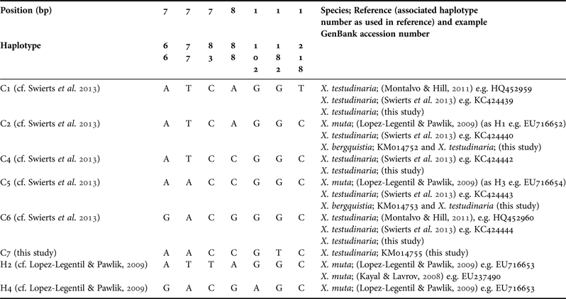
We obtained partial cox1 sequences (I3-M11 partition) of 211 specimens of Xestospongia testudinaria and six of X. bergquistia resulting in an alignment of 544 base pairs with six different haplotypes (Table 1). No amplification product for the neotype of X. testudinaria BMNH 1881.10.21.266, and the type material of X. muta MCZ PORa-6449 and MCZ PORa-6450 could be obtained. Five haplotypes were identical to haplotypes reported from Swierts et al. (Reference Swierts, Peijnenburg, de Leeuw, Cleary, Hörnlein, Setiawan, Wörheide, Erpenbeck and de Voogd2013) (C1, C2, C4–C6), whose labelling system is followed here; one novel haplotype was discovered and was subsequently named C7.
Haplotypes recovered from the Xestospongia barrel sponge alignment. Positions refer to the full cox1 sequence from Xestospongia muta EU716653 (Kayal & Lavrov, Reference Kayal and Lavrov2008); C1–C7, haplotype annotation follows Swierts et al. (Reference Swierts, Peijnenburg, de Leeuw, Cleary, Hörnlein, Setiawan, Wörheide, Erpenbeck and de Voogd2013); H1–H4, follows the labelling of Lopez-Legentil & Pawlik (Reference Lopez-Legentil and Pawlik2009).

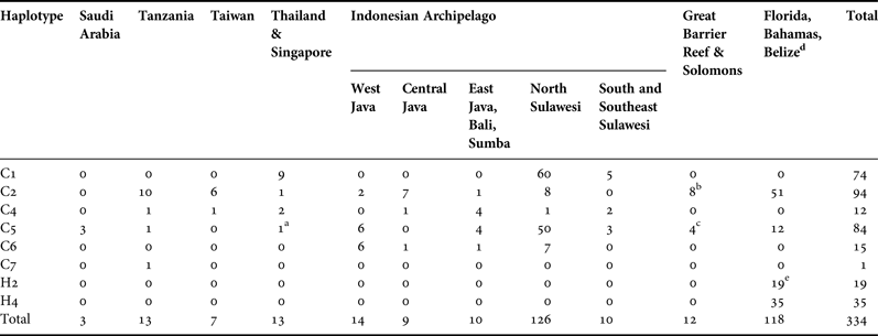
All six haplotypes were present in X. testudinaria (Figure 1). Two haplotypes are shared between X. testudinaria and X. bergquistia (Haplotype C2 and C5). The holotype of X. bergquistia (QM G25018) possessed the C5 haplotype and X. testudinaria BMNH 1881.10.21.267 possessed the C2 haplotype. These sequences differed by an uncorrected p-distance of 0.0092. The inclusion of previously published Xestospongia cox1 sequences from GenBank revealed that two haplotypes (C2 and C5) are shared between the Indo-Pacific and Caribbean barrel sponge species. The 116 specimens of the Caribbean barrel sponge (Xestospongia muta) had a total of four haplotypes. Two of these haplotypes (H2 and H4, as defined by Lopez-Legentil & Pawlik, Reference Lopez-Legentil and Pawlik2009), are not shared with the Indo-Pacific specimens (see Tables 1 & 2).
Haplotype distribution of Xestospongia barrel sponge sequences used in this study. The size in the pie charts is proportional to the sample size. Different haplotypes are colour- and pattern-coded (see Tables 1 & 2 for further details). Inset: Samples from the Caribbean (published by Kayal & Lavrov, Reference Kayal and Lavrov2008; Lopez-Legentil & Pawlik, Reference Lopez-Legentil and Pawlik2009). C1–C7 haplotype annotations follow Swierts et al. (Reference Swierts, Peijnenburg, de Leeuw, Cleary, Hörnlein, Setiawan, Wörheide, Erpenbeck and de Voogd2013), whereas H1–H4 follow Lopez-Legentil & Pawlik (Reference Lopez-Legentil and Pawlik2009).
Haplotype distribution of Xestospongia testudinaria and other barrel sponges according to their geographic localities. Haplotype annotation follows Swierts et al. (Reference Swierts, Peijnenburg, de Leeuw, Cleary, Hörnlein, Setiawan, Wörheide, Erpenbeck and de Voogd2013) and Lopez-Legentil & Pawlik (Reference Lopez-Legentil and Pawlik2009)
a One Xestospongia bergquistia specimen from Singapore.
b Including one specimen of X. bergquistia from the Great Barrier Reef and one Ridley (1881) specimen of X. testudinaria (BMNH 1881.10.21.267).
c Four specimens (including one holotype) of X. bergquistia.
d Taken from Lopez-Legentil & Pawlik (Reference Lopez-Legentil and Pawlik2009).
e Including full mt genome sequence of X. muta (see Kayal & Lavrov, Reference Kayal and Lavrov2008).
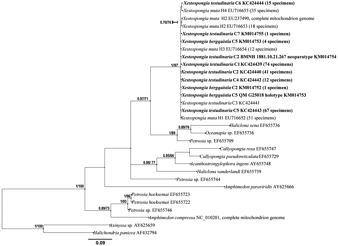
A close genealogical relationship among haplotypes was observed (Figure 2). All haplotypes were connectable with a maximum of one inferred mutational step. Haplotype C1 is the most abundant haplotype among the Indo-Pacific samples, comparable with H1 in the Caribbean (cf. Lopez-Legentil & Pawlik, Reference Lopez-Legentil and Pawlik2009) – with one mutational step separating those two major haplotypes. Haplotypes C5 and C1 were the dominant haplotypes in the Indonesian archipelago, from which the majority of specimens of X. testudinaria used in this study were collected. Haplotype C7 was exclusively found in the 13 samples from Tanzania, which in total displayed four different haplotypes. Among the three samples from the Red Sea, Saudi Arabia, only C5 was found (Figures 1 & 2). In the reconstructed phylogram the barrel sponges are recovered as monophyletic with high support (Figure 3). However, as expected from the overlapping haplotypes, the cox1 fragment fails to support the monophyly of any of the three barrel sponge species.
Haplotype network of the Xestospongia barrel sponge specimens with respect to their sampling location (see Tables 1 & 2 for details). Xestospongia muta sequences: (Kayal & Lavrov, Reference Kayal and Lavrov2008; Lopez-Legentil & Pawlik, Reference Lopez-Legentil and Pawlik2009). C1–C7 haplotype annotations follow Swierts et al. (Reference Swierts, Peijnenburg, de Leeuw, Cleary, Hörnlein, Setiawan, Wörheide, Erpenbeck and de Voogd2013), whereas H1–H4 follow Lopez-Legentil & Pawlik (Reference Lopez-Legentil and Pawlik2009). Numbers indicate the number of sequences analysed.
Bayesian inference phylogram of Xestospongia barrel sponge haplotypes included in the analysis and other marine haplosclerid taxa. New sequences are given in bold and followed by the number of sequences obtained in this study, including GenBank accession numbers. The haplotype annotations follow Swierts et al. (Reference Swierts, Peijnenburg, de Leeuw, Cleary, Hörnlein, Setiawan, Wörheide, Erpenbeck and de Voogd2013) and Lopez-Legentil & Pawlik (Reference Lopez-Legentil and Pawlik2009). Numbers on the branches represent posterior probabilities (PP)/bootstrap proportions (BP) of maximum likelihood analyses. Scale bar indicates the number of substitutions/site.
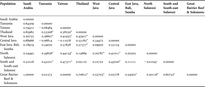
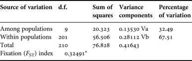
The nucleotide diversity (π) value of X. testudinaria was 0.0029 and Tajima's D 1.65911 (P > 0.1), which indicated neutral evolution of the I3-M11 fragment. Furthermore, samples from four regions (Thailand, West Java, North Sulawesi and GBR + Solomon Islands) displayed significantly different population structures from the other localities (see Table 3). Moreover, the hierarchical AMOVA analysis revealed that 32.49% of the genetic variation was found among populations and 67.51% within populations (see Table 4). Likewise, the overall F ST value (0.32491, P < 0.05) indicated the presence of genetic structuring of X. testudinaria in the Indo-Pacific area.
Pairwise F ST values between populations of X. testudinaria
*Significant values at P < 0.005 after Bonferroni corrections.
Hierarchical analysis of molecular variance (AMOVA) based on the I3-M11 region of X. testudinaria
*Significant values at P < 0.005 after Bonferroni corrections.
DISCUSSION
Cytochrome oxidase 1 and barrel sponge species differentiation
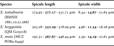
Currently the three barrel sponge species Xestospongia testudinaria, X. muta and X. bergquistia cannot be differentiated using the I3-M11 partition of cox1, which was suggested as a candidate marker for species level phylogenies other than the standard barcoding marker (Erpenbeck et al., Reference Erpenbeck, Hooper and Woerheide2006b), or by using any morphological or ecological parameter alone. The three barrel sponge species therefore can be regarded as ‘Gray zone species’ (de Queiroz, Reference de Queiroz2007) given our current understanding of them. The concept of Gray zone species arises when there are differences in the views of species concepts, which then lead to conflicts in species delimitation (de Queiroz, Reference de Queiroz2007). Nevertheless, despite the overlapping haplotypes observed here, the three Xestospongia taxa cannot constitute a single species as evident from their morphological and biochemical differences. No unique character distinguishing the three species has yet been found. The measured spicules of all three type specimens revealed overlapping spicule dimensions (see Table 5). However, with respect to the two Indo-Pacific species, the magnitude of spongin reinforcement in the skeletons (Fromont, Reference Fromont1991) and microsatellite data (Bell et al., Reference Bell, Smith, Hannan, Haris and Thomas2014b) supported species separation.
Spicule dimensions of the barrel sponge type specimens
Minimum-mean-maximum.
In terms of ecological parameters, the three species of barrel sponges constitute different species based on reproductive behaviour (Fromont, Reference Fromont and Choat1988, Ritson-Williams et al., Reference Ritson-Williams, Becerro and Paul2005), and by their wide geographic separation (Montalvo & Hill, Reference Montalvo and Hill2011). Contact zones between the Indo-Pacific and the Caribbean barrel sponges do not exist. While gene flow between barrel sponge populations in the Caribbean and the Indo-Pacific via ballast water from ships or vessels travelling through the Panama Canal is possible (Ruiz et al., Reference Ruiz, Torchin, Grant, Lang, MacIntyre and Rützler2007), it is unlikely to cause or explain the haplotype patterns observed in this study. No stepping stones bridging the Atlantic or the Pacific populations have been found yet that would facilitate a worldwide distribution of a single species. In addition, sponge larvae are known to typically have short lifespans (see Maldonado, Reference Maldonado2006). There are no data on the present or past existence of barrel sponges in the Eastern Pacific, which might potentially explain the haplotype overlaps as an historical consequence of gene flow between both regions (see in this context Knowlton et al., Reference Knowlton, Weigt, Solorzano, Mills and Bermingham1993; Knowlton & Weigt, Reference Knowlton and Weigt1998).
Why does the cytochrome oxidase 1 display overlapping haplotypes? We conclude that Xestospongia mitochondrial systematics suffers from factors that mask the distinctiveness of the individual lineages. This can occur if divergence time for the cytochrome marker is longer than the divergence time of the two sibling species (Charlesworth, Reference Charlesworth2010). In other words, the polymorphisms occurred before descendants split into two separate species. These so-called ancient polymorphisms explain the overlapping haplotype distribution among the widely distributed barrel sponge populations rather than the presence of a single cosmopolitan barrel sponge species, although additional gene loci are required to test this hypothesis (see e.g. Wu, Reference Wu1991; Takahata & Satta, Reference Takahata and Satta1997; Rannala & Yang, Reference Rannala and Yang2003; Zhou et al., Reference Zhou, Zeng, Wu, Chen, Yang, Shi and Wu2007). The distribution of morphological, ecological and biochemical features of the three barrel sponge species strongly suggest the presence of ancient polymorphisms and explain the presence of identical haplotypes for morphotypes in both X. muta and X. testudinaria barrel sponges (Lopez-Legentil & Pawlik, Reference Lopez-Legentil and Pawlik2009). These ancient polymorphisms, in combination with the slow mitochondrial substitution rates in poriferans, prevent the formation of monophyletic species based on cox1 data and falsely suggest the presence of cosmopolitan lineages, which is a concept successively rejected in the literature on sponges and other marine taxa (see Palumbi et al., Reference Palumbi, Grabowsky, Duda, Geyer and Tachino1997; Solé-Cava & Boury-Esnault, Reference Solé-Cava and Boury-Esnault1999; Wörheide et al., Reference Wörheide, Epp and Macis2008). This is particularly evident since microsatellites, which are faster evolving independent markers, clearly show the distinction between X. bergquistia (including the type material) and X. testudinaria samples (Bell et al., Reference Bell, Smith, Hannan, Haris, Jompa and Thomas2014a, b). These results imply that the I3-M11 cannot be regarded as universally suitable for detecting species boundaries in all demosponges.
The suitability of cytochrome oxidase 1 to resolve barrel sponge intra-specific relationships
Nucleotide diversity (π) is a measure of genetic variation, which is applied to calculate the degree of polymorphism in a population. Therefore, the higher the π value, the more suitable is a marker in assessing intraspecific genetic diversity. The low variation in mitochondrial genomes observed in several non-bilaterian taxa (see e.g. Shearer et al., Reference Shearer, van Oppen, Romano and Wörheide2002; Huang et al., Reference Huang, Meier, Todd and Chou2008) generally restricts the use of mitochondrial proteins as markers for population analyses in demosponges. However, a higher π value on the 3′ (I3-M11) partition in mitochondrial DNA would suggest that this marker is more suitable to resolve genetic diversity compared with the 5′ partition. For X. muta, Lopez-Legentil & Pawlik (Reference Lopez-Legentil and Pawlik2009) found a nucleotide diversity of π = 0.00058 in the 5′ partition, contrasting the much higher nucleotide diversity of π = 0.0039 in the I3-M11 partition, which restricts the use of the 5′ partition for intraspecific analyses and highlights the use of the I3-M11 partition for these purposes (see additional examples in Uriz & Turon, Reference Uriz and Turon2012). Among the Indo-Pacific X. testudinaria six haplotypes were sampled over a range of more than 10,000 km extending from the Red Sea to Melanesia with π = 0.00290 in the I3-M11 fragment. Five of these haplotypes were detected in specimens from the Indonesian archipelago in an area of 2300 km distance (π = 0.00302 west Java to north Sulawesi). Nevertheless, our analysis clearly highlights that the application of I3-M11 in barrel sponges is only suitable when the correct boundaries of the target species are defined. Among Indo-Pacific sponges all haplotypes of X. bergquistia, of which the I3-M11 fragment has been sequenced for the first time, overlap with haplotypes of X. testudinaria. Likewise 50% of the haplotypes of the Caribbean X. muta found by Lopez-Legentil & Pawlik (Reference Lopez-Legentil and Pawlik2009) are shared with Indo-Pacific haplotypes. Consequently, prior to the application of I3-M11 on the intraspecific relationships of barrel sponges, the correct assignment to either barrel sponge species is mandatory.
Implications for the genetic diversity and population structure of Xestospongia testudinaria
In Xestospongia testudinaria we recognize a major difference in common haplotypes among regions. For instance, C2 is the most common haplotype in Tanzania, Taiwan and the Great Barrier Reef. On the other hand, C1 is the most common haplotype in Sulawesi and Thailand, and is also only present in those areas. One haplotype is found in Tanzania (Indian Ocean) only. The occurrence of major haplotypes has been suggested to be influenced by environmental factors such as currents or anthropogenic effects (Lopez-Legentil & Pawlik, Reference Lopez-Legentil and Pawlik2009; Swierts et al., Reference Swierts, Peijnenburg, de Leeuw, Cleary, Hörnlein, Setiawan, Wörheide, Erpenbeck and de Voogd2013), and shows an eco-geographic heterogeneity in the Indo-Pacific. In addition, the hierarchical AMOVA and overall F ST results display genetic structuring for X. testudinaria, which also corroborated the results of the geographically narrow scale studies of north Sulawesi (Swierts et al., Reference Swierts, Peijnenburg, de Leeuw, Cleary, Hörnlein, Setiawan, Wörheide, Erpenbeck and de Voogd2013) and south-east Sulawesi (Bell et al., Reference Bell, Smith, Hannan, Haris, Jompa and Thomas2014a) populations.
The existence of different external morphotypes in X. testudinaria with distinct haplotypes, which likely represent reproductively isolated rather than ecophenotypic varieties, implies the presence of a X. testudinaria species complex composed of several sympatric species (Swierts et al., Reference Swierts, Peijnenburg, de Leeuw, Cleary, Hörnlein, Setiawan, Wörheide, Erpenbeck and de Voogd2013) (see also Bell et al., Reference Bell, Smith, Hannan, Haris, Jompa and Thomas2014a). Likewise, in X. muta, two morphotypes (smooth and digitate) were found to have different genotypes although the authors assume that hybridization prevents their separation (Lopez-Legentil & Pawlik, Reference Lopez-Legentil and Pawlik2009). The presence of different X. muta lineages is also evident from different sterol compositions in three specimens of this species examined by Fromont et al. (Reference Fromont, Kerr, Kerr, Riddle and Murphy1994), although they did not assign the different chemotypes to morphotypes. Nevertheless, our understanding of Xestospongia barrel sponge species with respect to their global genetic structuring and diversity is still limited due to unbalanced sample sizes and insufficient numbers of sequences at the regional scale (e.g. in the Solomon Islands, Great Barrier Reef, Thailand, Taiwan, Tanzania, Red Sea), including potentially missed haplotypes in some regions. These should be addressed in future analyses. Because of high sample numbers from one specific area and low numbers from other areas, we might have missed the occurrence of other haplotypes. For this reason, a more balanced sample size is required in order to get more accurate results for the future study of spatial relationships in every barrel sponge species, particularly X. testudinaria and X. bergquistia in the Indo-Pacific. Likewise, the utilization of nuclear intron markers, such as ATPS-beta (Bentlage & Wörheide, Reference Bentlage and Wörheide2007; Wörheide et al., Reference Wörheide, Epp and Macis2008) could be beneficial to support our understanding of barrel sponge relationships.
SUPPLEMENTARY MATERIAL
To view supplementary material for this article, please visit http://dx.doi.org/10.1017/S0025315415001149.
ACKNOWLEDGEMENTS
ES would like to thank DAAD (German Academic Exchange Service) for the award of a PhD Fellowship. In addition, ES acknowledges a Martin Fellowship from the Naturalis Biodiversity Center. The laboratory assistance from Gabriele Büttner, Simone Schätzle and Astrid Schuster, constructive criticism from Sergio Vargas, Gaurav Shimpi (Molecular Palaeobiology research group at LMU Munich) and Rob van Soest (The Naturalis Biodiversity Center, the Netherlands), as well as samples provided by Ratih Aryasari (Biology Faculty, Gadjah Mada University Indonesia), Elisabeth Crovetto, Yusheng Huang (National Penghu University of Science and Technology, Taiwan), Merrick Ekins (Queensland Museum, Australia), Adam Baldinger of Harvard University (Museum of Comparative Zoology) and Emma Sherlock (BMNH), are highly appreciated. We also thank Jane Fromont and another anonymous reviewer for constructive criticism that improved the manuscript considerably.