Introduction
Fibro-osseous lesions are primarily classified as ossifying fibromas, fibrous dysplasia or cemento-osseous dysplasia. Ossifying fibromas include juvenile psammomatoid ossifying fibroma and juvenile trabecular ossifying fibroma, which show aggressive growth and invasion of surrounding structures at an early age.Reference Nwizu, Aguire and Chen1 The best-known fibro-osseous lesion is fibrous dysplasia, which can be monostotic or polyostotic and mainly affects the cranial bones or extremities. The best treatment for fibro-osseous lesions is surgical contouring of the affected bone after the patient's bony growth has arrested, with a few exceptions, such as in patients with Albright syndrome.Reference Kusano, Hirabayashi, Eguchi and Sugawara2 The growth arrest of fibrous dysplasia is reported to occur at 23 years of age on average. However, lesions frequently recur, necessitating multiple surgeries.Reference Kusano, Hirabayashi, Eguchi and Sugawara2
The surgeon's decisions about treatment strategy include not only when to perform surgery, but also to what extent bone should be removed during lesion contouring.Reference Kusano, Hirabayashi, Eguchi and Sugawara2, Reference Gui, Zhang, Shen, Wang, Bautista and Voss3 Deciding the optimal amount of lesion to remove, so that the patient receives the best cosmetic outcome while preserving critical orbital structures, such as the optic nerve, the trigeminal nerve, and the medial palpebral ligament, is challenging. These considerations lengthen surgery time for fibro-osseous lesions.
Recently, computer-assisted simulation and navigation systems, which allow for pre-operative planning and intra-operative visualisation, have been used effectively in the treatment of sinonasal lesions, resulting in improved surgical outcomes.Reference Oeken and Torpel4 However, navigation systems that use superficial skin registration cannot prevent subtle discrepancies, because soft tissue swelling, changes in body position, or tumour growth after image capturing can easily cause dislocation of the registered surface and intra-operative markers. Fixed registration of cranial bone that does not cause dislocation of the target area and markers is required. However, conventional registration with screw markers implanted in the alveolar bone is invasive.Reference Gui, Zhang, Shen, Wang, Bautista and Voss3
We herein report three cases of fibro-osseous disease treated surgically with a modified navigation system. We employed a non-invasive template method for registration, as previously reported for temporal bone surgery.Reference Cho, Matsumoto, Mori, Komune and Hashizume5–Reference Matsumoto8 The dentists in our team made patient-specific dental templates. We set the goals of surgery pre-operatively using the mirror image of the patient's unaffected side or an image of the affected side taken before symptoms arose. Our system achieved precise intra-operative navigation, so that the surgeon could drill towards the goal that was established during pre-operative simulation. A sound aid navigation system informed the surgeon of the proximity to critical nerves and to the preset goal, shortening the operative time and increasing the safety of the procedure.Reference Cho, Oka, Matsumoto, Ouchida, Hong and Hashizume9
Methods
Production of the dental template
After the patient was scheduled for surgery, the dentists in our image-guided surgery team made an impression of the patient's upper teeth during a routine dental check-up for general anaesthesia. The dentists produced a tight dental splint and tested its stability on the patient's upper teeth (Figure 1a). This procedure took about 1.5 hours in the dentist's office.
Preparation for cranial bone registration. (a) Dental template made for each patient by dentist. Arrow: fiducial point (b) The frame with fiducial markers (arrow). (c) Pre-operative computed tomography image showing the locations of the markers.

An engineer attached seven titanium pins and a registration frame to the splint. The registration frame contained an additional 20 titanium pins (Figure 1b).
Registration of the splint to computed tomography
Facial computed tomography (CT) scanning was performed with the registration frame attached to the patient's upper teeth. The only request to the radiologists was to include the titanium pins in the scanning area. The institutional protocol produced CT images of 512 × 512 pixels, with each pixel representing 0.469 mm and a slice pitch of 0.625 mm. The data were imported to an open-source image-guided surgery software program (3D Slicer, www.slicer.org).
The registration frame was registered to the CT image using the coordinates of the titanium pins (Figure 1c). Next, the registration frame was detached from the splint and replaced with a reference frame. By completing the above procedure, the splint-attached reference frame was already registered to the CT dataset, so that the registration process in the operating room was minimised, involving only fitting the splint on the patient's upper teeth.
Image setup
The CT dataset was imported into the 3D Slicer software program. The fibrous dysplasia lesion was segmented and coloured. In patients 1 and 2, the mirror image of the unaffected side of the maxilla was used as the goal of contouring. The same strategy could not be used for patient 3, who had bilateral lesions. Instead, we set the contouring goal using a CT dataset obtained three years previously at another hospital before the onset of symptoms. All surgeons shared the contouring simulation before surgery. This allowed pre-operative discussion of the procedure to avoid encountering critical structures such as the orbit and modification of the preset goal prior to surgery (Figure 4b).
An engineer prepared the warning navigation interface, which monitored the distance between the drill tip and the surface of the preset goal, and generated audible signals that changed in tone according to distance.Reference Cho, Oka, Matsumoto, Ouchida, Hong and Hashizume9 Thus, the surgeon could drill the dysplastic bone and know when to stop drilling without watching the image-guided surgery screen.
Operating room procedure
The pre-registered dental splint with the reference frame was attached to the upper teeth and/or gums after the patient was orally intubated in the clean field. The registration of the CT image to the patient was completed with this attachment procedure, which required less than 1 minute. Next, sub-cilial and alveolar incisions were made to expose the surface of the lesion. The contouring of the cranial bone was performed using a drill with attached optical tracking balls, so that the location of the drill tip was continuously monitored. The surgeon drilled the lesion without injuring critical structures, based on macroscopic findings and information from the sound aid system. Three different audible warning tones were generated as the drill tip moved closer to the planned preset goal. The tones were set at 300, 600 and 900 Hz, corresponding to distances of 6, 4 and 2 mm, respectively. Using this navigation system, the surgeon stopped drilling when he heard the highest tone of 900 Hz.
Cases and results
The cases of three patients with fibro-osseous lesions who underwent contouring surgery using this non-invasive navigation system between 2012 and 2014 are presented (Table I).
Patient list

Case 1: A 25-year-old woman with fibrous dysplasia of the right maxilla
A second contouring operation using this navigation system was performed in 2012 after regrowth of the lesion had stopped (Figure 2a, c).
Case 1. (a) Photograph taken before surgery. (b) Photograph taken after surgery, showing improvement of malar lesion swelling. (c, d) Post-operative computed tomography. Blue line: pre-operative contour; red line: the preset goal.
The preset goal for contouring the maxillary bone was determined by creating a mirror image of the patient's unaffected side (Figure 2c, d). Sub-cilial and alveolar incisions were made. The surgery lasted 218 minutes, and estimated blood loss was 100 ml. Although the infra-orbital nerve was preserved, facial nerve palsy of the malar branch was detected post-operatively, along with hyperaesthesia of the malar region. The symmetry of the facial contour line was improved and remained stable without regrowth three years after surgery (Figure 2b). We compared the three-year post-operative CT images with the pre-operative surgical plan, and found that the pre-operative plan was achieved within acceptable discrepancy (Figure 2c, d).
Case 2: A 58-year-old woman with a fibro-osseous lesion affecting both orbits and the frontal sinus
The patient presented with a protrusive left malar lesion, dislocation of the left globe, and diplopia. In 2012, she had undergone endonasal and endoscopic sinus surgery with an external approach (Killian incision) to contour the fibro-osseous lesion around her left orbit. Two years later, she presented with bulging of her right malar lesion and vertical and horizontal diplopia. Revision surgery at our hospital was recommended, and a contouring procedure was performed in 2014 (Figure 3a, c).
Case 2. (a) Photograph taken before surgery. (b) Photograph taken after surgery. The dislocation of the right globe has decreased. (c) Pre-operative computed tomography (CT). (d) Post-operative CT. Blue line: pre-operative contour; red line: the preset goal. Although contouring of the posterior-inner portions of the orbit is minimal, most of the target line of the orbit is realised.
The dentist created a template of the patient's hard palate because she did not have any upper teeth. As the lesion affected both orbits, we could not use the mirror image of the unaffected side to determine goals of contouring. We therefore obtained CT datasets from her previous hospital. We used CT images taken in 2011, before the onset of symptoms, to set the goal for surgery. Sub-cilial and alveolar incisions were made and contouring performed using the navigation system and sound aid system as described above. That hard palate template remained fixed during the operation. The supra-orbital nerve was preserved, and operation time was 218 minutes. The previous operation without a navigation system had lasted 389 minutes. The facial contour line remained improved four months post-operatively (Figure 3b). The projection of the right globe and the horizontal diplopia had improved. However, the patient experienced hyperaesthesia of the frontal region. Results were assessed by superimposing the four-month post-operative CT images onto the pre-operative CT images. The planned preset goal was closely realised, except in the posterior-inner parts of the orbit (Figure 3d), and the build-up of the malar lesion had decreased. No bone regrowth was seen. Pathological evaluation did not reveal further classification details about this fibro-osseous lesion, indicating that further bone growth is likely. The preset goal used for this operation can be used for future procedures.
Case 3: A 28-year-old woman with fibrous dysplasia of the right maxilla
The patient had swelling of a right malar lesion, dislocation of the right globe and diplopia (Figure 4a, c). Contouring surgery using the navigation system was performed in 2013 to reduce the extreme build-up of the malar lesion.
Case 3. (a) Photograph taken before surgery. (b) Photograph taken after surgery, showing improvement in right globe dislocation. (c) Pre-operative 3D computed tomography (CT). Blue: pre-operative contour; red: desired preset goal. (d) Post-operative 3D CT, showing that most of the blue target area is contoured.
The preset goal for contouring the maxillary bone was determined by creating a mirror image of the patient's unaffected side. Surgery was performed via sub-cilial and alveolar incisions with the navigation system and sound aid system, as described above. The infra-orbital nerve was preserved using this navigation system. The lateral diplopia, facial contour line and globe dislocation remained improved seven months post-operatively (Figure 4c). Results were assessed by superimposing the seven-month post-operative CT images onto the pre-operative CT images. The planned preset goal was closely realised, and the build-up of the malar lesion had decreased (Figure 4d).
Discussion
Reports have described contouring for fibrous dysplasia using a navigation system with skin surface registration.Reference Gui, Zhang, Shen, Wang, Bautista and Voss3, Reference Yu, Shen, Wang, Zhang and Zhang10 However, the use of minimally invasive cranial bone registration for fibrous dysplasia has not been previously described. We report good results using this accurate and minimally invasive registration system. This modified navigation system had the following advantages: (1) Using a template fitted to the upper teeth (or hard palate) enabled minimally invasive cranial bone registration. (2) The mirror image or image taken before onset of symptoms was useful to determine the preset goal for the navigated surgery. (3) The system allowed pre-operative surgical simulation. (4) The sound aid navigation system improved safety, compared with conventional navigation systems.
Accuracy is challenging when performing registration of the skin surface, because the target point may move by the time of surgery. High system accuracy in the 1.0–2.0-mm range is mandatory for rhinological procedures. Highly precise surgery is recommended, especially at the anterior skull base.Reference Grauvogel, Grauvogel, Arndt, Berlis and Maier11 Grauvogel et al. reported the accuracy of non-invasive cranial bone registration using a headset.Reference Grauvogel, Grauvogel, Arndt, Berlis and Maier11 We used a more accurate and minimally invasive registration system reported by Matsumoto.Reference Matsumoto, Oka, Cho, Hong, Jinnouchi and Ouchida6, Reference Matsumoto8 We achieved good results using this minimally invasive cranial bone registration system. Preset goals were determined based on a mirror image of the patient's unaffected side or on a previous image before the onset of symptoms, pre-operative simulation was performed, and a sound aid navigation system was used.
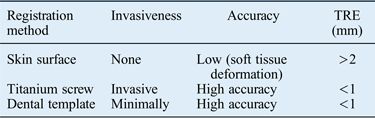
A dentist created a dental template fitted to the upper arch for each patient. We registered the cranial bone using this attachment. Although registration of the skin surface is useful and widely applicable in the navigation of sinus surgery, skin registration often results in discrepancies because of soft tissue dislocation and swelling, tumour growth and differences in head position between the time when the CT images are taken and the time of surgery. Surgery for tumours near the facial surface and for lesions resulting from trauma tend to have greater error. As a more accurate alternative, we registered the cranial bone using a dental template. This method is minimally invasive compared with using alveolar screws and is equally accurate. The target registration error using a dental template was less than 1 mm, the same error reported for titanium screws (Table II).Reference Matsumoto, Oka, Cho, Hong, Jinnouchi and Ouchida6–Reference Matsumoto8, Reference Grauvogel, Grauvogel, Arndt, Berlis and Maier11 With this dental template method, the registration time at surgery is less than 1 minute, and no dislocation or collapse of dental templates was observed during the procedures. One potential problem with using dental templates is difficulty with edentulous patients. We made a hard palate template for patient 3. The accuracy of this modification was acceptable, and no dislocation or collapse occurred. The dental and hard palate template method was accurate enough to be used in the surgical treatment of traumatic injuries.
• Conventional navigation systems using superficial skin registration cannot prevent subtle discrepancies during surgery for orbital fibro-osseous lesions
• The non-invasive cranial bone registration system reported by Matsumoto with patient-specific, dentist-designed dental templates maintained exact registration
• Using a mirror image of the unaffected side for unilateral lesions or an image taken prior to the onset of symptoms for bilateral lesions allowed easy determination of the preset goal
• A sound aid navigation system provided alerts regarding proximity to critical structures and to the desired preset goal
The invasiveness and accuracy of each registration method
TRE: - target registration error
The creation of mirror images from the unaffected side for planning the ideal preset goal has been previously reported.Reference Gui, Zhang, Shen, Wang, Bautista and Voss3 We used this method for cases 1 and 2. However, this method is not possible in patients with bilateral fibrous dysplasia. Imaging and drawing a preset goal without a reference is difficult and is associated with low accuracy.Reference Gui, Zhang, Shen, Wang, Bautista and Voss3 As an alternative, we used CT data obtained three years earlier at another hospital, when the patient was asymptomatic on the right side. We easily created a planned preset goal using that CT data. This method could be very useful in bilateral fibrous dysplasia.
Previous reports have described pre-operative simulation using navigation systems.Reference Nowinski, Messo, Hedlund and Hirsch12, Reference Osada, Iwasawa and Tanaka13 Navigation systems enable clear pre-operative surgical images and allow simulation of important steps, such as avoidance of the intracranial space, the nasal cavity and other critical structures. Simulation promotes the education of junior surgeons by stimulating discussion of the surgical procedure among several surgeons before surgery.
We used a sound aid system that alerted the surgeon with three different tones corresponding to the distance between the tip of the probe or burr and the desired preset goal. This sound aid system has been previously described,Reference Voormolen, Woerdeman, van Stralen, Noordmans, Viergever and Regli14, Reference Wegner and Karron15 and allows the surgeon to know the distance from the desired line without watching the image-guided surgery screen. The system enabled enhanced safety and a shorter operation time. In case 3, the repeat surgery, which used this navigation system, was 171 minutes shorter than the initial operation without the navigation system. Continuous navigation with a sound aid system alerts surgeons to unexpected proximity to critical structures, and increases operator comfort during the procedure.
Overall, this minimally invasive navigation system is a useful and safe method for facilitating the surgical treatment of fibro-osseous lesions of the orbital area.
Conclusion
We used a minimally invasive registration and navigation system to surgically treat fibro-osseous lesions of the orbital area. Non-invasive registration was performed using a dental template fitted to the upper teeth or hard palate. The desired preset goal was determined using mirror images of the patient's unaffected side or CT data taken before the onset of symptoms. Pre-operative simulation was performed to assist in avoiding complications. We used a sound aid navigation system to enhance the safety and speed of surgery. The non-invasive navigation system incorporating these concepts resulted in good outcomes following contouring surgery for fibro-osseous lesions of the orbital area.
Financial support
The author have no commercial association or financial disclorures that might pose.
Conflicts of interest
The author create no conflict of interest with the information presented in this article.
Ethical standards
The authors assert that all procedures contributing to this work comply with the ethical standards of Kyushu University's guidelines on human experimentation and with the Helsinki Declaration of 1975, as revised in 2008.