Introduction
Ménière’s disease (MD) is a chronic condition, presenting with episodic vertigo, hearing loss, tinnitus and aural fullness.Reference Lopez-Escamez, Carey, Chung, Goebel, Magnusson and Mandalà1 Delays in diagnosis frequently occur due to the fluctuating nature of symptoms and reliance on patients meeting clinical criteria, which can take up to 5 years in 20 per cent of patients.Reference Pyykkö, Nakashima, Yoshida, Zou and Naganawa2 Currently, diagnosis remains based on the American Academy of Otolaryngology - Head and Neck Surgery (AAO-HNS) criteria for stratification of patients into definite or probable MD, which was proposed in 2015.Reference Lopez-Escamez, Carey, Chung, Goebel, Magnusson and Mandalà1 However, patients frequently present with symptoms which fall outside of these criteria and may take many years to fully present.Reference Nakashima, Pyykkö, Arroll, Casselbrant, Foster and Manzoor3 Further complicating the presentation, symptoms may be permanent in the late stages of the disease.Reference Bernaerts, Vanspauwen, Blaivie, van Dinther, Zarowski and Wuyts4
In order to aid in the diagnosis, many clinicians refer patients for audio-vestibular assessment. However, there is no definitive gold standard objective diagnostic test.Reference Nakashima, Pyykkö, Arroll, Casselbrant, Foster and Manzoor3 In part, this is likely due to the fluctuating nature of the disease and a poor understanding of the pathophysiology of MD.Reference Gürkov, Pyykö, Zou and Kentala5 MD is attributed to the presence of endolymphatic hydrops (EH),Reference Gürkov, Pyykö, Zou and Kentala5 but the presence of EH does not appear to fully explain the symptoms of MD and is not exclusive to the condition, having been reported to be present in other vestibular diseases.Reference Van, Van, de Pont, Vijlbrief, Bommeljé and Koopman6
Imaging has long played a role in excluding pathology involving the retro-cochlear pathways, most commonly vestibular schwannoma. However, advancements in magnetic resonance imaging (MRI) hardware and pulse sequences have led to the adoption of MRI to identify EH in vivo, initially using delayed MRI about 24 hours after intratympanic administration of dilute gadolinium-based contrast agents (GBCA), but now most commonly using three-dimensional fluid-attenuated inversion recovery (3D FLAIR) and 3D true (ie phase-sensitive) inversion recovery (IR) sequences 4 hours after intra-venous (IV) GBCA administration.Reference Bernaerts7 Both the intra-tympanic and IV methods rely on the uptake of contrast into the perilymph on delayed MRI, allowing identification of the non-enhancing endolymphatic spaces, specifically the cochlear duct, utricle and saccule.
There is currently very little Australian data regarding MRI for MD, apart from one recent study in South Australia.Reference Nguyen, Lim and Chawla8 We aimed to evaluate the sensitivity and specificity of delayed contrast MRI for patients with suspected MD and assess the correlation between clinical diagnosis and presence of hydrops on MRI in a Western Australian context.
Methods
Approval for this retrospective study was sought from the North Metropolitan Health Service Quality Improvement Department (49176).
Patients
Between December 2022 and July 2023, 38 patients presenting with audio-vestibular symptoms to a single otologist were referred for MRI with hydrops protocol at Perth Radiological Clinic.
Clinical assessment
Patients were reviewed by a single otologist following referral for audio-vestibular symptoms. Age, comorbidities, presence of previous ear disease, laterality and nature of symptoms were collected from patient notes. Results of tuning fork tests, audiometry and vestibular function (caloric testing, electrocochleography, cervical vestibular evoked-myogenic potentials, virtual head impulse test) were also recorded. Patients were grouped as Definite MD or Probable MD according to the AAO-NHS Criteria.Reference Lopez-Escamez, Carey, Chung, Goebel, Magnusson and Mandalà1 A third group was included of Undifferentiated patients not meeting the above criteria.
Imaging
All MRI was performed on a 3T MRI scanner using a 32-channel phased array coil (Philips Ingenia Elition). Initial standard MRI of the brain was performed, including a volumetric high resolution heavily T2-weighted sequence and volumetric T1-weighted sequences immediately following IV gadobutrol (Gadovist 1.0; Bayer) using double the standard dose of contrast (ie 0.2 mmol/kg). Following a 4-hour delay after contrast administration, two dedicated sequences targeted to the temporal bones were performed for assessment of EH and abnormal perilymph enhancement: (1) a 3D FLAIR sequence (parameters: repetition time [TR] 6000 ms; echo time [TE] 178 ms; inversion time [TI] 2250 ms; echo train length 40; flip angle 90 degrees; slice thickness 1.6 mm with 0.8-mm separation) and (2) a 3D true IR sequence (parameters: TR 7000 ms; TE 300 ms; TI 1650 ms, echo train length 123; flip angle 90 degrees; slice thickness 1.3 mm with 0.65-mm separation).
Image interpretation
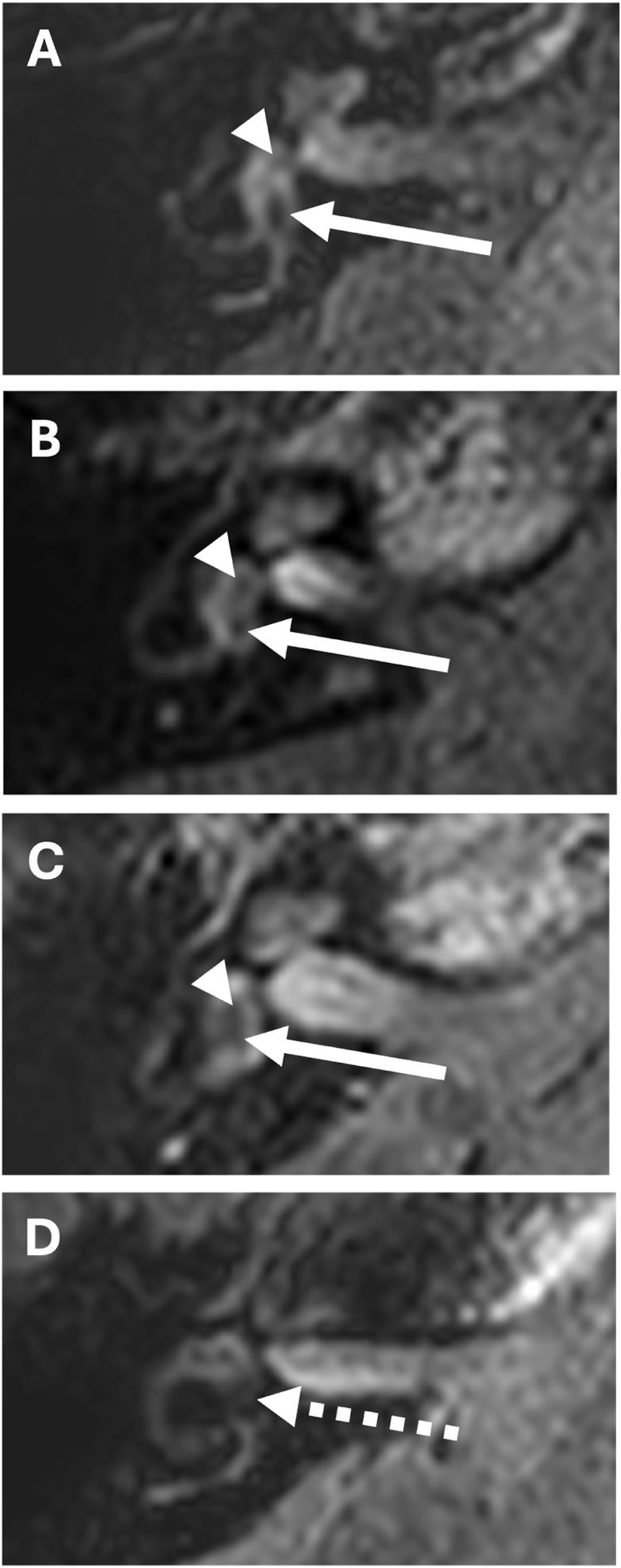
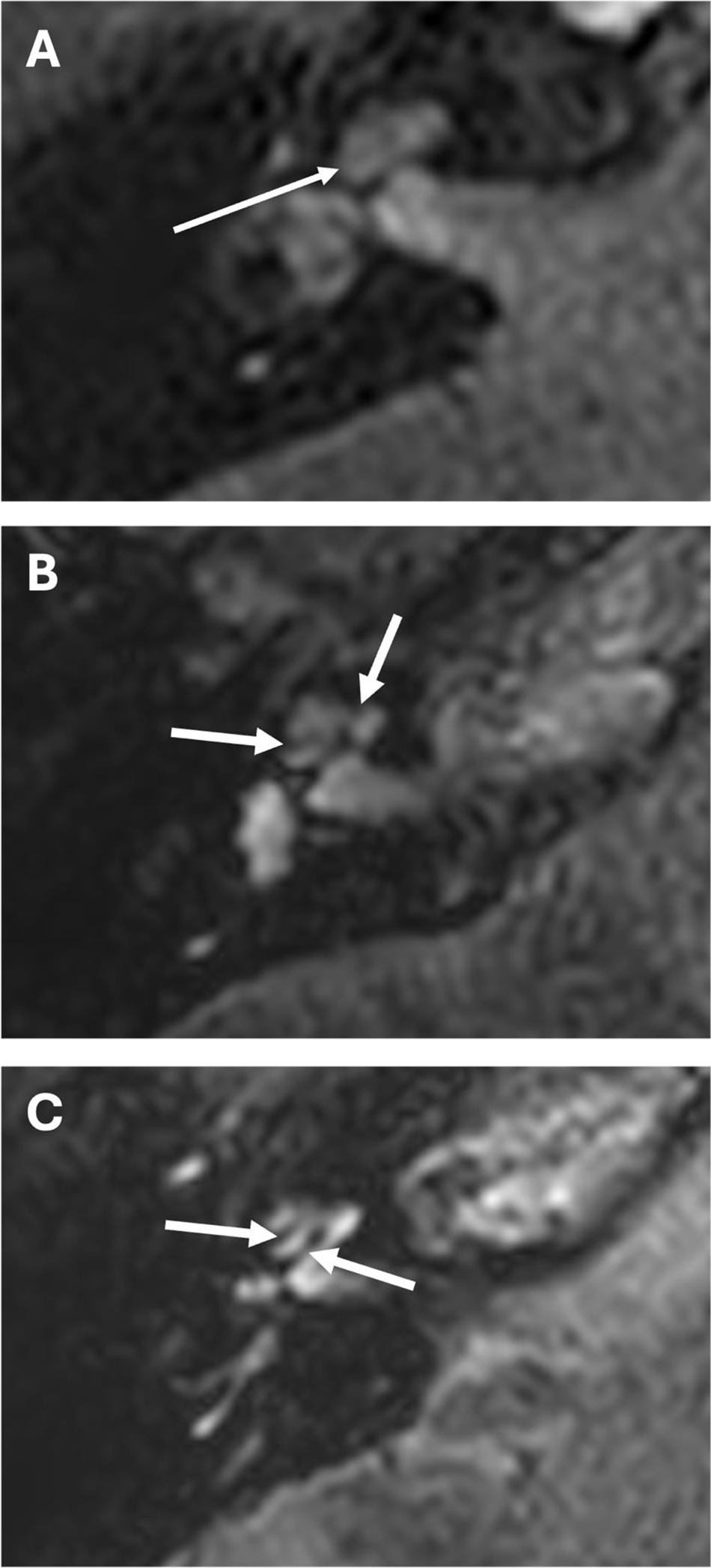
Images were interpreted by two head and neck radiologists each with 19 years’ experience in head and neck imaging, blinded to the clinical information. Images were assessed for the presence of abnormal perilymphatic enhancement and for the presence and severity of cochlear and vestibular EH. If present, cochlear and vestibular hydrops were graded on a three- or four-point grading system, respectively, according to the system described by Bernaerts et al. (Figures 1 and 2). Interpretation was repeated at four months post initial readings to allow for intra-observer analysis.Reference Bernaerts, Vanspauwen, Blaivie, van Dinther, Zarowski and Wuyts4 Inter-observer discrepancies were reviewed, and consensus was reached.
3D FLAIR-weighted MR images of the right inner ear acquired 4 hours after intra-venous administration of gadolinium-based contrast, illustrating Bernaerts et al. grading of vestibular hydrops.Reference Bernaerts, Vanspauwen, Blaivie, van Dinther, Zarowski and Wuyts4 (A) Normal smaller saccule (arrowhead) anterior and slightly medial to the larger utricle (arrow), on an axial image through the inferior part of the vestibule. The saccule and utricle appear as non-enhancing endolymphatic “filling defects” surrounded by enhancing perilymph. (B) Grade 1 vestibular hydrops with mild enlargement of the saccule (arrowhead), of similar size to the utricle (arrow) but remaining separated by enhancing perilymph. (C) Grade 2 vestibular hydrops with confluence of the saccule (arrowhead) and utricle (arrow), with surrounding perilymph still visible. (D) Grade 3 vestibular hydrops with confluence of the saccule and utricle (dotted arrow) and with effacement of the surrounding enhancing perilymph in the vestibule.

3D FLAIR-weighted MR images of the right inner ear acquired 4 hours after intra-venous administration of gadolinium-based contrast, illustrating Bernaerts et al. grading of cochlear hydrops.Reference Bernaerts, Vanspauwen, Blaivie, van Dinther, Zarowski and Wuyts4 (A) Normal cochlear duct (arrow) barely visible as a thin non-enhancing line between the enhancing perilymph within scala vestibuli and the scala tympani. (B) Grade 1 cochlear hydrops with dilatation of the cochlear duct appearing as indenting non-enhancing hypo-intense nodules peripherally (arrows). (C) Grade 2 cochlear hydrops with band-like hypo-intensities (arrows) of the markedly dilated cochlear duct.

Statistical analysis
Inter-observer and intra-observer reliability was assessed using a Cohen’s kappa test. Correlation between clinical diagnosis of MD with both vestibular function results (caloric weakness, elevated cochlear summating potential/auditory nerve action potential (SP/AP) ratio, reduced or absent cervical vestibular evoked myogenic potential [cVEMP] and abnormal video head impulse test [vHIT]) and presence of hydrops on MRI was assessed using Fishers exact test. Specificity and sensitivity was calculated across three groupings of clinical diagnosis: (1) Definite/Probable vs Undifferentiated, (2) Definite vs Undifferentiated and (3) Definite vs Probable/Undifferentiated.
Results
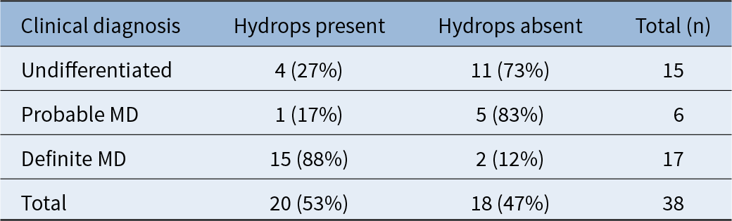
Thirty-eight patients (18 males and 20 females) were assessed over the period of 8 months. The mean age was 58.5 years (range 25–89); four had reported previous ear disease. Of the 38 patients, 17 were diagnosed as Definite MD, 6 as Probable MD and 15 as Undifferentiated. Hydrops was found in 20 patients (53 per cent), of which 3 had bilateral disease (Table 1). Using a Fisher’s exact test, there was no significant correlation between clinical diagnosis and vestibular function results supporting a diagnosis of MD (caloric weakness [p = 0.11], Eastern Cooperative Oncology Group [ECOG] with elevated SP/AP ratio [p = 0.61], absent or reduced cVEMP [p = 0.43], abnormal vHIT (p = 1)).
Presence of hydrops on MRI vs clinical diagnosis of Ménière’s disease

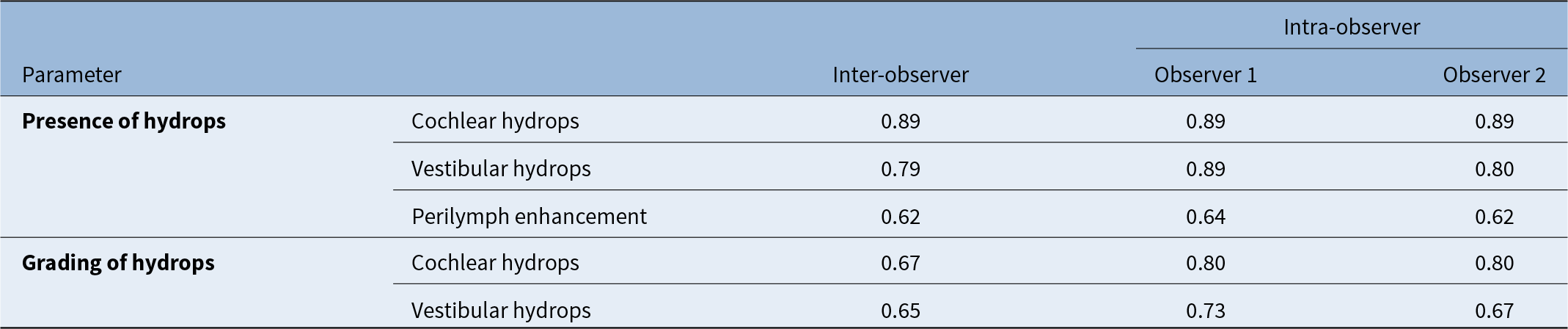
Inter-observer agreement was calculated using a Cohen’s kappa test (Table 2). Agreement for presence of cochlear and vestibular hydrops was 0.89 and 0.79, respectively. Agreement for presence of perilymph enhancement was 0.62. Agreement for presence of cochlear and vestibular hydrops irrespective of grade was 0.67 and 0.65, respectively.
Cohen’s kappa coefficient for inter-observer and intra-observer agreement for presence of cochlear hydrops, vestibular hydrops and perilymph enhancement

Similarly, intra-observer agreement was calculated using a Cohen’s kappa test (Table 2). Agreement for presence of cochlear hydrops was 0.89 and 0.89 for Observer 1 and 2, respectively, and 0.89 and 0.80 for presence of vestibular hydrops. Agreement for presence of perilymph enhancement was 0.64 and 0.62 for Observer 1 and 2, respectively. Agreement for cochlear hydrops grading was 0.80 and 0.80, and 0.73 and 0.67 for grading vestibular hydrops for Observer 1 and 2, respectively.
Fifteen of 17 (88 per cent) patients with Definite MD, 1 of 6 (17 per cent) with Probable MD and 4 of 15 (27 per cent) of Undifferentiated patients had hydrops on MRI (Table 1). Fisher’s exact test for correlation between clinical diagnosis and presence of hydrops was 0.00035 (p < 0.05).
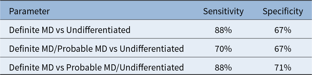
Sensitivity and specificity were calculated at 88 per cent and 67 per cent, respectively, when comparing only Definite MD and Undifferentiated patients. This decreased to 70 per cent and 67 per cent, respectively, when grouped as Definite MD/Probable MD and Undifferentiated patients, or increased to 88 per cent and 71 per cent, respectively, when comparing Definite MD and Probable MD/Undifferentiated patients (Table 3).
Sensitivity and specificity for MRI detection of hydrops by clinical diagnosis of Ménière’s disease, for patient cohort groups of (1) Definite MD vs Undifferentiated, (2) Definite MD/Probable MD vs Undifferentiated and (3) Definite MD vs Probable MD/Undifferentiated

Discussion
MD commonly presents with episodic vertigo, hearing loss, tinnitus and aural fullness.Reference Lopez-Escamez, Carey, Chung, Goebel, Magnusson and Mandalà1 Patients regularly undergo a range of vestibular testing, although these have failed to demonstrate adequate sensitivity.Reference Orimoto, Vartanyan and O’Leary9 Therefore, patients presenting with variable or atypical symptoms frequently experience a delay in diagnosis due to reliance on meeting clinical criteria and lack of definitive diagnostic test. Differentiating between MD and alternative vestibular diagnoses becomes important when initiating and continuing management.
The pathophysiology of MD remains poorly understood. The presence of EH has been recognised as the pathologic correlate, but can also be present in asymptomatic patients and other ear diseases.Reference Bernaerts, Vanspauwen, Blaivie, van Dinther, Zarowski and Wuyts4, Reference Neri, Tartaro and Neri10, Reference Baráth, Schuknecht, Naldi, Schrepfer, Bockisch and Hegemann11 Previously, identification of hydrops was only possible at post-mortem.Reference Pyykkö, Nakashima, Yoshida, Zou and Naganawa2, Reference Yamakawa12 Outside of Australia, MRI with administration of contrast has been used to visualise hydrops in situ.Reference Bernaerts, Vanspauwen, Blaivie, van Dinther, Zarowski and Wuyts4, Reference Van, Van, de Pont, Vijlbrief, Bommeljé and Koopman6, Reference Neri, Tartaro and Neri10, Reference Baráth, Schuknecht, Naldi, Schrepfer, Bockisch and Hegemann11, Reference Nakashima, Naganawa, Sugiura, Teranishi, Sone and Hayashi13, Reference Naganawa, Sugiura, Kawamura, Fukatsu, Sone and Nakashima14 This technique relies on the increased permeability of the blood-labyrinth barrier associated with MD and hydrops, allowing increased contrast ingress into the membranous labyrinth which can then be assessed at MRI.Reference Naganawa and Nakashima15 Commonly, MRI is performed using a 3D FLAIR sequence or 3D true IR sequence,Reference Bernaerts, Vanspauwen, Blaivie, van Dinther, Zarowski and Wuyts4 with either intra-tympanic or IV gadolinium.Reference Bernaerts, Vanspauwen, Blaivie, van Dinther, Zarowski and Wuyts4, Reference Naganawa and Nakashima15–Reference de Pont, van Steekelenburg, Verbist, van Buchem, Blom and Hammer17 IV dosing has the benefit of reduced time between gadolinium administration compared to intra-tympanic administration, as well as being less invasive and allowing simultaneous imaging of both ears.Reference Bernaerts, Vanspauwen, Blaivie, van Dinther, Zarowski and Wuyts4, Reference Baráth, Schuknecht, Naldi, Schrepfer, Bockisch and Hegemann11 The protocol evaluated employed a heavily T2 weighted, 3D FLAIR and 3D true IR sequences, following IV gadolinium administration.Reference Bernaerts, Vanspauwen, Blaivie, van Dinther, Zarowski and Wuyts4
Intra-observer and inter-observer agreement
There have been multiple proposed criteria for interpreting imaging findingsReference Liu, Pyykkö, Naganawa, Marques, Gürkov, Yang and Duan18; this study used the system developed by Bernaerts et al.Reference Bernaerts, Vanspauwen, Blaivie, van Dinther, Zarowski and Wuyts4 Bernaerts et al. outline a four-point system for vestibular hydrops (Figure 1), a three-point system for cochlear hydrops (Figure 2) and assesses presence of perilymphatic enhancement.Reference Bernaerts, Vanspauwen, Blaivie, van Dinther, Zarowski and Wuyts4 As seen in the current study, intra- and inter-observer was substantial to near perfect across groups when using this system and comparable to previously published data.Reference Van, Van, de Pont, Vijlbrief, Bommeljé and Koopman6 Previous systems have been proposed, including evaluating the ratio of the endolymph spaceReference Nakashima, Naganawa, Pyykkö, Gibson, Sone, Nakata and Teranishi19 and use of three-point grading of vestibular and cochlear hydrops;Reference Baráth, Schuknecht, Naldi, Schrepfer, Bockisch and Hegemann11 however, the addition of a low-grade vestibular hydrops and evaluating perilymph enhancement improved sensitivity without compromising specificity.Reference Bernaerts, Vanspauwen, Blaivie, van Dinther, Zarowski and Wuyts4 It is also worth noting that Bernaerts et al. assessed inter-observer reliability in clinical evaluation and radiological diagnosis and found a greater inter-observer reliability in radiological diagnosis, as compared to clinical diagnosis using the AAO-HNS 2015 criteria.Reference Bernaerts, Vanspauwen, Blaivie, van Dinther, Zarowski and Wuyts4
Hydrops on MRI
Presence of EH in clinically definite MD was 88 per cent (15/17), which is comparable to other published data.Reference Van, Van, de Pont, Vijlbrief, Bommeljé and Koopman6, Reference Baráth, Schuknecht, Naldi, Schrepfer, Bockisch and Hegemann11 A significant correlation was also noted between clinical diagnosis as Definite, Probable or Undifferentiated and presence of EH on MRI. Higher rates of hydrops were reported in the Undifferentiated group (27 per cent or 4/15) compared to other studies.Reference Baráth, Schuknecht, Naldi, Schrepfer, Bockisch and Hegemann11 Notably, many of these studies compared contralateral asymptomatic ears, patients with alternative vestibular diagnoses or healthy patients with patients with clinically diagnosed Definite MD, thus accounting for a wide variation in reported rates of hydrops in patients not diagnosed with MD.Reference Nakashima, Pyykkö, Arroll, Casselbrant, Foster and Manzoor3, Reference Van, Van, de Pont, Vijlbrief, Bommeljé and Koopman6, Reference Baráth, Schuknecht, Naldi, Schrepfer, Bockisch and Hegemann11 The current study’s patient population consisted entirely of patients who were offered the MRI as part of evaluation for MD. It is also reported that EH may be a common endpoint for multiple pathological processes.Reference Baráth, Schuknecht, Naldi, Schrepfer, Bockisch and Hegemann11 The presence of EH therefore does not provide definitive conclusions regarding pathophysiological sequence, disease development or disease progression, nor has EH been established as the cause of symptoms in MD. This makes interpretation of the significance of hydrops being present in patients with clinically alternative diagnoses challenging.Reference Bernaerts, Vanspauwen, Blaivie, van Dinther, Zarowski and Wuyts4, Reference Neri, Tartaro and Neri10, Reference Baráth, Schuknecht, Naldi, Schrepfer, Bockisch and Hegemann11
Sensitivity and specificity
The current study reported sensitivity and specificity of 88 per cent and 67 per cent for clinically Definite MD compared to the Undifferentiated group. The reported sensitivity is comparable to other published data;Reference Bernaerts, Vanspauwen, Blaivie, van Dinther, Zarowski and Wuyts4, Reference Van, Van, de Pont, Vijlbrief, Bommeljé and Koopman6, Reference Ito, Inoue, Inui, Miyasaka, Yamanaka and Kichikawa20 however, the current study’s specificity is lower. Possible explanations for this include the current study’s patient population including only those patients being evaluated for MD. Van Steekelenburg et al. also compared clinical diagnosis and MRI findings in patients presenting with suspected MD.Reference Van, Van, de Pont, Vijlbrief, Bommeljé and Koopman6 Following assessment by three otolaryngologists, patients were diagnosed as Probable MD, Definite MD, other Vertigo-associated inner ear pathology or Asymptomatic. All patients subsequently underwent MRI assessing for EH, and sensitivity and specificity were reported at 92 per cent and 93 per cent, respectively. These figures were calculated from the Definite MD and other Vertigo-associated inner ear pathology groups only as opposed to the current study comparing patients who have all been suspected of MD during their clinical course. This same difference in patient population is present in other studies reporting on sensitivity and specificityReference Bernaerts, Vanspauwen, Blaivie, van Dinther, Zarowski and Wuyts4, Reference Van, Van, de Pont, Vijlbrief, Bommeljé and Koopman6, Reference Ito, Inoue, Inui, Miyasaka, Yamanaka and Kichikawa20 and may account for this current study’s lower specificity. Due to the good sensitivity but moderate specificity, the current study supports use of MRI as an aid to diagnosis, particularly in patients suspected of having MD but who may not give clear histories.
Strengths of this study include the evaluation of both intra- and inter-observer agreement across all elements of image interpretation, as well as cohort size. A key limitation, however, is the inability to include asymptomatic patients as a control group for imaging.
• Ménière’s disease is attributed to the presence of endolymphatic hydrops but diagnosis remains clinical through use of the AAO-NHS criteria
• Endolymphatic hydrops was previously diagnosed at post-mortem examination but improvements in technology have seen increasing use of MRI enhanced by intra-venous gadolinium to visualise hydrops in vivo
• Use of MRI in assessing Ménière’s disease has not yet been studied in Western Australia with the Perth Radiological Clinic protocol
• The current study found presence of radiographic hydrops correlates significantly to clinical diagnosis, with good intra- and inter-observer agreement for presence and grading of radiographic hydrops and sensitivity and specificity was 88 per cent and 67 per cent
• The use of MRI may be of greatest value to aid diagnosing Ménière’s disease in patients with unclear presentation.
Conclusion
The use of MRI following the current protocol of a heavily T2 weighted sequence, 3D FLAIR and 3D true IR sequences 4 hours post double-dose IV gadolinium is useful in detecting hydrops in vivo, with good sensitivity. Images can be interpreted with substantial intra- and inter-observer agreement, and presence of hydrops is significantly correlated to a clinical diagnosis of Definite MD. Limitations exist due to reduced specificity in the population studied. We suggest the utility of the hydrops protocol studied lies in the use of MRI to aid diagnosis of patients with suspected MD, but with unclear history or not meeting the AAO-HNS 2015 criteria.
Funding statement
No conflict of interest to declare. No funding received.