Introduction
Several parasites that affect humans invade the central nervous system (CNS) (Abdel Razek et al., Reference Abdel Razek, Watcharakorn and Castillo2011), particularly if humans serve as intermediate or non-permissive hosts (Lv et al., Reference Lv, Zhang, Steinmann, Zhou and Utzinger2010). Although parasitological infections of the CNS are rare, their public health implications cannot be neglected (Lv et al., Reference Lv, Zhang, Steinmann, Zhou and Utzinger2010). Common clinical features of CNS parasitoses include headache, epilepsy, mental decline, impaired consciousness, visual impairment (reduced visual acuity or visual field defects) or weakness (S.M. Wang et al., Reference Wang, Yang, Huang, Shi and Weng2009). Parasites known to cause involvement of the CNS include cestodes (Taenia solium (neurocysticerciasis), Echinococcus granulosus (cerebral cystic echinococcosis), E. multilocularis (cerebral alveolar echinococcosis), Spirometra mansoni (neurosparganosis)), nematodes (Toxocara canis and T. cati (neurotoxocariasis), Trichinella spiralis (neurotrichinelliasis), Angiostrongylus cantonensis or A. costaricensis (neuroangiostrongyliasis) or Gnathostoma spinigerum (neurognathostomiasis)), trematodes (Schistosoma mansoni (cerebral bilharziosis) and Paragonimus westermani (neuroparagonimiasis)) or protozoa (Toxoplasma gondii (neurotoxoplasmosis), Acanthamoeba spp. or Balamuthia (granulomatous amoebic meningo-encephalitis (GAE)), Naegleria (primary amoebic meningo-encephalitis (PAM)), Entamoeba histolytica (amoebic brain abscess), Plasmodium falciparum (cerebral malaria), Trypanosoma brucei gambiense/rhodesiense (sleeping sickness) or Trypanosoma cruzi (cerebral Chagas disease)) (table 1). Diagnosing CNS parasitoses is challenging in cases where antibodies against helminths are absent in the serum or cerebrospinal fluid (CSF), if the infection is self-limiting (e.g. Angiostrongylus costaricensis) or if the infection remains asymptomatic (one-quarter of neurocysticerciasis cases) (Lv et al., Reference Lv, Zhang, Steinmann, Zhou and Utzinger2010). Risk factors for CNS parasitoses include poverty, low social status, living in slums, living in tropical areas, tourism or migration, the latter posing particular challenges to clinicians in non-endemic areas (Lv et al., Reference Lv, Zhang, Steinmann, Zhou and Utzinger2010). The aim of the present review was to summarize and discuss recent findings concerning the pathogenesis, clinical presentation, diagnosis and treatment of cerebral parasitoses. The following description of single parasitoses follows the order: infections by helminths (1. cestodes (tapeworms), 2. nematodes (roundworms), 3. trematodes) followed by infections by protozoa. The description of each disease includes the features aetiology, clinical manifestations, diagnosis and treatment.
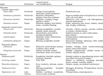
Parasitoses of the central nervous system.

GAE, granulomatous amoebic meningo-encephalitis; PAM, primary amoebic encephalitis.
Neurocysticerciasis
Neurocysticerciasis is caused by encysted larval stages of the tapeworm Taenia solium (Abdel Razek et al., Reference Abdel Razek, Watcharakorn and Castillo2011). Ingestion of eggs from the faeces of tapeworm-carriers is followed by their spread to neural, muscular or ocular tissue (Abdel Razek et al., Reference Abdel Razek, Watcharakorn and Castillo2011). Neurocysticerciasis is the most common helminthic disease of the CNS (Khade et al., Reference Khade, Lemos and Toussaint2011; Qi et al., Reference Qi, Ge, Yang, Bi and Li2011), particularly in endemic areas where the parasite exchange between humans and pigs is maintained (Nishimura & Hung, Reference Nishimura and Hung1997). Neurocysticerciasis results from direct invasion of the primary larvae (oncospheres) into the CNS and transformation into secondary, cystic larvae (cysticerci) (Abdel Razek et al., Reference Abdel Razek, Watcharakorn and Castillo2011). Cysticerci may be localized within the parenchyma, particularly the corticomedullary junction, subarachnoid space or intraventricularly (isolated or together with parenchymal disease) (Abdel Razek et al., Reference Abdel Razek, Watcharakorn and Castillo2011). Parenchymal neurocysticerciasis is divided into four stages (vesicular, colloidal–vesicular, granular–nodular and nodular–calcified). Larval death causes an inflammatory reaction resulting in a perifocal oedema, which consecutively damages the parenchyma (Qi et al., Reference Qi, Ge, Yang, Bi and Li2011). Cysts within the ventricle are particularly problematic since they carry the risk of ependymitis, ventriculitis or acute obstructive hydrocephalus. Complications of the subarachnoid form include vasculitis, occurring in 50% of cases, calcification of arteries or arachnoiditis (Abdel Razek et al., Reference Abdel Razek, Watcharakorn and Castillo2011). The cerebrum is much more frequently affected than the spinal cord (1–5% of the patients with neurocysticerciasis). The spinal form is divided into an extra-medullary (leptomeningeal) and an intramedullary form (Abdel Razek et al., Reference Abdel Razek, Watcharakorn and Castillo2011).
Cerebral manifestations of neurocysticerciasis include epilepsy (50–70%); headache (40%), which may be due to increased intracranial pressure (Sinha & Sharma, Reference Sinha and Sharma2012) or due to liberation of antigens to the CNS parenchyma during remodelling of cysticercotic calcifications (Del Brutto & Del Brutto, Reference Del Brutto and Del Brutto2012); hydrocephalus (33%); meningitis; aneurysm formation or stroke. Ischaemic stroke occurs in 4–12% of the neurocysticerciasis patients (Marquez & Arauz, Reference Marquez and Arauz2012). Neurocysticerciasis is considered to be the most common cause of symptomatic epilepsy worldwide (Takayanagui & Odashima, Reference Takayanagui and Odashima2006). Seizures result from perilesional inflammation, stroke, vasculitis or calcified granulomas (Abdel Razek et al., Reference Abdel Razek, Watcharakorn and Castillo2011). Encephalitic cysticerciasis develops if multiple lesions are simultaneously in the granular stage or if there is overwhelming inflammation around the cyst (Abdel Razek et al., Reference Abdel Razek, Watcharakorn and Castillo2011). Intramedullary cysticerciasis is rare and may manifest as paraparesis, paraspasticity, bowel or bladder incontinence, or sexual dysfunction (Qi et al., Reference Qi, Ge, Yang, Bi and Li2011).
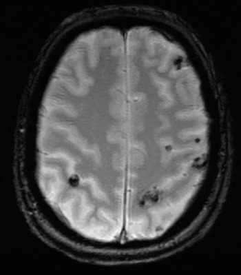
Neurocysticerciasis is diagnosed according to the clinical presentation, the epidemiological background, blood and CSF investigations, and imaging studies. Specific IgG antibodies can be detected in the serum or CSF. Other CSF findings include elevated protein, low or normal glucose, moderate lymphocytic pleocytosis or eosinophilia, which is present only in about one-third of the patients (Qi et al., Reference Qi, Ge, Yang, Bi and Li2011). Serological or CSF tests for specific IgG antibodies against the parasite include the enzyme-linked immune-electrotransfer blot (ELITB), the enzyme-linked immunosorbent assay (ELISA) or the dot-blot test (Singhi & Singhi, Reference Singhi and Singhi2009). Sensitivity/specificity of these tests, however, varies depending on the presence of single/multiple lesions, parenchymal/extra-parenchymal lesions and serum/CSF samples. Magnetic resonance imaging (MRI) may show T2-hyperintense, homogeneously enhancing vesicular, colloid–vesicular, granular–nodular or nodular–calcified lesions in the cortical or subcortical parenchyma, white matter, cisterns, ventricles or spinal cord (fig. 1) (Chang & Han, Reference Chang and Han1998). A rare manifestation of neurocysticerciasis is the formation of aneurysms of intracerebral arteries, with cysts within the aneurysm (Agapejev et al., Reference Agapejev, Parra-Marinello, Bazan, Ueda and Zanini2011; Kashyap et al., Reference Kashyap, Das, Jain, Agarwal, Kaushik and Kaur2012). Within the spinal cord the parasite is most frequently located in the thoracic spine, which supports the hypothesis that the cysticercus comes from the blood circulation because the thoracic medulla receives much more blood supply than the other parts of the medulla (Qi et al., Reference Qi, Ge, Yang, Bi and Li2011). Differential diagnoses of neurocysticerciasis include echinococcosis, toxoplasmosis, tuberculoma, microabscess, fungal lesion, glioma and metastasis (Abdel Razek et al., Reference Abdel Razek, Watcharakorn and Castillo2011).
Neurocysticerciasis. T2-weighted cerebral MRI showing multiple cystic lesions in the cortex and the subcortical white matter.

The most effective treatment of neurocysticerciasis is the combination of praziquantel with albendazole (Palomares et al., Reference Palomares, Palencia, Ambrosio, Ortiz and Jung-Cook2006; Garcia et al., Reference Garcia, Lescano, Lanchote, Pretell, Gonzales, Bustos, Takayanagui, Bonato, Horton, Saavedra, Gonzalez and Gilman2011). Both drugs have limited solubility and extensive metabolism, resulting in highly variable inter-individual plasma levels (Jung-Cook, Reference Jung-Cook2012). Surgical resection of isolated cysts either within the parenchyma or the ventricles is a therapeutic option but should be followed by postoperative therapy with albendazole and praziquantel (Palomares et al., Reference Palomares, Palencia, Ambrosio, Ortiz and Jung-Cook2006; Garcia et al., Reference Garcia, Lescano, Lanchote, Pretell, Gonzales, Bustos, Takayanagui, Bonato, Horton, Saavedra, Gonzalez and Gilman2011). In cases of intraventricular neurocysticerciasis resection of the cyst may be necessary to reduce or eliminate the mass effect, CSF obstruction or cysts within the fourth ventricle (Sinha & Sharma, Reference Sinha and Sharma2012). Recently, spontaneous resolution of parenchymal cysticerciasis with a benign outcome has been reported (Takayanagui & Odashima, Reference Takayanagui and Odashima2006).
Cerebral cystic echinococcosis (cystic hydatidosis)
Cerebral cystic echinococcosis is caused by infection with larvae of Echinococcus granulosus, a tapeworm which requires canines as definitive hosts and herbivores or humans as intermediate hosts (Scarlata et al., Reference Scarlata, Giordano, Saporito, Marasa, Li Pani, Odierna, Scaglione, Di Carlo and Romano2011). A cyst is surrounded by three layers, the outer layer (pericyst), the middle laminated membrane (ectocyst) and the inner layer (endocyst) (Abdel Razek et al., Reference Abdel Razek, Watcharakorn and Castillo2011). According to their viability, cysts are classified as active fertile (CE1, CE2), transitional (CE3) or degenerated (CE4, CE5).
Cystic echinococcosis is a self-limiting but potentially fatal disease, with CNS involvement in only 1–3% of cases (Altas et al., Reference Altas, Aras, Serarslan, Davran, Evirgen and Yilmaz2010). Generally, cysts are located within the parenchyma, the subarachnoid space or the ventricles. In cases of cerebral location of the cysts, patients present with headache, vomiting, hemiparesis and other focal neurological deficits, or seizures (Wani et al., Reference Wani, Kousar, Gojwari, Robbani, Singh, Ramzan, Khan, Kirmani and Wani2011). Patients in whom cysts are located within the spinal cord, which is a rare finding, may develop acute paraplegia (Scarlata et al., Reference Scarlata, Giordano, Saporito, Marasa, Li Pani, Odierna, Scaglione, Di Carlo and Romano2011) or transverse syndrome (Druschky et al., Reference Druschky, Niederstadt, Jourdan, Stoltze and Heckl1995).
The diagnosis is established by clinical, serological, CSF and imaging findings. Antibodies against E. granulosus in the serum or CSF can be detected by Western blot or polymerase chain reaction (PCR). After removal of cerebral cysts, histological investigations may confirm the diagnosis (Potapov et al., Reference Potapov, Goriaĭnov, Okhlopkov, Kravchuk, Zakharova, Podoprigora, Kurnosov, Khit', Panina and Eliseeva2011). Cerebral computed tomography (CT) may show single or multiple intra-axial spherical or ovaloid cysts, which are entirely surrounded by cerebral parenchyma, and may cause a mass effect (Wani et al., Reference Wani, Kousar, Gojwari, Robbani, Singh, Ramzan, Khan, Kirmani and Wani2011). The wall of the cysts is usually smooth and thin, non-enhancing and without calcification.
Therapy is based on medical treatment and surgical removal of the cysts (Altas et al., Reference Altas, Aras, Serarslan, Davran, Evirgen and Yilmaz2010). The drug of choice is albendazole (Baykaner et al., Reference Baykaner, Doğulu, Oztürk, Edali and Tali2000). Surgical decompression of the cerebrum or the spinal cord is indicated in cases of acute, severe neurological deficit due to a mass effect (Prabhakar et al., Reference Prabhakar, Acharya, Modi and Jadav2005). If possible, cerebral cysts should be removed without rupture by means of a microsurgical technique with neuronavigation, and intraoperative neurophysiological and ultrasound monitoring (Potapov et al., Reference Potapov, Goriaĭnov, Okhlopkov, Kravchuk, Zakharova, Podoprigora, Kurnosov, Khit', Panina and Eliseeva2011).
Cerebral alveolar echinococcosis
Cerebral alveolar echinococcosis is caused by infection with larvae of Echinococcus multilocularis, a tapeworm which requires canines, herbivores or humans as hosts. Invasion of the CNS by larvae of E. multilocularis has been only occasionally reported (Senturk et al., Reference Senturk, Oguz, Soylemezoglu and Inci2006).
Patients initially present with manifestations of increased intracranial pressure, most frequently headache, papillary oedema, vomiting or vertigo (Tappe et al., Reference Tappe, Weise, Ziegler, Müller, Müllges and Stich2008).
The diagnosis is established by clinical, serological, CSF and imaging findings. Antibodies against E. multilocularis in the serum or CSF can be detected by Western blot or PCR. Imaging may show solid or multi-focal cystic masses, which are calcified and surrounded by oedema. In single cases, heterogeneous ring-like, or nodular enhancement has been reported (Tunaci et al., Reference Tunaci, Tunaci, Engin, Ozkorkmaz, Ahishali and Rozanes1999). There may also be alveolar structures of numerous irregular cysts with a diameter of 1–20 mm, which are not sharply delineated from the surrounding tissue (Abdel Razek et al., Reference Abdel Razek, Watcharakorn and Castillo2011). Central cavities are due to necrosis of the inner part of the cyst. The cyst wall is irregularly thickened and partially calcified.
The treatment of choice is surgery together with albendazole or mebendazole (J. Wang et al., Reference Wang, Cai and You2009). A complication of treatment can be hydrocephalus (J. Wang et al., Reference Wang, Cai and You2009). Cerebral alveolar echinococcosis is lethal if untreated (J. Wang et al., Reference Wang, Cai and You2009). A major differential diagnosis is cerebral malignancy, due to the invasive and destructive nature of the cyst growth (Ozdol et al., Reference Ozdol, Yildirim, Daglioglu, Divanlioglu, Erdem and Belen2011).
Neurosparganosis
Sparganosis is a rare but underestimated parasitic, extra-intestinal infection, caused by the larvae of the tapeworms Spirometra mansoni or S. proliferum (Deng et al., Reference Deng, Xiong and Qian2012). The common route of infection is either ingestion of contaminated water or of an intermediate host (frog, snake), or contact between an open wound and an intermediate host (Duggal et al., Reference Duggal, Mahajan, Duggal and Hans2011). Plerocercoid larvae, known as spargana, migrate to muscle, subcutis, the eye, urogenital tract, gut and, rarely, to the cerebrum, resulting in a space-occupying lesion with mass effect (Duggal et al., Reference Duggal, Mahajan, Duggal and Hans2011). Sparganosis is uncommon in Europe and is most frequently seen in South-East Asia (Walker et al., Reference Walker, Cooper and Brandis2007).
Most patients develop headache, epilepsy, focal weakness or sensory disturbances (Abdel Razek et al., Reference Abdel Razek, Watcharakorn and Castillo2011; Deng et al., Reference Deng, Xiong and Qian2011). Single patients may develop cerebral vasculitis complicated by ischaemic stroke (Han et al., Reference Han, Park, Kim and Son2001). Some patients may experience intracerebral haemorrhage as the initial manifestation of the infection (Jeong et al., Reference Jeong, Bae, Hwang, Kim and Lee1998).
For years a detailed protocol for the diagnosis of cerebral sparganosis was lacking (Deng et al., Reference Deng, Xiong and Qian2011). Recently, however, a stereotactic aspiration technique complemented by microsurgery has been proposed for diagnosis (Deng et al., Reference Deng, Xiong and Qian2011). Additionally, antibodies against S. mansoni can be detected in the serum and CSF. An ELISA, which is available only in selected laboratories, is useful to reach the correct diagnosis. Imaging in the acute stage may show a single mass lesion with perifocal oedema, resembling a metastatic brain tumour, and ventricular dilatation (Okamura et al., Reference Okamura, Yamamoto, Ohta, Matsuoka and Uozumi1995). A ‘tunnel sign’, characterized by a T1-hypointense and T2-hyperintense column- or fusiform-shaped tunnel with shallow enhancement, may be visible (Abdel Razek et al., Reference Abdel Razek, Watcharakorn and Castillo2011). There may also be conglomerated ring- or bead-shaped enhancement representing inflammatory granuloma (Abdel Razek et al., Reference Abdel Razek, Watcharakorn and Castillo2011). In the chronic stage, cortical atrophy, dilation of the ipsilateral ventricle or punctate calcifications are apparent (Abdel Razek et al., Reference Abdel Razek, Watcharakorn and Castillo2011).
Treatment of cerebral sparganosis is surgical (Walker et al., Reference Walker, Cooper and Brandis2007). Larvae should be directly removed by stereotactic aspiration or, in case of failure, stereotactic microsurgery (Deng et al., Reference Deng, Xiong and Qian2011). Surgeons must avoid injuring the larva or leaving behind any larval residue (Deng et al., Reference Deng, Xiong and Qian2011).
Neurotoxocariasis
Toxocariasis is caused by ingestion of eggs of the roundworm Toxocara canis or T. cati living in dogs, puppies or cats (Garcia-Pedrique et al., Reference Garcia-Pedrique, Diaz-Suarez and Estevez2004; Roldán et al., Reference Roldán, Cavero, Espinoza, Jiménez and Gutiérrez2010). Occasionally, humans become infected after ingesting embryonated eggs from the soil, or after exposure to contaminated hands or food (Abdel Razek et al., Reference Abdel Razek, Watcharakorn and Castillo2011). Although T. canis is known to cross the blood–brain barrier (Liao et al., Reference Liao, Cho, Kao, Su, Lin and Fan2008), CNS infection is rare (Abdel Razek et al., Reference Abdel Razek, Watcharakorn and Castillo2011). Toxocara larvae are metabolically active and produce an array of molecules that cause tissue damage and a marked inflammatory reaction (Abdel Razek et al., Reference Abdel Razek, Watcharakorn and Castillo2011). The CNS is mainly affected by a granulomatous process.
CNS manifestations of toxocariasis include eosinophilic meningitis, encephalitis, meningo-encephalitis, ependymitis, arachnoiditis, vasculitis, meningo-myelitis, meningo-radiculitis, epilepsy, dementia or optic neuritis (Duprez et al., Reference Duprez, Bigaignon, Delgrange, Desfontaines, Hermans, Vervoort, Sindic and Buysschaert1996; Dauriac-Le Masson et al., Reference Dauriac-Le Masson, Chochon, Demeret and Pierrot-Deseilligny2005; Finsterer & Auer, Reference Finsterer and Auer2007).
Neurotoxocariasis is diagnosed on the basis of the clinical presentation, the epidemiological background, eosinophilia in the serum or CSF, presence of IgG antibodies on ELISA against T. canis excretory–secretory antigen in the CSF on Western blotting, or focal CNS lesions on imaging (Scheid et al., Reference Scheid, Tina Jentzsch and Schroeter2008). There are, however, also cases with blood eosinophilia and normal CSF findings (Vidal et al., Reference Vidal, Sztajnbok and Seguro2003), this is why a negative spinal tap does not rule out neurotoxocariasis. Cerebral MRI may show cortical or subcortical, multifocal, homogeneously enhancing T2-hyperintense lesions or a combination of circumscribed and diffuse changes (Ruttiner & Hadidi, Reference Ruttiner and Hadidi1991). These lesions are hypodense on cerebral CT and hypointense on T1-weighted MRI. In case of spinal cord involvement, imaging may reveal non-tumorous myelopathy mimicking transverse myelitis, single or multiple T2-hyperintense lesions with strong, focal nodular enhancement on posterior or posterolateral segments of the spinal cord, or migration of these lesions (Lee et al., Reference Lee, Kim, Oh, Kim, Kim, Jeon and Byun2010).
The most commonly applied drug for neurotoxocariasis is albendazole alone or in combination with steroids (Finsterer & Auer, Reference Finsterer and Auer2007). Other options include oxibendazole, flubendazole, mebendazole or thiabendazole, but albendazole penetrates best into the CSF and is less toxic (Finsterer & Auer, Reference Finsterer and Auer2007). A further alternative may be diethylcarbamazine together with steroids (Finsterer & Auer, Reference Finsterer and Auer2007). Antihelminthic treatment may result in a marked decrease in the size and number of lesions (Abdel Razek et al., Reference Abdel Razek, Watcharakorn and Castillo2011).
Neurotrichinelliasis
Trichinelliasis is an anthropozoonosis due to infection with Trichinella spiralis or other Trichinella species (Teunis et al., Reference Teunis, Koningstein, Takumi and van der Giessen2012). CNS involvement in trichinelliasis is said to occur in 10–15% of the cases (Knezević et al., Reference Knezević, Turkulov, Canak, Lalosević and Tomić2001; Gelal et al., Reference Gelal, Kumral, Vidinli, Erdogan, Yucel and Erdogan2005). Pathogenetic mechanisms responsible for the development of the disease include obstruction of cerebral arteries by larvae, cysts or granulomas; toxic vasculitis with secondary thrombosis or bleeding; granulomatous inflammation of the cerebrum; or allergic reaction (Nikolić et al., Reference Nikolić, Vujosević, Sasić, Poluga, Misić, Najdanović, Dulović, Dragojlović and Milosević1998).
Trichinelliasis starts gradually with non-specific flu-like symptoms and febrile gastroenteritis (Gallardo et al., Reference Gallardo, Mateos, Artieda, Wesslen, Ruiz, García, Galmés-Truyols, Martin, Hernández-Pezzi, Andersson, Gárate and Christensson2007). CNS manifestations start with symptoms and signs of diffuse encephalopathy (disorientation and somnolence) (Teunis et al., Reference Teunis, Koningstein, Takumi and van der Giessen2012). Later on, patients develop focal neurological deficits, such as hemiparesis (Teunis et al., Reference Teunis, Koningstein, Takumi and van der Giessen2012), representing the clinical manifestations of meningitis, encephalitis or polyradiculoneuritis (Nikolić et al., Reference Nikolić, Vujosević, Sasić, Poluga, Misić, Najdanović, Dulović, Dragojlović and Milosević1998). Rarely, neurotrichinelliasis may manifest with sinus venous thrombosis (el Koussa et al., Reference el Koussa, Chemaly, Fabre-Bou Abboud, Tamraz and Haddad1994).
The diagnosis is established by the clinical presentation, hypereosinophilia, presence of antibodies against trichinella in the serum or CSF by an immune-fluorescence test, microscopic identification of larvae or imaging studies (Nikolić et al., Reference Nikolić, Vujosević, Sasić, Poluga, Misić, Najdanović, Dulović, Dragojlović and Milosević1998). Occasionally, conventional CSF investigations are normal (Knezević et al., 2001). Imaging shows multifocal small lesions in the cortex and the white matter (Gelal et al., Reference Gelal, Kumral, Vidinli, Erdogan, Yucel and Erdogan2005). These lesions are believed to represent multifocal ischaemic lesions rather than inflammatory infiltrations of the brain (Gelal et al., Reference Gelal, Kumral, Vidinli, Erdogan, Yucel and Erdogan2005). Single cases may also present with multiple microinfarcts at the border zones of major vascular territories, in the periventricular white matter and the corpus callosum (Abdel Razek et al., Reference Abdel Razek, Watcharakorn and Castillo2011). These lesions are T2-hyperintense without enhancement. Single cases may also present with diffuse or focal, bilateral, enhancing T2-hyperintensities in the centrum semiovale (Gelal et al., Reference Gelal, Kumral, Vidinli, Erdogan, Yucel and Erdogan2005).
Neurotrichinelliasis is treated with albendazole or mebendazole (Bartuliene et al., Reference Bartuliene, Liausediene and Motiejuniene2009). In children, albendazole may be more effective than mebendazole (Neghina et al., Reference Neghina, Iacobiciu, Neghina and Marincu2011). In untreated patients mortality is about 50% (Nikolić et al., Reference Nikolić, Vujosević, Sasić, Poluga, Misić, Najdanović, Dulović, Dragojlović and Milosević1998).
Neuroangiostrongyliasis
Angiostrongyliasis is caused by the nematode Angiostrongylus cantonensis, transmitted by ingestion of uncooked snails (Kanpittaya et al., Reference Kanpittaya, Sawanyawisuth, Intapan, Khotsri, Chotmongkol and Maleewong2012). Angiostrongylus cantonensis is a specifically neurotopic nematode, which requires the CNS of mammalian hosts for its growth (Nishimura & Hung, Reference Nishimura and Hung1997). Particularly, third-stage larvae frequently invade the skeletal muscle and the CNS (Nishimura & Hung, Reference Nishimura and Hung1997). Ingestion of larvae can be prevented by avoiding consumption of meat from paratenic hosts. Another causative agent may be A. costaricencis (Mojon, Reference Mojon1994).
The most frequent clinical manifestation of neuroangiostrongyliasis is headache, pain in the trunk or the limbs, sensory disturbances, or visual impairment due to eosinophilic meningitis, usually without spinal cord involvement (Wang et al., Reference Wang, Zheng, Yin, Qi, Li, Diao, Wang, Ji, Feng and Guo2007; Kanpittaya et al., Reference Kanpittaya, Sawanyawisuth, Intapan, Khotsri, Chotmongkol and Maleewong2012). Patients with the encephalitic form may present with impaired consciousness (Kanpittaya et al., Reference Kanpittaya, Sawanyawisuth, Intapan, Khotsri, Chotmongkol and Maleewong2012). Some patients may also present with ventriculitis (Jin et al., Reference Jin, Ma, Ma, He, Ji and Yin2008).
CSF investigations may indicate eosinophilic meningitis (Wang et al., Reference Wang, Zheng, Yin, Qi, Li, Diao, Wang, Ji, Feng and Guo2007). ELISA tests, which are available only in single laboratories, may reveal IgG antibodies against A. cantonensis antigen (Wang et al., Reference Wang, Zheng, Yin, Qi, Li, Diao, Wang, Ji, Feng and Guo2007). Cerebral CT may be normal or may show meningeal enhancement (Jin et al., Reference Jin, Ma, Ma, He, Ji and Yin2008; Kanpittaya et al., Reference Kanpittaya, Sawanyawisuth, Intapan, Khotsri, Chotmongkol and Maleewong2012). Patients with myeloencephalitis may show enhancing cerebral lesions, iso- or hypointense on T1 and T2-hyperintense (Jin et al., Reference Jin, Ma, Ma, He, Ji and Yin2008). The encephalitic form may be complicated by communicating hydrocephalus (Kanpittaya et al., Reference Kanpittaya, Sawanyawisuth, Intapan, Khotsri, Chotmongkol and Maleewong2012). Occasionally, such lesions may be also found on spinal MRI (Jin et al., Reference Jin, Ma, Ma, He, Ji and Yin2008). Most lesions resolve within a few weeks (Jin et al., Reference Jin, Ma, Ma, He, Ji and Yin2008).
The common treatment is either albendazole or praziquantel (Wang et al., Reference Wang, Huang, Dong, Lin, Wang, Li, Nawa and Yoshimura2002). Outcome is usually fair with only minimal remaining neurological deficits (Wang et al., Reference Wang, Zheng, Yin, Qi, Li, Diao, Wang, Ji, Feng and Guo2007).
Neurognathostomiasis
Human gnathostomiasis is caused by larvae of the nematode Gnathostoma spinigerum. Gnathostomiasis is most commonly seen in South-East Asia.
The most frequent CNS manifestation of the infection is myelitis. Myelitis is often associated with radiculitis but may also progress to encephalitis (Boongird et al., Reference Boongird, Phuapradit, Siridej, Chirachariyavej, Chuahirun and Vejjajiva1977). Ten times more rarely, intracranial bleeding can be found. Most frequently bleeding is intracerebral, but some patients may also develop subdural, subarachnoid or intraventricular bleeding (Sawanyawisuth et al., Reference Sawanyawisuth, Chlebicki, Pratt, Kanpittaya and Intapan2009; Kanpittaya et al., Reference Kanpittaya, Sawanyawisuth, Intapan, Khotsri, Chotmongkol and Maleewong2012). Intracerebral or subarachnoid bleeding results from rupture of mycotic aneurysms, which are typical for CNS gnathostomiasis (Germann et al., Reference Germann, Schächtele, Nessler, Seitz and Kniehl2003).
Neurognathostomiasis is diagnosed upon the clinical presentation, serological tests, CSF investigations and cerebral imaging, including angiography. Confirmation of the infection is achieved by the verification of specific antibodies in the serum or CSF by an immunoblot assay, which is available only in selected laboratories. CSF eosinophilia is highly typical for the disease (Kanpittaya et al., Reference Kanpittaya, Sawanyawisuth, Intapan, Khotsri, Chotmongkol and Maleewong2012). Cerebral MRI may be normal or may show scattered, deep intracerebral bleeding with diffuse white matter lesions with nodular enhancement (Sawanyawisuth et al., Reference Sawanyawisuth, Tiamkao, Kanpittaya, Dekumyoy and Jitpimolmard2004). MRI of the spinal cord may show fusiform dilatation of the cord and diffuse T2-hyperintensity of the grey–white matter regions without evidence of bleeding (Sawanyawisuth et al., Reference Sawanyawisuth, Tiamkao, Kanpittaya, Dekumyoy and Jitpimolmard2004; Bunyaratavej et al., Reference Bunyaratavej, Pongpunlert, Jongwutiwes and Likitnukul2008; Kanpittaya et al., Reference Kanpittaya, Sawanyawisuth, Intapan, Khotsri, Chotmongkol and Maleewong2012). Angiography may reveal mycotic aneurysms, which are typical for CNS gnathostomiasis (Germann et al., Reference Germann, Schächtele, Nessler, Seitz and Kniehl2003).
No drug is available for gnathostomiasis although albendazole has been tried with little success and ivermectin has been suggested. To prevent severe cerebral oedema, steroids (prednisolone) should be given before starting albendazole or ivermectin (Sithinamsuwan & Chairangsaris, Reference Sithinamsuwan and Chairangsaris2005).
Neuroschistosomiasis (bilharziosis)
Neuroschistosomiasis is caused by haematogenic invasion of the CNS with eggs from the blood-vessel-dwelling trematode Schistosoma mansoni (Vale et al., Reference Vale, Marques, de Sousa-Pereira and Lambertucci2011), S. japonicum (Ferrari & Moreira, Reference Ferrari and Moreira2011) or S. haematobium (Imai et al., Reference Imai, Koibuchi, Kumagai, Maeda, Osada, Ohta, Koga, Nakamura, Miura, Iwamoto and Fujii2011). Other routes of CNS invasion include retrograde venous spread by way of Batons's plexus or migration of worms through the brain (Abdel Razek et al., Reference Abdel Razek, Watcharakorn and Castillo2011).
CNS involvement may occur at any time of the infection. Both the brain and the spinal cord can be affected (Ferrari & Moreira, Reference Ferrari and Moreira2011). Schistosoma mansoni and S. haematobium usually cause myelopathy, whereas S. japonicum usually causes encephalitis (Ferrari & Moreira, Reference Ferrari and Moreira2011). Single cases with encephalomyelitis have been reported (Vale et al., Reference Vale, Marques, de Sousa-Pereira and Lambertucci2011). Patients with CNS involvement present with manifestations of a space-occupying lesion, comprising headache, mental disturbances, seizures, sensory abnormalities, weakness, papillary oedema or cerebellar ataxia (Ferrari & Moreira, Reference Ferrari and Moreira2011). Border-zone infarctions have been also reported (Ferrari & Moreira, Reference Ferrari and Moreira2011; Wu et al., Reference Wu, Wu, Tian, Chen, Liu, Chen, Wang, Cai, Ji, Wang, Zhang, Ruan, Zhu and Guo2012). In the chronic stage, patients may present with tumoral encephalic schistosomiasis or spinal cord schistosomiasis (Ferrari & Moreira, Reference Ferrari and Moreira2011). Tumoral encephalic schistosomiasis presents with variable focal neurological signs depending on the site of the lesion (Ferrari & Moreira, Reference Ferrari and Moreira2011). The most common manifestations include headache, motor deficits, visual disturbances, seizures, altered mental state, vertigo, sensory disturbances or ataxia. Spinal cord affection manifests as back pain, paraparesis, bladder dysfunction, sensory disturbances, acute transverse myelitis, lumbosacral radiculopathy or cauda syndrome (Ferrari & Moreira, Reference Ferrari and Moreira2011). Clinical presentation and outcome of the neurological abnormalities is highly variable, depending on the phase of the disease and the type of Schistosoma (Ferrari & Moreira, Reference Ferrari and Moreira2011).
Neuroschistosomiasis is often underdiagnosed or misdiagnosed because of the heterogeneity of clinical manifestations (Wu et al., Reference Wu, Wu, Tian, Chen, Liu, Chen, Wang, Cai, Ji, Wang, Zhang, Ruan, Zhu and Guo2012). Blood tests may reveal specific antibodies against Schistosoma, and urine analysis may reveal macrohaematuria. The CSF may be normal or may show non-specific abnormalities (Ferrari & Moreira, Reference Ferrari and Moreira2011). The diagnosis is confirmed by PCR of the CSF, which may be positive even if serum antibodies are absent or show cross-reaction with other helminthes, and in cases where no ova are excreted via the urine or faeces (Imai et al., Reference Imai, Koibuchi, Kumagai, Maeda, Osada, Ohta, Koga, Nakamura, Miura, Iwamoto and Fujii2011). In acute schistosomal encephalopathy, imaging may show cerebral oedema and multiple, small, contrast-enhancing lesions in the frontal, temporal or occipital lobes, or the brainstem (Ferrari & Moreira, Reference Ferrari and Moreira2011). In the chronic stage, imaging shows non-specific tumour-like lesions, with oedema, mass effect and heterogeneous contrast behaviour (Ferrari & Moreira, Reference Ferrari and Moreira2011). Spinal MRI may show non-specific T2-hyperintensities, spinal cord thickening, spinal root thickening and heterogeneous enhancement pattern (Ferrari & Moreira, Reference Ferrari and Moreira2011). The most important differential diagnosis of the tumour type of neuroschistosomiasis is glioma (Wu et al., Reference Wu, Wu, Tian, Chen, Liu, Chen, Wang, Cai, Ji, Wang, Zhang, Ruan, Zhu and Guo2012).
The most effective compound in the treatment of neuroschistosomiasis is praziquantel (Liu et al., Reference Liu, Dong, Guo, Zhao and Jiang2011). According to a meta-analysis of more than 50 studies, the therapeutic effect of praziquantel can be enhanced if combined with either artemether or artesunate (Liu et al., Reference Liu, Dong, Guo, Zhao and Jiang2011). Drug treatment does not improve the condition but may prevent spreading of the disease.
Neuroparagonimiasis
Paragonimiasis is a rare parasitosis caused by ingestion of eggs of Paragonimus westermani or numerous other species occurring in crayfish or crabs (Choo et al., Reference Choo, Suh, Lee, Lee, Song, Shin and Lee2003).
CNS manifestations of paragonimiasis include generalized seizures, headache, dizziness, spastic hemiplegia, hemianopsia, mental retardation or gait disturbance (Choo et al., Reference Choo, Suh, Lee, Lee, Song, Shin and Lee2003). In single cases, neuroparagonimiasis may manifest as haemorrhagic stroke, manifesting with headache, hemiparesis or dysarthria (Chen et al., Reference Chen, Zhu, Lin, Wu and Feng2008), or as meningitis (Oh, Reference Oh1968).
Paragonimiasis is diagnosed upon the clinical presentation, the epidemiological background, blood and CSF investigations for antibodies against Paragonimus (ELISA), which is available only in single laboratories, and imaging studies (Chen et al., Reference Chen, Zhu, Lin, Wu and Feng2008). Cerebral CT may show cystic intracerebral lesions (Miyazaki et al., Reference Miyazaki, Mazabel and Grados1974) or multiple conglomerated, calcified masses or nodules in the cerebral parenchyma (grape-cluster appearance) with surrounding oedema in one hemisphere (Cha et al., Reference Cha, Chang, Cho, Han, Kong, Suh, Choi, Kang and Kim1994). Cerebral MRI may show multiple conglomerated T1- and T2-isointense or hypointense round nodules with a rim of peripheral enhancement (Kaw & Sitoh, Reference Kaw and Sitoh2001; Choo et al., Reference Choo, Suh, Lee, Lee, Song, Shin and Lee2003). More rarely, ring-like lesions, irregularly enhancing lesions, or focal bleeding with or without enhancing lesions may be found (Cha et al., Reference Cha, Chang, Cho, Han, Kong, Suh, Choi, Kang and Kim1994).
Chemotherapy with praziquantel has been shown to be beneficial. Surgical removal of calcified masses is a therapeutic option with a beneficial effect in single cases (Choo et al., Reference Choo, Suh, Lee, Lee, Song, Shin and Lee2003).
Neurotoxoplasmosis
Neurotoxoplasmosis is caused by the obligate intracellular protozoal parasite Toxoplasma gondii, which enters the human body via the oral or transplacental route (Patil et al., Reference Patil, Patil, Rajmane and Raje2011). Common sources of human infection are consumption of undercooked meat from lamb or pork that contains viable tissue cysts or direct ingestion of oocytes from contaminated soil, water, goat's milk or unwashed vegetables (Patil et al., Reference Patil, Patil, Rajmane and Raje2011).
In adults most T. gondii infections are subclinical but severe clinical manifestations occur in immune-compromised patients (Patil et al., Reference Patil, Patil, Rajmane and Raje2011). Involvement of the cerebral grey or white matter in these patients results in encephalitis or a CNS mass lesion, which manifests clinically as headache, epilepsy, hemiparesis, psychosis, cognitive dysfunction or adynamia (Kongsaengdao et al., Reference Kongsaengdao, Samintarapanya, Oranratnachai, Prapakarn and Apichartpiyakul2008). The basal ganglia, thalamus and cortico-medullary junction are most frequently affected. Toxoplasmic encephalitis is particularly common in human immunodeficiency virus (HIV)-infected patients and can result in tissue destruction or brain abscess formation (Kongsaengdao et al., Reference Kongsaengdao, Samintarapanya, Oranratnachai, Prapakarn and Apichartpiyakul2008).
Neurotoxoplasmosis is diagnosed upon clinical, serological, CSF or imaging investigations. The highest sensitivity can be achieved by application of PCR methods (multilocus, nested) in the serum or CSF, or specific IgG-antibodies in the CSF (Adurthi et al., Reference Adurthi, Mahadevan, Bantwal, Satishchandra, Ramprasad, Sridhar, Shankar, Nath and Jayshree2011). Additionally, ELISA or immunoblotting of the CSF can be helpful, particularly in areas with high prevalence of latent toxoplasmosis (Meira et al., Reference Meira, Vidal, Costa-Silva, Frazatti-Gallina and Pereira-Chioccola2011). Typical findings on imaging include single or multiple nodular or ring-enhancing lesions with mass effect in the cerebral cortex, subcortical white matter, basal ganglia, cerebellum or the brain stem, with perifocal oedema (Pradhan et al., Reference Pradhan, Yadav and Mishra2007; Ho et al., Reference Ho, Sun, Chen, Hsieh, Sheng and Chang2008). Lesions are characterized by three zones, a central zone, an intermediate zone and the peripheral zone. On T2-images lesions may show a target sign with central hyperintensity due to necrosis, a hypointense rim due to inflammation and peripheral hyperintensity due to oedema (Abdel Razek et al., Reference Abdel Razek, Watcharakorn and Castillo2011). The most important differential diagnosis is primary CNS lymphoma (Masamed et al., Reference Masamed, Meleis, Lee and Hathout2009). If the corpus callosum is affected, cerebral toxoplasmosis may imitate multiform glioblastoma.
Toxoplasmic encephalitis is most successfully treated with a combination of pyrimethamine and sulfadiazidine plus folinic acid (Kongsaengdao et al., Reference Kongsaengdao, Samintarapanya, Oranratnachai, Prapakarn and Apichartpiyakul2008). Other agents given for cerebral toxoplasmosis include cotrimoxazol, or sulphonamide/clindamycin plus folinic acid (Pradhan et al., Reference Pradhan, Yadav and Mishra2007; Patil et al., Reference Patil, Patil, Rajmane and Raje2011). In immune-compromised patients cerebral toxoplasmosis is associated with a high mortality if left untreated (Patil et al., Reference Patil, Patil, Rajmane and Raje2011).
CNS infections by free-living amoebae
Free-living amoebae associated with amoebic CNS disease include Acanthamoeba spp. and Balamuthia mandrillaris, which cause granulomatous amoebic meningo-encephalitis (GAE), Naegleria fowleri, which causes primary amoebic meningo-encephalitis (PAM), and Entamoeba histolytica, which causes cerebral amoebic abscess (Visvesvara, Reference Visvesvara2010). Encephalitis may be also caused by an amoeba of the genus Sappinia (Sappinia pedata) (Visvesvara, Reference Visvesvara2010).
Granulomatous amoebic meningo-encephalitis (GAE)
Granulomatous amoebic meningo-encephalitis (GAE) is caused by Acanthamoeba spp. or Balamuthia mandrillaris (Visvesvara, Reference Visvesvara2010; Abdel Razek et al., Reference Abdel Razek, Watcharakorn and Castillo2011). GAE due to these parasites is frequently associated with nasopharyngeal infection, as well as disseminated infection of the skin, sinuses, lungs, prostate or uterus (Visvesvara, Reference Visvesvara2010). Acanthamoeba spp. also cause infection of the cornea (Acanthamoeba spp. keratitis).
GAE is a rare and almost always fatal encephalitis, which usually occurs in the setting of immune-compromise (HIV, transplant) or chronic disease (Akpek et al., Reference Akpek, Uslu, Huebner, Taner, Rapoport, Gojo, Akpolat, Ioffe, Kleinberg and Baer2011). Amoebic infection initially involves the skin or lungs and becomes fatal once it crosses the blood–brain barrier (Akpek et al., Reference Akpek, Uslu, Huebner, Taner, Rapoport, Gojo, Akpolat, Ioffe, Kleinberg and Baer2011). In addition to GAE, these protozoa may cause the development of brain abscesses (Visvesvara, Reference Visvesvara2010).
GAE is usually diagnosed post-mortem owing to a lack of awareness and delayed diagnosis (Akpek et al., Reference Akpek, Uslu, Huebner, Taner, Rapoport, Gojo, Akpolat, Ioffe, Kleinberg and Baer2011). Recurrent multiple panniculitis-like subcutaneous nodules associated with eosinophilia and a history of chronic rhinitis or sinusitis suggest amoebic infection (Akpek et al., Reference Akpek, Uslu, Huebner, Taner, Rapoport, Gojo, Akpolat, Ioffe, Kleinberg and Baer2011). Histopathological examination, indirect immuno-fluorescence, multiplex real-time PCR or cytokine studies from tissue and cerebrospinal fluid are the main devices for diagnosis of GAE (Silva et al., Reference Silva, Araújo Sde, Avellar, Pittella, Oliveira and Christo2010). Imaging may show solitary mass-like lesions with linear and superficial gyriform enhancement, multifocal patterns or their combination (Singh et al., Reference Singh, Kochhar, Vashishta, Khandelwal, Prabhakar, Mohindra and Singhi2006). The multifocal pattern is hyperintense on T2 and shows a heterogeneous or ring-like enhancement (Singh et al., Reference Singh, Kochhar, Vashishta, Khandelwal, Prabhakar, Mohindra and Singhi2006). Autopsy may show cerebral oedema, multifocal parenchymal lesions representing focal cerebritis, necrotizing angiitis, intralesional bleeding or leptomeningitis (Healy, Reference Healy2002).
Although several different microbials are applied in patients with GAE, no drug has been found to be really useful (Visvesvara, Reference Visvesvara2010). Single successful outcomes may be achieved in GAE due to Acanthamoeba spp. with a combination of multiple drugs, including fluoconazole, itraconazole, ketoconazole, pentamidine, trimethoprim, sulfadiazine and flucytosine (Visvesvara, Reference Visvesvara2010; Akpek et al., Reference Akpek, Uslu, Huebner, Taner, Rapoport, Gojo, Akpolat, Ioffe, Kleinberg and Baer2011). In addition to multimodal antimicrobial chemotherapy, hyperbaric oxygen therapy may be beneficial (Maritschnegg et al., Reference Maritschnegg, Sovinz, Lackner, Benesch, Nebl, Schwinger, Walochnik and Urban2011). Surgical removal of granulomatous mass lesions followed by chemotherapy may provide a chance of survival. Abscess formation may require neurosurgical intervention (Doyle et al., Reference Doyle, Campbell, Fuller, Spelman, Cameron, Malham, Gin and Lewin2011). GAE due to B. mandrillaris may respond to a combination of pentamidine, sulfadiazine, azithromycin, sulfadiazine and fluoconazole (Visvesvara, Reference Visvesvara2010).
Naegleriasis (primary amoebic meningo-encephalitis (PAM))
Primary amoebic encephalitis (PAM) is a rare cerebral manifestation of an infection with Naegleria fowleri. Non-immune-compromised children and young adults are affected. Entry of the organism is via the olfactory tract during contact with contaminated water or by inhalation of contaminated soil or dust (Abdel Razek et al., Reference Abdel Razek, Watcharakorn and Castillo2011). After penetration of the nasal epithelium, amoebae migrate along the olfactory nerves to the cerebrum (Visvesvara, Reference Visvesvara2010).
Onset of PAM is acute, with typical clinical manifestations, such as fever, headache and neck stiffness. Progression of clinical manifestations is rapid and the outcome is almost always fatal (Abdel Razek et al., Reference Abdel Razek, Watcharakorn and Castillo2011).
PAM is usually diagnosed post-mortem due to a lack of awareness and delayed diagnosis. Neuropathological features include congestion of meningeal vessels, cortical oedema, cerebral and cerebellar herniation, and a purulent leptomeningeal exudate with bleeding and necrosis (Abdel Razek et al., Reference Abdel Razek, Watcharakorn and Castillo2011). During life, culture and real-time PCR may identify the parasite. Imaging is non-specific and may show cerebral oedema and hydrocephalus, obliteration of the cisterns and basilar exudates. Smaller or larger ischaemic lesions may occasionally be seen.
The drug of choice in the treatment of PAM is amphotericin B. Outcome may be improved if amphotericin B is given simultaneously intravenously and intrathecally. A synergistic effect may be provided by miconazole (Visvesvara, Reference Visvesvara2010; Abdel Razek et al., Reference Abdel Razek, Watcharakorn and Castillo2011).
Entamoebiasis (brain abscess)
Entamoebiasis is a rare CNS infection due to Entamoeba histolytica (Abdel Razek et al., Reference Abdel Razek, Watcharakorn and Castillo2011), which mainly manifests as brain abscess.
Patients may present with focal neurological deficits, epilepsy, impaired consciousness and signs of increased intracerebral pressure.
The diagnosis is established by the clinical presentation, presence of antibodies against Entamoeba histolytica in the serum or CSF, and imaging studies.
Treatment is based on surgery with drainage of the abscess, antibiotics and antiepileptic therapy.
Cerebral malaria
A complication of malignant tertian malaria is cerebral malaria. Cerebral malaria is caused by Plasmodium falciparum, of which the asexual stages develop within red blood cells (Buffet et al., Reference Buffet, Safeukui, Deplaine, Brousse, Prendki, Thellier, Turner and Mercereau-Puijalon2011). Parasites multiply in the liver and later in erythrocytes. Development of cerebral malaria is explained by a number of pathomechanisms (Grau & Craig, Reference Grau and Craig2012). Sequestration of infected erythrocytes to the cerebral vasculature and adhesive interaction between infected erythrocytes and host cells are believed to best explain the pathogenesis (Grau & Craig, Reference Grau and Craig2012). Additionally, infected erythrocytes occlude cerebral arteries and cause diffuse petechial haemorrhage (Abdel Razek et al., Reference Abdel Razek, Watcharakorn and Castillo2011). There may also be a vascular pathology due to accumulation of proinflammatory cells (leucocytes, thrombocytes) or denuded endothelium (Dorovini-Zis et al., Reference Dorovini-Zis, Schmidt, Huynh, Fu, Whitten, Milner, Kamiza, Molyneux and Taylor2011). Cerebral malaria occurs in 2% of the patients infected with P. falciparum (Abdel Razek et al., Reference Abdel Razek, Watcharakorn and Castillo2011). In children from India it has recently been shown that cerebral malaria may also be caused by Plasmodium vivax (Tanwar et al., Reference Tanwar, Khatri, Sengar, Kochar, Kochar, Middha, Tanwar, Khatri, Pakalapati, Garg, Das and Kochar2011).
Cerebral malaria is the most severe complication of malignant tertian malaria. It is clinically defined as unarousable coma in the absence of other potential causes but presence of malaria-infected erythrocytes. Impaired consciousness with coma may occur spontaneously or may develop at least 1 h after termination of a seizure, following 1–3 days of fever, or after correction of hypoglycaemia (Carter et al., Reference Carter, Mung'ala-Odera, Neville, Murira, Mturi, Musumba and Newton2005; World Health Organization, 2008). Some patients develop headache, delirium or coma following progressive weakness or prostration due to cerebral oedema manifesting as brainstem signs or retinal changes, often associated with systemic complications (Newton et al., Reference Newton, Hien and White2000; Carter et al., Reference Carter, Mung'ala-Odera, Neville, Murira, Mturi, Musumba and Newton2005). Some patients develop cortical infarcts or sinus venous thrombosis (Idro et al., Reference Idro, Marsh, John and Newton2010). Survivors have an increased risk of long-term complications such as cognitive impairment, speech and language problems, behavioural difficulties or epilepsy (Carter et al., Reference Carter, Mung'ala-Odera, Neville, Murira, Mturi, Musumba and Newton2005; Idro et al., Reference Idro, Marsh, John and Newton2010).
The diagnosis is based on the clinical presentation and blood smear tests. Imaging may show cerebral oedema (thalamic and white matter hypodensities on CT, and focal or diffuse T2-hyperintense lesions on MRI), cortical or subcortical strokes, and multiple microbleeds (Abdel Razek et al., Reference Abdel Razek, Watcharakorn and Castillo2011). Whether pontine or extra-pontine myelinolysis is a manifestation of cerebral malaria or secondary to treatment or other causes, remains speculative (Abdel Razek et al., Reference Abdel Razek, Watcharakorn and Castillo2011). Cerebral malaria may be also seen on ophthalmoscopy as patchy retinal whitening or focal changes of vessel colour, or retinal bleeding (Molyneux et al., Reference Molyneux, Taylor, Wirima and Borgstein1989; Beare et al., Reference Beare, Lewallen, Taylor and Molyneux2011).
Treatment of choice for cerebral malaria in sub-Saharan Africa is quinine (Dondorp et al., Reference Dondorp, Fanello, Hendriksen, Gomes, Seni, Chhaganlal, Bojang, Olaosebikan, Anunobi, Maitland, Kivaya, Agbenyega, Nguah, Evans, Gesase, Kahabuka, Mtove, Nadjm, Deen, Mwanga-Amumpaire, Nansumba, Karema, Umulisa, Uwimana, Mokuolu, Adedoyin, Johnson, Tshefu, Onyamboko, Sakulthaew, Ngum, Silamut, Stepniewska, Woodrow, Bethell, Wills, Oneko, Peto, von Seidlein, Day, White and AQUAMAT group2010). In a recent study on >5000 African children with cerebral malaria, however, it has been shown that those taking artesunate had the better outcome. They experienced coma, seizures and deterioration of coma score less frequently, and developed fewer neurological sequelae and lower mortality than those receiving quinine (Dondorp et al., Reference Dondorp, Fanello, Hendriksen, Gomes, Seni, Chhaganlal, Bojang, Olaosebikan, Anunobi, Maitland, Kivaya, Agbenyega, Nguah, Evans, Gesase, Kahabuka, Mtove, Nadjm, Deen, Mwanga-Amumpaire, Nansumba, Karema, Umulisa, Uwimana, Mokuolu, Adedoyin, Johnson, Tshefu, Onyamboko, Sakulthaew, Ngum, Silamut, Stepniewska, Woodrow, Bethell, Wills, Oneko, Peto, von Seidlein, Day, White and AQUAMAT group2010). This is why it was recommended to replace quinine by artesunate in the treatment of cerebral malaria (Dondorp et al., Reference Dondorp, Nosten, Stepniewska, Day and White2005). Adjunctive therapies for cerebral malaria include albumin, levamisole, rosiglitazone, arginine or erythropoetin (John et al., Reference John, Kutamba, Mugarura and Opoka2010). Mannitol and dexamethasone have been proven deleterious. The case fatality rate ranges from 8% in children from Africa to 15% in adults from South-East Asia (Grau & Craig; Reference Grau and Craig2012). Applying new agents to patients with cerebral malaria appears to reduce the incidence of cerebral malaria (Mayor, Reference Mayor2008).
Sleeping sickness (African trypanosomiasis)
African trypanosomiasis is either caused by Trypanosoma brucei gambiense, which occurs mainly in Central and Western Africa (West African form), or by T. b. rhodesiense, which occurs mainly in Eastern Africa (East African form) (Abdel Razek et al., Reference Abdel Razek, Watcharakorn and Castillo2011). Trypanosoma brucei invades the brain either by seeding in the choroid plexus and secondary passage into the CSF or via direct passage into the cerebral capillaries (Abdel Razek et al., Reference Abdel Razek, Watcharakorn and Castillo2011). Trypanosoma brucei is transmitted by the bite of the tsetse fly.
African trypanosomiasis follows two stages. In stage one the parasite proliferates within the haemolymphatic system, followed by stage two, in which the parasite invades the CNS, resulting in meningo-encephalitis with progressive neurological dysfunction and a disrupted sleep–wake pattern (Tapie et al., Reference Tapie, Buguet, Tabaraud, Bogui, Doua and Bert1996; Barrett et al., Reference Barrett, Boykin, Brun and Tidwell2007). Patients are sleepy by day but awake and restless by night (Buguet et al., Reference Buguet, Bert, Tapie, Tabaraud, Doua, Lonsdorfer, Bogui and Dumas1993). Encephalopathy may progress to an apallic syndrome/vegetative state/unresponsive wakefulness (Checkley et al., Reference Checkley, Pepin, Gibson, Taylor, Jäger and Mabey2007).
Cerebral African trypanosomiasis is diagnosed by clinical, serological, CSF and imaging findings. The diagnosis is mainly based on white blood cell count and the detection of the parasite in the CSF (Courtioux et al., Reference Courtioux, Pervieux, Bisser and Bouteille2008). CSF analysis will remain the only method of disease staging until reliable non-invasive methods are developed, but there is no consensus as to what exactly defines CNS disease biologically or which specific CSF findings should justify drug therapy for late-stage involvement (Kennedy, Reference Kennedy2008). Cerebral CT in the late stages may show meningeal enhancement, hypodense areas in the centrum semiovale, periventricularly and in the basal ganglia (Gill et al., Reference Gill, Chatha and Carpio-O'Donovan2003). Cerebral MRI may show meningeal thickening, multiple, symmetric T2-hyperintense lesions in the central grey matter, internal capsule, cerebellar peduncles, corpus callosum or cortex (Gill et al., Reference Gill, Chatha and Carpio-O'Donovan2003). In single cases microbleeds have been reported (Checkley et al., Reference Checkley, Pepin, Gibson, Taylor, Jäger and Mabey2007). Follow-up investigations may show reduction of white matter lesions and cerebral atrophy (Gill et al., Reference Gill, Chatha and Carpio-O'Donovan2003). The electroencephalogram may be highly abnormal but does not show hypersomnia (Tapie et al., Reference Tapie, Buguet, Tabaraud, Bogui, Doua and Bert1996). There is mainly disorganization of circadian alteration of sleeping and waking with no or little alteration in the states of vigilance (Buguet et al., Reference Buguet, Bert, Tapie, Tabaraud, Doua, Lonsdorfer, Bogui and Dumas1993).
African trypanosomiasis due to infection with T. brucei gambiense is best treated by a combination of nifurtimox and eflornithine (Gill et al., Reference Gill, Chatha and Carpio-O'Donovan2003; Kennedy et al., 2008; Jacobs et al., Reference Jacobs, Nare and Phillips2011) and trypanosomiasis due to infection with T. brucei rhodesiense is best treated with melarsoprol, pentamidine, suramin, diminazene or isometamidium (Burchmore et al., Reference Burchmore, Ogbunude, Enanga and Barrett2002, Kennedy et al., 2008; Paine et al., Reference Paine, Wang, Generaux, Boykin, Wilson, De Koning, Olson, Pohlig, Burri, Brun, Murilla, Thuita, Barrett and Tidwell2010). If left untreated, African trypanosomiasis is fatal (Kennedy et al., 2008).
Cerebral Chagas disease (American trypanosomiasis)
American trypanosomiasis is caused by Trypanosoma cruzi, which occurs only in Latin America (Rodgers, Reference Rodgers2010; Abdel Razek et al., Reference Abdel Razek, Watcharakorn and Castillo2011).
In the acute stages, Chagas disease presents mainly with myocarditis, and with mega-oesophagus and megacolon in the chronic stages (Lury & Castillo, Reference Lury and Castillo2005). CNS involvement manifests as acute meningitis, which is a hallmark of the disease, or as multifocal, nodular encephalitis due to reactivation (Py, Reference Py2011), occasionally complicated by a brain abscess (Córdova et al., Reference Córdova, Maiolo, Corti and Orduña2010). Also, patients with cardio-embolic stroke have been described (Py, Reference Py2011). Isolated cases with CNS involvement may also present with dementia, confusion, or sensory or motor deficits (Córdova et al., Reference Córdova, Maiolo, Corti and Orduña2010). Cerebral Chagas disease is frequently associated with HIV.
American trypanosomiasis is diagnosed by clinical, serological, CSF and imaging findings. Chagas disease is confirmed by serological and CSF tests for T. cruzi and histological examination of cerebral biopsies. In cases of Chagas meningo-encephalitis, appropriate CSF abnormalities can be found. Chagas meningo-encephalitis manifests as multiple, T2-hyperintense lesions with nodular or annular enhancement in the corpus callosum, periventricular white matter, subcortical regions, the cerebellum or spinal cord (Pagano et al., Reference Pagano, Segura, Di Lorenzo, Garau, Molina, Cahn, Perez, Vítolo, Grondona, Piedimonte, Giannaula, Ramia, Miranda, Sierra and Sica1999; Lury & Castillo, Reference Lury and Castillo2005; Cohen et al., Reference Cohen, Ceballos, Rodríguez, González, Marciano, Dackiewicz and Berberian2010). The most important differential diagnoses on imaging include cerebral toxoplasmosis and lymphoma.
CNS manifestations are treated with nifurtimox or benzidazole (Cohen et al., Reference Cohen, Ceballos, Rodríguez, González, Marciano, Dackiewicz and Berberian2010). To prevent cardio-embolic stroke, appropriate cardiac treatment is necessary. In patients at risk for cardio-embolic stroke from Chagas myocarditis, oral anticoagulation should be considered (Prost et al., Reference Prost, Romero Villanueva, Morikone, Polo and Bosch2000).
Conclusions
CNS manifestations of parasite infections in Europe are most frequently seen with T. canis, T. solium, E. granulosus, or T. gondii. Parasitic CNS infections due to other agents are more prevalent in non-European areas but should not be neglected here. Due to tourism, migration, economic and military links, parasitoses not endemic in Europe are increasingly seen. Since cerebral parasitoses may be easily missed or misinterpreted, knowledge about these disorders should be more widely distributed and those involved in the management of affected patients should be familiarized with the clinical presentation, diagnoses and treatment. It is also essential that diagnostic tools, particularly methods to directly or indirectly detect the parasite in the serum or CFS, should be improved. Cerebral MRI is generally superior to cerebral CT in the evaluation of most CNS parasitoses and, particularly in endemic areas, nearly diagnostic (Chang & Han, Reference Chang and Han1998). Application of contrast medium is essential not only for the specific diagnosis but also for the assessment of inflammatory activity (Chang & Han, Reference Chang and Han1998). Since the outcome of cerebral parasitoses is dependent on early and effective treatment, diagnostic procedures need to be optimized and made available, particularly in endemic areas. Parasitoses are still a worldwide health problem, of which the solution requires the conjoint effort of global, regional and local health authorities, but also the education of those at risk of becoming infected.