Case report
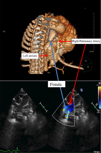
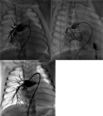
We report the case of a newborn with a prenatal diagnosis at 23 weeks of gestation of a fistula between the right pulmonary artery and the right pulmonary vein; the fetal echocardiography found a dilatation of the left cavities due to the right–left shunt but without heart failure. The newborn was delivered naturally at 39 weeks and 5 days with an initial difficult adaptation (Apgar 1/4/6/10) requiring a transitory invasive ventilation before being admitted to our unit specialising in paediatric cardiology. At the admission, the weight was 2.9 kg and the saturation was 75% at rest. The postnatal echocardiography revealed a fistula between the right pulmonary artery and the posterior part of the left atrium, near the right inferior pulmonary vein (S-Video S1). The Doppler showed a continuous flow about 3 m/s through this right–left shunt responsible for cyanosis; the ductus arteriosus was present but small when it arrived on the left pulmonary artery. A cardiac CT was performed confirming the diagnosis (Fig 1). From 24 hours of life, the newborn presented profound desaturations until 50% requiring non-invasive ventilation with a maximum of oxygen and nitric oxide therapies in order to improve the pulmonary perfusion. The fistula was closed after 48 hours of life by an Amplatzer duct occluder II 6/4 mm after selective pulmonary angiograms showing a direct communication from the right pulmonary artery to the left atrium (fistula type 1); the procedure (Fig 2) was successful with a normalisation of the saturation after stopping oxygen and nitric oxide therapies (S-Video S2–S3). Note that the angiography found varicose right pulmonary veins. The post-procedural echocardiography confirmed the closure of the fistula without a residual shunt, non-obstruction of the pulmonary artery and the pulmonary venous return, and showed a normalisation of the left cavities size.
Image above: cardiac CT of the fistula; image below: small-axis ultrasound section of the fistula.

Image above: selective pulmonary angiograms showing a direct communication from the right pulmonary artery to the left atrium; image below: fistula closed by an Amplatzer duct occluder II 6/4 mm.

The newborn was discharged home 5 days after the procedure with a saturation of 100% and without any treatment.
Discussion
The diagnosis of fistula between the right pulmonary and the left atrium continues to be rare, but unlike the fistula between the left pulmonary artery and the left atrium, remains the most common subtype and accounts for more than 95% of the pulmonary artery to left atrium fistula case.Reference Thakkar, Raval and Oswal1 There is a 3:1 male predominance and must be differentiated from a right pulmonary arteriovenous malformation with shunting that is similar to right pulmonary artery to left atrium communication, but it is strongly associated with hereditary haemorrhagic telangiectasia manifesting with an autosomal dominant inheritance pattern.Reference Ide, Miyoshi, Matsuyama, Kurosaki and Yoshimatsu2 Since the first surgical repair by Blalock, as described by Friedlich and colleagues in 1950,Reference Friedlich, Bing and Blount3 less than 100 cases have been reported in the literature.
In 1973, Nelson et al categorised them into three general types: an anomalous vessel connecting the posterior aspect of the right pulmonary artery and draining into the left atrium with normal pulmonary venous connections, or an anomalous communication draining into the left atrium in place of the absent right lower pulmonary vein, and finally the pulmonary veins draining into the anomalous vessel.Reference De Souza e Silva, Giuliani, Ritter, Davis and Pluth4
In 1978, Ohara et al added a fourth classification which looks like the first type, but without dilated anomalous vessel but on the contrary, an aneurysmal dilatation of the left atrium.Reference Ohara, Ito and Kohguchi5
We report the case of a prenatal diagnosis, which is uncommon. Indeed, in the literature, there were 10 cases reported only (including the present report) with 5 survived, 3 terminations of pregnancy, and 2 neonatal deaths.Reference Ide, Miyoshi, Matsuyama, Kurosaki and Yoshimatsu2 In fetal life, the fistula is responsible for an enlargement of the left heart chambers, a congestive heart failure, and intrauterine growth retardation. After birth, the patient presents cyanosis with the risk of serious complications such as heart failure, systemic embolism, infective endocarditis, and brain abscesses. Amongst the approximately 92 cases of RPA-to-LA fistula described in the literature as of 2020, only 18 (including the present study) were diagnosed in neonates.
The timing of the onset of symptoms is primarily dependent on the percentage of blood that passes through the fistula. Surgical treatment has been the method of choice for most of the reported cases, but was associated with a significant morbidity and mortality as high as one in five;Reference Boudjemline, Ladouceur and Bonnet6 especially, at the start, when the surgical repair was planned later in the infants when they weighed approximately 10 kg and that the surgical approach was almost exclusively by right lateral thoracotomy.Reference Jimenez, Fournier and Choussat7,Reference Cheatham, Barnhart and Gutgesell8 In the current era, the surgical repair is done quickly, even in neonates, and the surgical approach by median sternotomy provides an excellent exposure of the fistula and an easy application of the cannulation sites of the cardiopulmonary bypass.Reference Mongé, Russell, Popescu and Robinson9 Since a few years, some reports of success of transcatheter closure were reported: exactly 16 whom 5 were neonates (with our case-report).Reference Boudjemline, Ladouceur and Bonnet6,Reference Mongé, Russell, Popescu and Robinson9 In patients who undergo transcatheter closure, the duration of hospital stay after the procedure is much shorter compared with surgery, and complications such as phrenic nerve paralysis are avoided. However, all the forms of fistula cannot be closed by transcatheter and potential complications are possible as device dislocation or migration, device erosion through the thin-walled aneurysmal fistula, incomplete closure, recanalisation, and access-related complications. Sometimes, at least 6 months of anticoagulant use to be considered to minimise the risk of systemic embolism.Reference Yakut, Varan and Ozkan10
Conclusion
Transcatheter closure of a right pulmonary artery to left atrium fistula in a neonate using an Amplatzer duct occluder is a safe option to avoid the need for surgical repair.
Acknowledgements
None.
Financial support
This research received no specific grant from any funding agency, commercial, or not-for-profit sectors.
Conflicts of interest
None.
Ethical standards
None.
Supplementary material
To view supplementary material for this article, please visit https://doi.org/10.1017/S1047951121000068.