I. INTRODUCTION
In the past decade, research on morphologically controlled synthesis of metal nanostructures has been very active because the unique optical, catalytic, electronic, and magnetic properties of nanomaterials are largely dependent on their size and shape.Reference Hao, Li, Bailey, Zou, Schatz and Hupp1–Reference Wiley, Chen, McLellan, Xiong, Li, Ginger and Xia5 In particular, silver nanostructures have attracted much attention recently due to their far-ranging applications.Reference Schwartzberg, Grant, Wolcott, Talley, Huser, Bogomolni and Zhang6–Reference Haes and Van Duyne10 So far, many methods have been reported to synthesize silver nanostructures with diverse well-defined morphologies, such as spheres,Reference Huang, Ni, Loy, Chew, Tan, Loh, Deng and Xu11, Reference Morones and Frey12 rods,Reference Cepak and Martin13, Reference Guo, Dong and Wang14 plates,Reference Cui, Li, Bao, Zheng, Zang, Ooi and Guo15 cubes,Reference Zhu, Kan, Zhu, Wan, Han, Zhao, Wang and Wang16–Reference Wiley, Sun and Xia18 polymer fibers,Reference Wang and Qi19, Reference Saquing, Manasco and Khan20 and wires.Reference Wiley, Sun and Xia18, Reference Sun, Yin, Mayers, Herricks and Xia21–Reference Korte24 Among these nanostructures, silver-nanoparticle-embedded polymer nanofibers have received great interest owing to their significant applications in various areas, such as catalysis, sensing, microelectronics, and biomedicine.Reference Grunlan, Choi and Lin25–Reference Li, Hoa, Cho, Kim and Kim29
Previous studies have demonstrated that nanofibrous structures can provide much higher cell attachment compared to other macro- or nanostructures. Therefore, it is considered a promising antimicrobial scaffold.Reference Kong and Jang30 In addition, numerous reports have verified that silver nanoparticles possess toxicity to microorganisms and strong biocidal effects on as many as 16 species of bacteria including Escherichia coli.Reference Sondi and Salopek-Sondi31 From this point of view, it is notably important to develop a simple and convenient method to fabricate silver-impregnated polymer nanofibers as a new class of biomedical materials by releasing biocidal silver with a protective polymer barrier against infection.
Until now, there are only few approaches, e.g., electrospinning (ES) and sacrificial template strategy reported to incorporate noble metals (e.g., Au and Ag) into nanofibrous polymer structures.Reference Wang and Qi19, Reference Saquing, Manasco and Khan20 The ES method is not accessible to many laboratories due to its expensive experimental setup, a complicated procedure, and the requirement of an extremely high voltage. The sacrificial template strategy requires an additional step of fabricating nanofiber templates and produces a limited kind of nanofiber structures. In yet another approach, the Xia group utilized a soft and solution-phase approach (polyol methodReference Zhang, Grass, Kuhn, Tao, Habas, Huang, Yang and Somorjai32–Reference Pierre-Yves, Rondaldo, Nicolas, Venugopal and Kamar Tekaia35) for the large-scale synthesis of uniform silver nanostructures.Reference Wiley, Sun and Xia18, Reference Sun, Yin, Mayers, Herricks and Xia21 In the polyol process, silver nitrate is reduced by ethylene glycol (EG) in the presence of poly(vinyl pyrrolidone) (PVP), which works as the capping molecule to kinetically control the growth rates of various faces on the silver crystal. Although the reaction conditions are relatively mild and the shape of the product can be readily manipulated via adjusting the concentration of the anisotropic medium, the polyol approach has been widely explored to fabricate diverse anisotropic silver nanostructures ranging from silver cube to silver prism. Other types of polymers [e.g., poly(2-ethyl-2-oxazoline) (PEtOx)] have also been utilized to fabricate silver nanoparticles via simple wet chemical method.Reference Mayer and Mark36–Reference Kim, Wong and Kang39 However, so far, there are no reports on synthesizing nanofibrous polymer structures with PVP or PEtOx with either the polyol or the wet chemical method. Inspired by these methodologies, herein, we report a facile way to fabricate length-controllable and water-dispersed silver-nanoparticle-embedded polymer nanofibers with PEtOx through the polyol process for the first time.
In this work, water soluble silver-nanoparticle-embedded PEtOx nanofibers with an average diameter of 40 nm were obtained through adjusting the reaction time t, AgNO3/PEtOx molar ratio R, and heating temperature T. Transmission electron microscopy (TEM), high-resolution transmission electron microscopy (HRTEM), energy dispersive spectrum (EDS), x-ray diffraction (XRD), and inductive coupled plasma-mass spectrometry (ICP-MS) were used to characterize their physical and chemical properties. The high surface roughness of the nanofiber indicates its potential application as a highly efficient surface enhanced Raman spectroscopy (SERS) substrate for biosensing or other optical devices. Because PEtOx has been previously demonstrated to be biocompatible and water soluble,Reference Adams and Schubert40 the functional nanocomposite reported here could be an effective antibacterial material. In addition, since there are no additional seed nanoparticles or metal ions introduced during the polyol process, the purity of this nanofiber is very high. No post-purification procedure is needed. Compared with the ES and sacrificial template approaches, our procedure shows higher reproducibility as well as simplified experimental steps. Finally, the high yield, mild reaction conditions, and low cost of this approach signify that it could be potentially considered as a large-scale industrial nanoparticle-embedded polymer nanofibers fabrication route for antibacterial material or optical sensor substrate.
II. EXPERIMENTAL PROCEDURE
A. Preparation of the silver-nanoparticle-embedded PEtOx nanofibers
Silver nitrate (AgNO3) and EG are obtained from Sinopharm Chemical Reagent Co. Ltd (Shanghai, China). PEtOx (molecular weight 50,000) is purchased from Sigma-Aldrich Chemical Co. (St. Louis, MO). All these reagents are used directly without further purification.
Silver-nanoparticle-embedded PEtOx nanofibers are synthesized by reducing AgNO3 with EG in the presence of PEtOx. First, 6 mL of EG was injected into a round-bottom flask and then heated in an oil bath set at 150 °C for 1 h. This step ensured the water dissolved in EG was completely removed. Then, 2 mL PEtOx (8 mg/mL) in EG was added into the flask within 10 s, and 2 mL EG solution of AgNO3 (68 mg/mL) was subsequently injected dropwise. Magnetic stirring was applied throughout the entire synthesis process. The reaction time for other control experiments is 1 h if not mentioned otherwise.
After PEtOx was injected, the solution was almost transparent. Then, the reactant mixture turned to a pale yellow and a bit turbid after the addition of a few drops of AgNO3 solution. Over a period of 1 h, the mixture appeared yellow and opaque, and a little precipitate was created in this process. For characterization, the product was washed with anhydrous ethanol for more than three times accompanied with ultrasonication and centrifuged to remove excess reagents and then dispersed in deionized water.
B. Characterization of the silver-nanoparticle-embedded PEtOx nanofibers
For TEM and HRTEM (JEM 3010; JEOL, Tokyo, Japan) characterization, a drop of washed product was placed onto carbon-coated copper grid and dried at room temperature. For XRD (SIEMENS D-5000; Siemens, Munich, Germany) measurement, the condensed product was dispersed on a glass substrate. ICP-MS (HP 4500 series; Agilent Technologies Inc., Santa Clara, CA) was used for the determination of silver contents of the nanofibers.
III. RESULTS AND DISCUSSION
A. Structure characterization
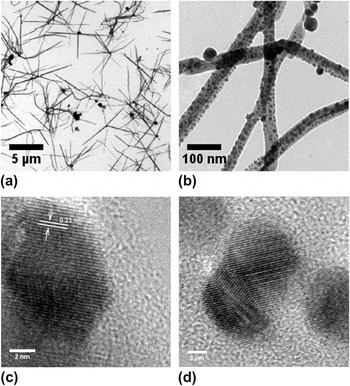
Based on the experimental results, we note that the optimum conditions to synthesize these silver-nanoparticle-embedded PEtOx nanofibers are: AgNO3/PEtOx molar ratio R = 5, reaction temperature T = 150 °C, and reaction time t = 1 h. Figure 1(a) shows the TEM image of the final product obtained from the conditions described above. One common feature of these nanofibers is that they are composed of many branches that are obviously different from the linear structure of the ES or sacrificial template product.Reference Wang and Qi19, Reference Saquing, Manasco and Khan20 The majority of these nanofibers are joined by a central nanoparticle, and their lengths are typically more than 1 μm. The magnified TEM image [Fig. 1(b)] displays that plenty of silver nanoparticles are embedded in the PEtOx nanofibers instead of being adsorbed on their surface. This is confirmed with the ICP-MS results where the measured content of silver element in the supernatant solution is less than 1% after extensive washing for one or two times.
Transmission electron microscopy (TEM) images of the final product synthesized at the conditions of [Ag+] = 80 mM, silver nitrate/poly(2-ethyl-2-oxazoline) (AgNO3/PEtOx) = 5, T = 150 °C, and t = 1 h. (a) Large-area TEM result of silver-nanoparticle-embedded PEtOx nanofibers. (b) A magnified TEM image. (c) and (d) High-resolution transmission electron microscopy images of silver nanoparticles on these polymer nanofibers.

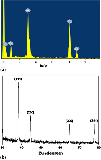
To further confirm whether these nanoparticles were made of silver, we conducted HRTEM characterization [Figs. 1(c) and 1(d)]. The fringe spacing of these silver nanoparticles on the nanofiber is about 0.23 nm, consistent with the lattice spacing of silver crystal. Some silver nanoparticles are heavily surrounded by a polymer layer. An EDS analysis revealed that the metal component from nanofibers consisted of silver [see Fig. 2(a), the copper element was originated from copper grid]. Figure 2(b) presents the typical XRD taken from a large quantity of the final product as shown in Fig. 1. The lattice constant determined from the XRD pattern is 4.0862 Å, identical to the reported data (a = 4.0862 Å, Joint Committee on Powder Diffraction Standards File 04-0783). The ICP-MS result showed that the yield of these silver nanofibers was about 73 ± 5%.
(a) Energy dispersive spectrum analysis of silver nanoparticles illustrated in Fig. 1. The copper element came from copper grid. (b) X-ray diffraction pattern of the product. All peaks could be indexed to face-centered cubic silver nanoparticles. Reaction conditions were the same as those indicated in Fig. 1.

B. Influence of reaction conditions
1. Effect of reaction temperature
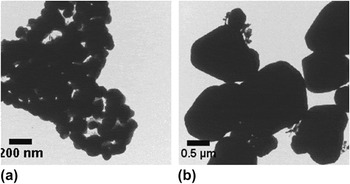
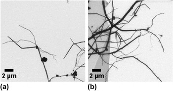
It is well known that temperature usually plays an important role in the formation of silver nanostructures in the polyol process. To explore the temperature effect on the morphology of the polymer nanofibers, a series of experiments were carried out at three reaction temperatures (120, 150, and 180 °C) while keeping other conditions fixed. When the mixture was heated at 120 °C, the resulting solution was clear with little precipitate in the bottom of the flask. The TEM image [Fig. 3(a)] shows that silver nanoparticles with a diameter of a few 100 nm are the dominant product, whereas no polymer nanofibers are formed. This may be attributed to the following two factors: (i) the rate of producing new silver nanoparticles is much slower than the fusion rate of the preformed small silver nanoparticles surrounded by PEtOx; (ii) at this temperature, the rate of generating fresh nucleic sites for silver nanoparticles is low, which would result in the high tendency of newly reduced silver atoms to attach on the surface of those preformed large silver nanoparticles. When the reaction temperature reached 150 °C, the color of the final reaction solution was pale yellow and there was some precipitate. The TEM images (Fig. 1) demonstrate that multibranched silver-nanoparticle-embedded PEtOx nanofibers were produced. At the even higher temperature of 180 °C, the resulting product was similar to that at 120 °C. The up-layer of the reaction solution was nearly transparent and a large amount of precipitate was formed, which was obviously different from the case at 150 °C. Irregular and micrometer size silver nanoparticles were the main product [see Fig. 3(b)]. These results suggest that the optimum reaction temperature for fibril growth is about 150 °C.
TEM images of the silver nanostructures synthesized under different reaction temperatures: (a) 120 °C and (b) 180 °C. The other conditions were [Ag+] = 80 mM, AgNO3/PEtOx = 5, and reaction time t = 1 h.

2. Effect of reaction time
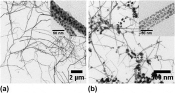
Figures 1, 4(a), and 4(b) are the TEM images of the product obtained at different reaction times of 1, 1.5, and 2 h, respectively, while the reaction temperature was maintained at 150 °C and the AgNO3/PEtOx molar ratio was kept constant at 5. At the beginning, the color of the reaction solution was slight yellow, without noticeable precipitate. Gradually, the solution became opaque gently and a small amount of precipitate was formed after 1 h (Fig. 1). The diameter of silver nanoparticles in this fibrous structure was estimated to be 7.3 ± 1.8 nm. As the reaction time was extended to 1.5 h, the color of the solution did not change significantly but more precipitate was formed. The polymer nanofibers were still the main product [Fig. 4(a)] and they were significantly longer than those reacted for 1 h [Fig. 1(a)], which suggested that the reduction rate of the unreacted silver ions in solution was much faster than the aggregation rate of the preformed small silver nanoparticles on the fiber before 1.5 h. Therefore, the number of reduced silver nanoparticles still kept on growing along the polymer when the reaction time was extended. Compared with that of 1-h product, the diameter of silver nanoparticles embedded in the nanofiber did not change obviously, i.e., 7.4 ± 1.5 nm [see the inset TEM image in Fig. 4(a)]. When the reaction time was prolonged to 2 h, the reaction solution turned transparent and large silver particles were found on the TEM image [see Fig. 4(b)]. The majority of them were dispersed individually in the solution. The length of the polymer nanofibers became much shorter. This result demonstrates that the dispersed silver nanoparticles in these nanofibers are prone to agglomerate into larger ones as long as the reaction time is long enough, which would seriously decrease the yield of the polymer nanofibers. Meanwhile, the diameter of silver nanoparticles embedded in these nanofibers became a bit larger than that of 1 h and 1.5 h [8.4 ± 1.8 nm; in the case of ellipsoidal shape, we used the length of short axis as the diameter of the nanoparticle, see the inset image in Fig. 4(b)]. Their structures also became much more irregular. Most of them showed ellipsoidal shape. This may be caused by the fusion process between neighboring nanoparticles. On the basis of these results, we conclude that the optimum reaction time for the nanofiber growth is between 1 and 1.5 h, depending on the required length of the polymer nanofibers.
TEM images of silver-nanoparticle-embedded PEtOx nanofibers synthesized at different reaction time: (a) 1.5 h and (b) 2 h. The other conditions were [Ag+] = 80 mM, AgNO3/PEtOx = 5, and reaction temperature T = 150 °C.

3. Effect of molar ratio of reactants and the order of injection
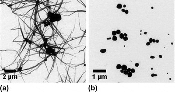
The parameter of the AgNO3/PEtOx molar ratio R also significantly affects the size and yield of the product. Figures 5(a) and 5(b) show the TEM images of these nanofibers obtained at three AgNO3/PEtOx molar ratios: R = 5 (Fig. 1), R = 1 [Fig. 5(a)], and R = 0.2 [Fig. 5(b)], with all other reaction conditions fixed. At all three molar ratios, the reaction solutions were opaque and yellow. Very little precipitate was observed. After the reaction was completed, we extracted the same volume reaction solution from different molar ratios and washed extensively. We found that the highest yield was achieved when R = 5. From the TEM picture in Fig. 5(b), it can be easily found that the diameter of the polymer nanofibers becomes much larger when the ratio decreases to 0.2. This strongly verifies that PEtOx acts as template in this reaction.
TEM images of the obtained silver-nanoparticle-embedded nanofibers under the conditions of (a) the ratio of AgNO3/PEtOx R = 1 and the temperature T = 150 °C, (b) R = 0.2 and T = 150 °C.

After exploring the influence of molar ratio on the yield and morphology of nanofibers, we also discovered that the injection order of reactants played an important role on determining the nanofiber morphology. Setting the temperature at 150 °C and R at 5, when we firstly added template PEtOx to the reaction solution, and then injected AgNO3 dropwise 10 s later, the well-defined silver-nanoparticle-embedded PEtOx nanofibers [Fig. 1 (b)] were readily obtained. However, when the order of injection was reversed, in other words, AgNO3 was added first and followed by adding PEtOx, the particles on the center of the nanofibers became much bigger [Fig. 6(a)]. Dropwise injecting AgNO3 solution into the well-dispersed PEtOx solution permitted the silver ions to be embedded more homogeneously in the polymer template. Thus, well-dispersed silver-nanoparticle-embedded polymer nanofiber with small particle sizes could be readily formed (Fig. 1). However, when the steps were reversed, the PEtOx polymer encapsulated large amounts of silver ions immediately after they were dropped into the AgNO3 solution. At high temperature, the energetic EG molecules would show pronounced reduction capacity. As a result, those highly localized silver ions could be quickly reduced into silver atoms and they were inclined to form larger silver nanoparticles [Fig. 6(a)] to reduce the surface energy.
(a) TEM image of resulted silver-nanoparticle-embedded PEtOx nanofiber when AgNO3 was injected first. (b) The obtained silver nanostructure when PEtOx was substituted by poly(vinyl pyrrolidone). The other conditions were the same as those described in Fig. 1.

4. Comparison between PVP and PEtOx
In the polyol method, PVP participates in the synthesis process as a common template and most of these works concern the fabrication of different silver nanostructures. The application of PVP as a polymeric capping reagent to kinetically control the growth rates of various faces of silver crystal is the key factor to generate anisotropic nanostructures within a highly isotropic medium.Reference Sun, Mayers, Herricks and Xia22 To investigate the formation mechanism of the silver-nanoparticle-embedded PEtOx nanofibers, we performed similar experiment under the optimum reaction conditions (t = 1 h, T = 150 °C, and R = 5) with PVP instead of PEtOx. The result is displayed in Fig. 6(b). The main product was irregular silver nanoparticles, and there were no nanofibers formed. When synthesizing silver nanowires with PVP through the polyol process, the introduction of a seeding step to serve as nuclei for the subsequent growth of silver nanowire is important.Reference Sun, Yin, Mayers, Herricks and Xia21 Since our optimum conditions did not involve an additional seeding step, this well explains why there was no silver nanowire formed when replacing PEtOx with PVP. Surprisingly, when using PVP as the template, there was no precipitate in the reaction solution while some precipitate was found when using PEtOx. This indicates that PVP has much better protection capability than PEtOx at special silver crystal faces, but it cannot guide the silver nanoparticles to grow along the longitudinal direction. Thus, only irregular silver nanoparticles could be formed. This also demonstrates that the PEtOx should have different template effect in the polyol process in comparison with PVP.
C. The growth mechanism of the silver-nanoparticle-embedded PEtOx nanofibers
Previously, the formation mechanism of silver nanostructures in polyol process was extensively studied.Reference Sun, Yin, Mayers, Herricks and Xia21 Similarly, at a high reaction temperature, EG can be oxidized as follow:

Therefore, silver atoms can be formed through the reduction of AgNO3 with EG. These silver atoms will experience a homogeneous process because there are no additional nuclei in solution. Through the homogeneous nucleation process, silver atoms yield small silver nanoparticles. It is well known that the surface energy of larger particles is lower than that of smaller ones. When the dispersed silver nanoparticles are heated at high temperatures, the smaller nanoparticles progressively disappear to the benefit of larger ones via a process known as Ostwald ripening. As the reaction progresses for an extended period of time, the smaller particles are no longer stable in solution and they begin to agglomerate and contribute to the growth of larger ones. At the same time, with the assistance of PEtOx, the polymers in solution can act as template to induce the silver nanoparticle to grow in the fibrous structure. If PVP is used as a template, as shown in Fig. 6(b), only irregular silver nanoparticles are formed. This phenomenon indicates that PEtOx is a good template guide in forming silver-nanoparticle-embedded PEtOx nanofiber.
IV. CONCLUSIONS
We successfully synthesized well-dispersed and lengthy mutibranched silver-nanoparticle-embedded polymer nanofibers using PEtOx as a template through the polyol process. The rough surfaces of PEtOx nanofibers loaded with silver nanoparticles signify that they can serve as good SERS substrates for biosensing due to the strong near-field coupling effect between neighboring silver nanoparticles. Meanwhile, owing to the highly efficient antibacterial effect of silver nanoparticles and hydrophilic property of PEtOx, this novel kind of silver-nanoparticle-embedded PEtOx nanofibers also can be potentially used as an antibacterial agent in biomedicine. Our strategy shows significant advantages over the previously reported polymer nanofiber fabrication methods, such as mild reaction conditions, simplified experimental procedures, and low cost; therefore, this method could be considered as a large-scale industrial fabrication route for silver-nanoparticle-embedded polymer nanofibers.
ACKNOWLEDGMENTS
We greatly thank Song Chen (Hunan Normal University) for the TEM measurement and Dr. Yongjun Li for valuable discussions. L-H. Xiao is thankful for partial support from the Scholarship Award for Excellent Doctoral Student from Ministry of Education, China. This work was supported by Hunan University 985 fund.