INTRODUCTION
Molluscs are the preferred hosts of many obligately parasitic/symbiotic ciliate groups, typically the orders Rhynchodida (e.g. Sphenophrya and Ancistrocoma), Thigmotrichida (e.g. Ancistrum and Boveria) and Mobilida (e.g. Trichodina and Urceolaria). In addition, many sessile peritrich ciliates (e.g. Epistylis and Scyphidia) occur as symbionts of numerous aquatic animals including crustaceans, fish, molluscs, etc. The data available suggest that marine molluscs host a high diversity of parasitic ciliates, which usually show higher host specificity than ciliates parasitizing marine fish (Xu et al., Reference Xu, Song and Warren1999, Reference Xu, Song and Warren2000). Although molluscs belong to the largest and most widely distributed invertebrate group in seas, only a small number of parasitic ciliates have been described from marine molluscs (Sprague, Reference Sprague1970; Lom & Dykova, Reference Lom and Dykova1992; Xu & Song, Reference Xu, Song, Song, Zhao, Xu, Hu and Gong2003a, Reference Xu, Song, Song, Zhao, Xu, Hu and Gongb). On the other hand, the molecular phylogeny of most parasitic ciliates has been only poorly investigated, partly due to the impediment of alpha taxonomy (Miao et al., Reference Miao, Wang, Li, Al-Rasheid and Song2009, Reference Miao, Wang, Song, Clamp and Al-Rasheid2010; Yi et al., Reference Yi, Song, Gong, Warren, Al-Rasheid, Al-Arifi and Al-Khedhairy2009; Pan et al., Reference Pan, Huang, Hu, Fan, Al-Rasheid and Song2010; Gao et al., Reference Gao, Fan, Yi, Strüder-Kypke and Song2010). In this paper we document a new rhynchodid and a new peritrich ciliate occurring mainly as gill parasites of marine molluscs and provide detailed descriptions based on live observation and silver impregnation.
MATERIALS AND METHODS
Specimens of the host mollusc bamboo clam Solen grandis Dunker were collected from marine culture beds of the Laoshan Bay on the Qingdao coast of the Yellow Sea, China (36°20′N 120°25′E) in December 1997. The water temperature was about 11°C and salinity 30–31‰ during sampling. The infection rate of Sphenophrya solinis was up to 80% (N = 10), and large quantities of ciliates occurred on the gills of individual mollusc specimens. Planeticovorticella paradoxa was first found on the gills and in the mantle cavity of the host mollusc Asiatic hard clam Meretrix meretrix (L.), which was collected from the culture beds on the Rizhao coast of the Yellow Sea, China (35°23′N 119°32′E) in April 1995. The water temperature was about 8°C and salinity at 22‰ during sampling. Later, it was observed from the same host mollusc collected with Solen grandis from the culture beds of the Laoshan Bay on the Qingdao coast of the Yellow Sea in December 1997. All five specimens of the host Meretrix meretrix examined were infected by Planeticovorticella paradoxa, and the intensity of infestation was moderate, that is, up to 100 trophonts/clusters on one host specimen.
Live ciliates were isolated directly from field samples with a micropipette and observed in vivo using a differential interference contrast (DIC) microscope Leica DM4500B (Fan et al., Reference Fan, Chen, Song, AL-Rasheid and Warren2010). Protargol impregnation (Wilbert, Reference Wilbert1975; Ji et al., Reference Ji, Xu, Choi, Warren and Song2009) was used to reveal the infraciliature and nuclear apparatus. The wet silver nitrate method, as described in Foissner (Reference Foissner1991), was used to reveal the silverline system of Planeticovorticella paradoxa. Counts and measurements on impregnated specimens were performed at a magnification of 1000×. In vivo measurements were conducted at magnifications of 100× to 1000×. Drawings of live cells were based on free-hand sketches; those of impregnated specimens were made with the help of a drawing device. Systematics follows Lynn (Reference Lynn2008). Terminology is mainly according to Raabe (Reference Raabe1970) and Corliss (Reference Corliss1979).
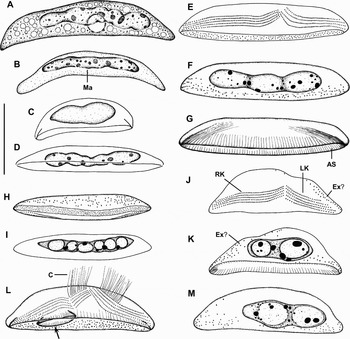
Sphenophrya solinis sp. nov. in vivo (A–D) and after protargol impregnation (E–M). (A) A representative specimen; (B–D) body variability of trophic cells; (E–G) holotype to show the ciliary and nuclear pattern. The adhesive sole is equipped with fibres more distinctly elongated in the ends forming an oral basket; (H & I) ventral view of same specimen to show the adhesive sole, the macronucleus and the granules which are probably extrusomes; (J & K) a stout specimen. The two systems of kineties form a usual pattern of 5 + 4 kineties with the right kineties longer than the left ones; (L & M) same specimen showing the early stage of budding. A bud is developing in the region where the two systems of kineties converge with the emergence of a new adhesive sole (arrow) for the parental cell. AS, adhesive sole; C, cilia; LK, left kineties; Ex, extrusomes; Ma, macronucleus; RK, right kineties. Scale bars: 30 µm.

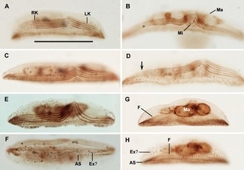
Sphenophrya solinis, trophic cells after protargol impregnation. (A–E) Specimens with usually four (A & B), occasionally five (C & D) and rarely three (E) kineties in the left systems of kineties. Arrow denotes the loosely spaced kinetosomes in the margin of right kineties; (F) ventral view to show the narrowly elongate adhesive sole; (G & H) same specimen at different focal planes to show the adhesive sole equipped with fibres. AS, adhesive sole; LK, left kineties; Ex, extrusomes; Ma, macronucleus; Mi, micronucleus; RK, right kineties. Scale bars: 30 µm.

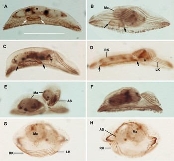
Sphenophrya solinis, cells budding after protargol impregnation. Arrows indicate the newly developed adhesive sole for parental cells. Arrowheads mark the developing adhesive sole for buds. (A) A new adhesive sole for the parental cell emerges first beneath the right side kineties; (B) the macronucleus is condensed to an ellipsoidal or globular mass, and the right kineties broken first at the top third region; (C & D) the adhesive sole for parental cells is growing (arrows) and a new sole for the bud is produced (arrowheads); (E) the developed bud is globular, and has a globular macronucleus and two systems of short ciliated kineties; (F) the parental cell after budding inherits the main part of the macronucleus, which is located in the region of the right side kineties; (G & H) a tomite with broadly fusiform adhesive sole in dorsal and ventral view. AS, adhesive sole; LK, left kineties; Ma, macronucleus; RK, right kineties. Scale bars: A–H, 30 µm.

Type Material
The holotype (QD-971204-01) and one paratype (QD-971204-02) as two slides of protargol-impregnated specimens have been deposited in the Laboratory of Protozoology, Ocean University of China, Qingdao, China.
TYPE HOST AND SITE
Solen grandis Dunker, gills.
TYPE LOCALITY
Found in culture beds of the Laoshan Bay, on the Qingdao coast of the Yellow Sea (36°20′N 120°25′E).
ETYMOLOGY
This species is named after the generic name of the host mollusc Solen grandis.
NOMENCLATURE
Sphenophrya solinis Xu & Song, 2003 in Song et al. (Reference Song, Zhao, Xu, Hu and Gong2003, p. 122) is not valid because it is not indicated as a new species (see Article 16.1 of the ICZN, 1999).
DIAGNOSIS
Marine Sphenophrya about 70 × 15 µm in vivo; body narrowly elongate boat-like with bluntly pointed ends, nearly triangular in cross-section. Macronuclear nodules are moniliform. Two systems of kineties converge in middle left body portion and extend to ends of adhesive sole; right kineties longer than left kineties with a length ratio about 1.4:1, composed of usually 5 rarely 6 equidistant rows; left kineties more loosely spaced than right kineties, composed of usually 4, occasionally 5, rarely 3 equidistant rows. Bud develops on the dorsal left of body during budding.
DESCRIPTION OF TROPHONTS
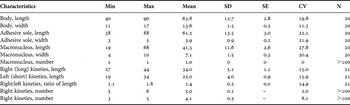
Size in vivo 50–100 × 12–20 µm, usually near 70 × 15 µm; in protargol impregnation the mean size decreases to about 64 × 14 µm, corresponding to shrinkage of about 10% (Table 1). Shape highly variable, usually narrowly elongate boat-like with bluntly pointed ends; nearly triangular in cross-section. Macronucleus located in middle two-thirds of body, more or less moniliform and about 42 × 7 µm in protargol-impregnated cells; usually with thin karyoplasm and several nucleoli 1–4 µm across. Likely a single spherical micronucleus near or attached to the macronucleus, often very faintly impregnated with protargol and thus difficult to discern (Figures 1A–D, F, I & 2B, G). Numerous minute extrusomes (mucocysts?) studded in cortex, mainly distributed in adhesive sole and on the side without kinetosomes, only a few occur on the side bearing kinetosomes; in impregnated specimens easily mistaken as kinetosomes because of their similar sizes (Figures 1E, H, J–M & 2E–H). A vacuole present in the middle of cell, but no contraction observed (Figure 1A). Cytoplasm is transparent at low magnification. Trophonts lack cilia and thus motionless, attaching to the gills of the host. Many of the ciliate individuals fell off and lay still on the bottom when the host gills were placed in a Petri dish with seawater, and cells died off in 12 hours.
Morphometric data on Sphenophrya solinis sp. nov. Data based on protargol-impregnated and randomly selected specimens. Measurements in µm.

CV, coefficient of variation in %; Max, maximum; Min, minimum; N, number of specimens investigated; SD, standard deviation; SE, standard error of mean.
The adhesive sole (= the sucker) with which cells attach to host gill epithelia is considered as the ventral surface of the body according to Chatton & Lwoff (Reference Chatton and Lwoff1950) and Raabe (Reference Raabe1970); stretching almost same length as body, concave and very narrowly elliptical (Figures 1E, G, H & 2F). The adhesive sole is likely the oral opening as it is equipped with fibres that are distinctly elongated in the ends of sole, forming an oral basket (Figures 1G & 2G, H). The dorsal side, which is opposite to the adhesive sole, is basically convex with an inconspicuous depression in middle portion.
Two systems of parallel and non-ciliated kineties converge in middle left of body apex and obliquely extend to pointed ends of adhesive sole; kinetosomes in the upper two-thirds of the systems more densely spaced than the margins (Figure 2A–E). The right kineties sensu Raabe (Reference Raabe1970), more or less distinctly longer than the left ones, length ratio of right to left kineties on average 1.4:1; composed of mostly 5 equidistant rows with an interspace of 0.9 (0.7–1.2) μm distant, a 6-row pattern occurred only in two out of 200 specimens analysed (Figure 2A–E; Table 1). The left kineties at the same level or slightly higher than the right kineties at the apex; more loosely spaced than the right kineties, with an interspace of 1.3 (1–1.8) μm distant; in 200 specimens analysed, 87.5% specimens have 4 equidistant rows, 10% with 5 rows, and only 2.5% with 3 rows (Figure 2A–E).
BUDDING
When budding commences, a new adhesive sole for parental cell emerges first beneath the top of the right side kineties; no change is recognizable in the infraciliature or the macronucleus (Figure 3A). Next, the cells become thicker (increase in height) and the macronucleus is condensed to an ellipsoidal or globular mass (Figure 3B). The new adhesive sole for the parental cell is gradually enlarged to an elliptical form under the right side kineties (arrows in Figure 3A–E). Meanwhile, a bud is gradually produced on the middle left of the apical surface, viz., in the region where the two systems converge. The kineties for the bud are obtained by the partition of the parental kineties, first in the top one-third region of the right kineties followed by the left kineties (Figure 3B–D). The cilia develop on the two new systems of parallel kineties (Figures 1L & 3D, E). Simultaneously, a new adhesive sole for the bud is produced underneath the apical surface of the bud (arrowheads in Figure 3C–E). Then the macronucleus slightly stretches and is unequally distributed to the bud (a small proportion) and the parental cell. The bud to be detached from the parental cell is globular, and has a globular macronucleus and two systems of short ciliated kineties (Figure 3E).
The just separated bud (= tomite) can swim away and attach with its adhesive sole. The parental cell has a distinct depression in the detachment region and a stout macronucleus in the region of the right side kineties. The parental left kineties near the sole are distinctly curved, indicating the proliferation of kinetosomes before budding division (Figure 3F). Tomites are broadly ellipsoidal and about 18–19 × 16 µm. The adhesive sole of the tomite is broadly fusiform in ventral view and gradually stretches to each end to form the adult elongated sole. The tomite becomes motionless when the somatic cilia are lost. Like those of trophic cells, the right kineties of the tomites are more or less distinctly longer than the left ones; both systems of kineties are extended with the proliferation of kinetosomes (Figure 3G, H).
REMARKS
Sphenophrya solinis belongs to the rhynchodid genus Sphenophrya Chatton & Lwoff, Reference Chatton and Lwoff1921, which is unique in having two systems of parallel kineties without cilia and an adhesive sole that cells use to attach on the gills of bivalves (Raabe, Reference Raabe1970; Lynn, Reference Lynn2008). Only six species of Sphenophrya have previously been described. Among these, Sphenophrya solinis most closely resembles S. dosiniae Chatton & Lwoff, Reference Chatton and Lwoff1921, S. cardii Chatton & Lwoff, Reference Chatton and Lwoff1950, S. minor Poljansky, Reference Poljansky1951 and S. sphaerii Mjassnikowa, Reference Mjassnikowa1930. Sphenophrya solinis differs distinctly from S. cardii and S. sphaerii by the pointed ends of body (versus rounded ends in both species) (Mjassnikowa, Reference Mjassnikowa1930; Chatton & Lwoff, Reference Chatton and Lwoff1950). Furthermore, S. cardii is nearly circular in cross-section (versus triangular in S. solinis), and S. sphaerii is a freshwater species (versus marine) with the left kineties separated into two parts forming a pattern of 2 + 2 or 2 + 3 kineties (versus equidistant kineties). Sphenophrya solinis differs from S. minor by the comparatively long and densely ciliated left kineties (versus very short and sparsely ciliated) and in having fewer right kineties (5–6 versus 7–9) (Poljansky, Reference Poljansky1951; Raabe, Reference Raabe1970).
Sphenophrya solinis strongly resembles S. dosiniae, a species that has been described in great detail from a number of marine bivalves in Europe and North America. While all known populations of S. dosiniae invariably possess three kineties in the left system (Chatton & Lwoff, Reference Chatton and Lwoff1921, Reference Chatton and Lwoff1950; Raabe, Reference Raabe1938, Reference Raabe1949, Reference Raabe1970; Fenchel, Reference Fenchel1965), a three-kinety pattern occurs only occasionally in S. solinis, most specimens having four, sometimes five, kineties. Nonetheless, S. solinis differs from S. dosiniae in that the right kineties are distinctly longer than the left ones (length ratio about 1.4:1) resulting in the convergence of the two systems of kineties in the dorsal left of the body. By contrast, in S. dosiniae the right kineties are shorter than the left ones and thus the convergence is located in the middle right of body (Chatton & Lwoff, Reference Chatton and Lwoff1921, Reference Chatton and Lwoff1950; Fenchel, Reference Fenchel1965; Raabe, Reference Raabe1970). The length difference in the kinety systems is a decisive feature because it is associated with a clearly different budding configuration in the two species. As a rule, during budding the bud in Sphenophrya is invariably formed in the convergent region of the kinety systems, with the main section of the dividing macronucleus of the parental cell located on the opposite side of the cell to the bud (Chatton & Lwoff, Reference Chatton and Lwoff1950; Fenchel, Reference Fenchel1965; Raabe, Reference Raabe1970). In S. dosiniae, the bud is formed near the right side of the body and the main section of the dividing macronucleus is located on the left side of parental cell. By contrast, the bud of S. solinis is invariably developed on the left side of the body, while the main section of the dividing macronucleus is distributed to the right side of parental cell (Figure 3D, E). The budding configuration of S. solinis is similar to that of S. sphaerii which, like S. solinis, has the right kineties longer than the left ones (Mjassnikowa, Reference Mjassnikowa1930; Raabe, Reference Raabe1949, Reference Raabe1970). Thus, we consider S. solinis as new to science.
Sphenophrya solinis has been found only once from the bamboo clam Solen grandis in China, indicating it is very rare. We did not detect it in China from a number of commercial bivalves (e.g. Dosinia japonica, Mactra chinensis, M. veneriformis and Mya arenaria). The occurrence of a large number of such ciliate resulted in no obvious host-response. Although no mortalities have been associated with sphenophyriid infections, the mass occurrence of Sphenophrya may influence the health of host as the ciliate lives only on host cells (Bower et al., Reference Bower, McGladdery and Price1994).
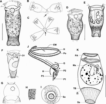
Planeticovorticella paradoxa sp. nov. in vivo (A–G) and after protargol (J, K) and wet silver nitrate impregnation (H & I). (A) A typical trophont; (B & C) trophont doublet and quadriad, cells of which are connected by very short, rigid stalk (arrows); (D & E) slightly contracted trophonts; (F & G) a developing and a mature telotroch; (H & I) silverline system in lateral and aboral views; (J) detailed structure of oral infraciliature. Arrowhead marks the separated row of the infundibular polykinety 2; (K) holotype showing the ciliary and nuclear pattern and the myoneme system. CV, contractile vacuole; EM, epistomial membrane; G, germinal row; H, haplokinety; Ma, macronucleus; P1–3, infundibular polykineties 1–3; PA, peristomial area; Po, polykinety; Sa, scopula; TB, trochal band. Scale bars: 30 µm.

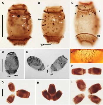
Planeticovorticella paradoxa after protargol (A–I) and wet silver nitrate impregnation (J). (A–C) Ciliary and nuclear apparatus. Arrows indicate the separated row 3 (based on conventional numbering) of the infundibular polykinety 2. Arrowheads mark the myoneme system; (D) a trophont with a short, non-contractile stalk indicated by the arrows; (E & F) trophont doublets each composed of two cells connected by a short stalk indicated by the arrows; (G–I) two triplets and one quadriad; (J) silverline system between trochal band and scopula. EM, epistomial membrane; G, germinal row; H, haplokinety; Ma, macronucleus; Mi, micronucleus; P1–3, infundibular polykineties 1–3; Sa, scopula; TB, trochal band. Scale bars: 30 µm.

TYPE MATERIAL
The holotype slide (RZ-950428-01) of protargol-impregnated specimens and one paratype slide (QD-950428-03) of wet silver impregnated specimens is deposited in the Laboratory of Protozoology, Ocean University of China, Qingdao, China.
TYPE HOST AND SITE
Meretrix meretrix (L.) have gills and mantle cavity.
TYPE LOCALITY
Found in culture beds on the Rizhao coast of the Yellow Sea (35°23′N 119°32′E).
ETYMOLOGY
The Latin adjective paradoxus (bizarre) refers to the curious appearance of the species.
NOMENCLATURE
Planeticovorticella paradoxa which was originally misplaced in the genus Epistylis as Epistylis paradoxa Xu & Song, 2003 in Song et al. (Reference Song, Zhao, Xu, Hu and Gong2003, p. 143) is not valid because it is not indicated as a new species (see Article 16.1 of the ICZN, 1999).
DIAGNOSIS
Marine epibiontic Planeticovorticella have motile trophonts about 75 × 35 µm in vivo, cylindroidal with one-layer peristomial lip. Macronucleus C-shaped, longitudinally oriented in mid-body. They have a contractile vacuole near ventral wall of infundibulum, with epistomial membrane, but lacking distal kinetal fragment. Haplokinety and polykinety complete one and a half circles of peristome before entering infundibulum. There are transverse silverlines numbering about 87 from oral area to trochal band and ~30 from trochal band to scopula.
DESCRIPTION
Trophonts are typically motile, most occurring individually and some as clusters. In 80 trophonts analysed, 81% occurred as single cells, 12.5% as doublets of two cells joined, 4% as triplets of three cells, and 2.5% as quadriads of four cells (Figure 5A–I). Cells of individual clusters (doublets, triplets and quadriads) were attached scopula to scopula by short, transparent and non-contractile stalk (Figures 4B, C & 5D). Trophonts of clusters were likely linked temporarily, as indicated by doublets with disconnecting stalk as well as certain single trophonts possessing a short stalk possibly inherited from parental stalk (Figure 5D, E). Cells of doublets were of similar size to trophont specimens (Figure 5E, F), although there was always a larger cell connected with two similar-sized smaller cells in triplets and two similar-sized larger and two similar-sized, slightly smaller cells in quadriads (Figure 5G–I). The distinct size differences in triplets and quadriads indicate the clusters are likely the products of binary fission (Figure 5G–I).
Single trophonts have usually no stalk, can swim slowly with their peristomial cilia or attach to substrate with their scopula. Stalk, when present, is transparent and non-contractile, shorter than one-quarter of body length, up to 10 µm thick, and surface without striation (Figures 4A, D & 5D). Scopula more or less distinctly concave with marginal indentations in living cells, but only slightly concave in protargol-impregnated specimens, where distal surface of scopula is studded with many kinetosome-like black dots (Figures 4A, D, E & 5A–C).
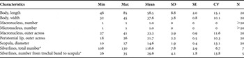
Size of individual trophonts 60-90 × 30–45 µm in vivo, usually about 75 × 35 µm, as calculated from some in vivo measurements and protargol-impregnated specimens, which showed more shrinkage in body length than width (Table 2). The shape of individual trophonts moderately variable, usually cylindroidal to elongate vase-like with maximum width in peristomial lip as well as in mid-body; slightly constricted below peristomial collar. The epistomial disc is dome-like, strongly elevated in expanded individuals and highly depressed in contracted individuals (Figure 4A–E). The peristomial collar is up to 7 µm in height and 2 µm in thickness, without circumferential fold (Figure 4A–E). Macronucleus longitudinally located in middle body portion, C-shaped, up to 40 µm across and 18 µm thick, contains many nucleoli. A spherical micronucleus is near or attached to the macronucleus. Single contractile vacuole near ventral wall of infundibulum, can expand to 10 µm across (Figure 4A–E). The pellicle striations are visible at low magnification, very distinct under high magnification; no cortical granules recognizable in vivo. Cytoplasm basically transparent in anterior three-quarters, frequently contains several globular to ellipsoidal lipid droplets 6–12 µm across and several food vacuoles up to 10 µm across, while the cytoplasm below the aboral trochal band is very dark due to numerous small, tightly packed body inclusions (Figure 4A, D, E).
Morphometric data on Planeticovorticella paradoxa sp. nov. Data based on protargol and wet silver impregnated (*) specimens. Measurements in µm.
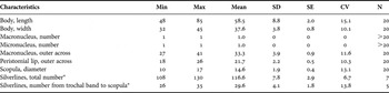
CV, coefficient of variation in %; Max, maximum; Min, minimum; N, number of specimens investigated; SD, standard deviation; SE, standard error of mean.
Infundibulum reaches mid-body. Epistomial membrane is located near opening of infundibulum as commonly seen in other peritrichs. Haplokinety and polykinety complete one and a half circles of peristome before entering infundibulum and a further turn within the infundibulum, with peristomial cilia about 18 µm (Figures 4J, K & 5A–C). Haplokinety composed of two rows of kinetosomes in zigzag pattern, parallel to germinal kinety (G) within upper half of infundibulum, while opposite infundibular polykineties on wall of lower half of infundibulum (Figure 4J). Germinal kinety located above haplokinety, hook-like, composed of kinetosomes that are loosely spaced in anterior half and densely packed in curved region. Polykinety composed of three rows of kinetosomes, parallel to haplokinety before entering the lower half of infundibulum, where it is differentiated to three polykineties (P1–3) each comprising three rows of kinetosomes. Infundibular polykinety 1 (P1) longer than polykinety 2 (P2), kinetal rows of P1 and P2 generally parallel and converge at anterior end except for row 3 (based on conventional numbering) of P2 which is directed anteriorly; shortest polykinety 3 (P3) about half length of P1, located near posterior ends of P1 and P2 (Figures 4J & 5A, B). Trochal band 1–2.5 µm thick, composed of two to many staggered rows of kinetosomes at posterior third of body (Figure 5A–C).
Silverline system consists of a total of about 117 pellicular striae; among these about 30 striae are distributed between trochal band and scopula, with an average distance between adjacent silverlines of 0.5 µm (Table 2). Numerous pellicular pores (i.e. argentophilic dots) associated with pellicular striae, 0.5–1 µm in diameter (Figures 4H, I & 5J). Myoneme system sparse, intensely stained with protargol only on periphery of epistomial area and in body portion between trochal band and scopula (Figures 4K & 5C).
Cilia form in the trochal band in the early stage of telotroch development; mature telotroch broadly ellipsoidal and distinctly concaved on aboral side (Figure 4F, G).
REMARKS
Planeticovorticella paradoxa has several peculiar morphotypes/features, including the typically stalkless trophont that swims by means of its peristomial cilia, the attachment of trophonts to a substrate via its scopula, and the occurrence of trophont doublets, triplets and quadriads, cells of which are linked temporarily by very short and non-contractile stalks. The features mentioned above suggest the generic assignment in the monotypic genus Planeticovorticella Clamp & Coats, Reference Clamp and Coats2000, which is characterized by the typically stalkless and free-swimming trophonts as well as the trophont doublets. Planeticovorticella paradoxa differs from its only congener, P. finleyi Clamp & Coats, Reference Clamp and Coats2000, by its body shape (cylindrical versus subconical), the location of the contractile vacuole (apical versus in mid-body), and the epibiontic (versus planktonic) life style. Clamp & Coats (Reference Clamp and Coats2000) reported also the occasional occurrence of sessile, Vorticella-like trophonts in P. finleyi with long and contractile stalk. However, such an unusual morphotype occurred only in cultures of P. finleyi when the ciliates had been subcultured several times at intervals of ~3–4 days. Clamp & Coats (Reference Clamp and Coats2000) clearly stated that the Vorticella-like stage had never been found in field samples. It is not known whether a similar stage occurs also in P. paradoxa, as no culture could be established for this epibiontic ciliate. To date, P. paradoxa has been found three times and only from the host clam Solen grandis, suggesting it may be host specific.
Without the knowledge of Planeticovorticella, Xu & Song (Reference Xu, Song, Song, Zhao, Xu, Hu and Gong2003a) misplaced P. paradoxa in the genus Epistylis as Epistylis paradoxa, an invalid species because it is not indicated as a new one (see Nomenclature). The free-swimming nature of the trophonts, and the ability to attach to a substrate via the scopula, in P. paradoxa preclude its assignment to either Rhabdostyla Kent, 1881 or Epistylis Ehrenberg, 1830, both of which are invariably sessile and attach to their substrate via a stalk (Ma & Overstreet, Reference Ma and Overstreet2006; Lynn, Reference Lynn2008). Nonetheless, Epistylis has been frequently reported as epibionts from molluscs, arthropods, fish and amphibians, and certain short-stalked Epistylis might look similar to P. paradoxa. Boshko (Reference Boshko1987) described an epibiontic epistylid, namely Epistylis borysthenicus, on the soft tissues of the freshwater mollusc genera Unio and Anodonta. This form comprises individual forms or colonies with two zooids with a very short stalk that bears a large bowl-like attaching disc. Nevertheless, Planeticovorticella paradoxa can clearly be separated from this form by its mobile (versus sessile) trophonts, marine habitat (versus freshwater) and differences in stalk structure. No other species within the genera Epistylis and Rhabdostyla look similar to the new species.
ACKNOWLEDGEMENTS
This work was supported by the National Science Foundation of China (Project No. 41076102), the Knowledge Innovation Program of the Chinese Academy of Sciences (No. KZCX2-YW-417) and a joint grant from the Centre of Excellence in Biodiversity, King Saud University.