Implications
Research on prebiotic substances has been done mostly on human subjects or small laboratory animals. And mostly only changes in gut bacteria populations were investigated disregarding effects on the intestine itself. Since prebiotics are claimed to be a possible alternative to antibiotics as growth promoters in livestock husbandry this study has been done on calves to further investigate potential beneficial effects on cattle health. To determine modes of action of the prebiotics inulin and lactulose changes of morphology in the small and large intestine and the adjoining immune defense tissues have been observed.
Introduction
In recent years the European Union introduced a ban on antibiotics as growth promoters in animal feed. Now alternatives have to be found to assure animal health and performance during meat or milk production. Possible beneficial feed additives may be prebiotics such as inulin or lactulose which can be able to modulate intestinal bacterial populations towards a healthier flora (Gibson and Roberfroid, Reference Gibson and Roberfroid1995).
Inulin is a natural constituent of a wide range of plants including many common vegetables and cereals in western diet (Van Loo et al., Reference Van Loo, Coussement, de Leenheer, Hoebregs and Smits1995). It consists of β-(2-1)-linked fructo-oligosaccharides of varying degrees of polymerization from 2 to 60 sugar units. In the intestine inulin leads to a shift in the bacterial flora towards more bifidobacteria regarded as beneficial for host health (Gibson et al., Reference Gibson, Beatty, Wang and Cummings1995). Additionally inulin-type fructans have been shown to have anti-carcinogenic properties (Hughes and Rowland, Reference Hughes and Rowland2001; Femia et al., Reference Femia, Luceri, Dolara, Giannini, Biggeri, Salvadori, Clune, Collins, Paglierani and Caderni2002). Fructo-oligosaccharides were also reported to enhance the performance of livestock including pigs and calves (reviewed by Van Loo, Reference Van Loo2007).
Lactulose (4-O-β-d-galactopyranosyl-d-fructose) is a semi-synthetically produced disaccharide that does not occur naturally (Schumann, Reference Schumann2002). It is fermented by lactobacilli and bifidobacteria, but also by other bacteria species such as Clostridium perfringens, Escherichia coli or Bacteroides spp. (Mitsuoka et al., Reference Mitsuoka, Hidaka and Eida1987). Lactulose is commonly used to treat constipation (Attar et al., Reference Attar, Lemann, Ferguson, Halphen, Boutron, Flourie, Alix, Salmeron, Guillemot, Chaussade, Menard, Moreau, Naudin and Barthet1999) and hepatic encephalopathy (Bircher et al., Reference Bircher, Müller, Guggenheim and Haemmerli1966) but also to enhance the animal performance (Fleige et al., Reference Fleige, Preißinger, Meyer and Pfaffl2007).
Until now most of the studies were done on human subjects or laboratory rats instead of livestock and mostly studied changes in bacterial flora. Comparably little research has been done on possible changes of intestinal morphology. Thus the object of this study was to provide information about the influences of inulin and lactulose feeding on performance, morphology of the intestinal mucosa and on mesenteric lymphoid nodes in pre-ruminant calves.
Material and methods
Animals, husbandry, feeding and experimental procedures
Forty-two male Holstein–Friesian calves were purchased from the Viehzentrum Waldkraiburg GmbH and housed at the experimental station Karolinenfeld (Bayerische Landesanstalt für Landwirtschaft – LfL, Institut für Tierernährung und Futterwirtschaft). The animals were subdivided into three experimental groups (n = 14 per group) with balanced weight (52.9 ± 6.2 kg) and age (22 ± 5 days).
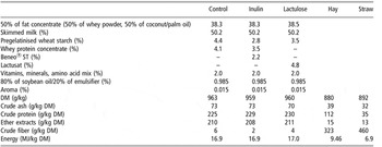
Compositions of the three diets are given in Table 1. Calves of the control group were fed with the milk replacer Milkibeef Top (Milkivit, Trouw Nutrition, Burgheim, Germany). The other groups were fed with the same milk replacer iso-energetically and iso-nitrogenically enriched with either 2% inulin (Beneo® ST, Orafti, Tienen, Belgium) or 2% lactulose (Lactusat, Milei GmbH, Leutkirch, Germany). Calves were fed individually by transponder automatic feeders (Förster Technik, Engen, Germany). During the experimental period the milk replacer concentration was rising from 125 g/l to 200 g/l with daily intake volumes rising from 6 l to 16 l. Calves had free group access to fresh drinking water and up to 300 g hay per day and animal. Since the calves were housed on straw a further uptake of roughage could not be excluded. The animals were slaughtered after 20 weeks.
Ingredients and analysis of nutrient and energy content of the diets (energy content of the milk replacer was estimated with the program Zifo (LfL, 2005))

DM = dry matter.
Histological sampling
Immediately after slaughtering the gastrointestinal tract was removed from the carcass. Sections of 0.5 to 1 cm of centre parts of jejunum, ileum and colon as well as mesenteric lymphoid nodes were collected. Tissue samples were washed in physiological NaCl solution and placed in neutral buffered of 3.7% formalin (Carl Roth GmbH, Karlsruhe, Germany) for 24 h. At the Landesuntersuchungsamt (LUA, Oberschleißheim) samples where embedded in paraffin. Sections of 4 to 6 μm were cut (Microtom LEICA RM2145, Leica, Wetzlar, Germany), deparaffinized and rehydrated before further treatment. All microscopic analyses were done randomly by one person without knowledge of treatment groups. Histological sections of ileum, jejunum and mesenteric lymphoid nodes were examined with the light microscope Axioskop 2 plus (Zeiss, Oberkochen, Germany), sections of the colon with the stereomicroscope Stemi 2000-C (Zeiss). Pictures were taken with the AxioCam MRc (Zeiss) and analyzed with the connected software AxioVision 3.1.
Histomorphometry
Histomorphometry was done on sections stained with Alcian blue/periodic acid Schiff’s reagent (AB-PAS) (see histochemistry) or haematoxylin (Carl Roth GmbH, Karlsruhe, Germany) and eosin yellowish solution (Fluka-Chemie AG, Buchs, Switzerland). Measurement techniques were adapted from Sehm et al. (Reference Sehm, Lindermayer, Dummer, Treutter and Pfaffl2006). Briefly villus length from lamina muscularis to tip and width were measured in sections of jejunum and ileum. Crypt depth from lamina muscularis to the crypt mouth and distance between crypts were examined in sections of the colon. Figure 1 gives an overview of conducted measurements on the intestinal mucosa. All were done on three well-defined villi or crypts of one section. In mesenteric lymphoid nodes and in ileal Peyer’s patches the number of lymph follicles in a defined area and areas of these follicles were measured.
Morphological measurements in the small and large intestine: Conducted measurements are shown as an overview for both small and large intestine (B) and as actual measurements in sections of jejunum (A) and colon (C). (1) lamina muscularis mucosae, (2) crypt of Lieberkühn, (3) villus; small intestine: (a) villus length, (b) villus width, (c) villus tip; large intestine (no villi existent), (d) crypt depth, (e) distance between crypts.

Histochemistry
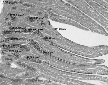
The AB-PAS staining method was used to investigate the number of goblet cells in jejunum and ileum. Briefly sections pre-treated with 3% acetic acid were treated with 1% Alcian blue in 3% acetic acid and subsequently with 0.5% periodic acid. Tissues were then treated with Schiff’s reagent (all chemicals from Carl Roth GmbH, Karlsruhe, Germany) and counterstained with haematoxylin. AB-PAS positive cells were counted individually for crypts and villi of jejunum and ileum. Goblet cell density was determined as number of AB-PAS positive cells per length of villus tip or crypt outline (Figure 2). Quantification of goblet cells in the colon was not possible due to the high density of AB-PAS stained cells in this area.
Quantitative analysis of goblet cells: goblet cell density was determined as number of goblet cells (x) per length of villus or crypt outline.

Immunohistochemistry
Detection of proliferative cells was done on six randomly selected samples of each treatment group with the antibody MIB-1 (Dako cytomation, Glostrup, Denmark) directed against the proliferation marker Ki-67. For antigen accessibility sections were incubated in boiling 0.01 M citrate buffer (pH 6) and all washing was done in phosphate buffered saline with 0.05% Tween (phosphate buffer saline (PBS)-T). To block endogenous peroxidase activity and unspecific antibody binding the slides were treated with 1% hydrogen peroxide and 10% goat serum (Dako cytomation), respectively. Binding of MIB-1 (1 : 50 in PBS) was done at 4°C over night. Afterwards sections were incubated with horse raddish peroxidase-labeled polyclonal goat anti-mouse antibody (1 : 50 in PBS, Dako cytomation). Visualization was done with 3,3′-diaminobenzidine solution (Sigma-Aldrich Chemie GmbH, Buchs, Switzerland). Tissue was then counterstained with haematoxylin. Quantification of MIB-1 positive cells was done similarly to AB-PAS positive cells in the crypts of jejunum, ileum and colon.
Statistical analysis
For each parameter the mean group values and the residual standard error of the mean were determined. Comparison of dietary treatments was done with the one-way ANOVA method of SigmaStat 3.0 (SPSS Inc., Chicago, IL, USA). Homoscedascity among compared groups was not formally tested as the test would pose a large risk of type II statistical error due to limited sample size (Altman, Reference Altman1985). However, the data was screened using a scatter plot. The individual contrasts were tested using the conventional Tukey–Kramer test for multiple comparisons. Nutritional effects were considered if P < 0.05. Tendency was considered if P < 0.1.
Results
During the current experiment general animal performance data also were collected. Animals fed inulin had significantly (P < 0.05) higher daily weight gains (1269 ± 101 g/day) than animals fed lactulose (1124 ± 178 g/day) while the control treatment led to intermediate values (1203 ± 152 g/day). Daily ingestion of milk replacer (dry matter) was very similar between inulin and control groups (1702 ± 128 g/day and 1709 ± 117 g/day, respectively) while calves of the lactulose group collected significantly less (P < 0.05) of their daily allowance (1581 ± 181 g/day). Since hay consumption was not determined for each individual animal, feed efficiency was not statistically analyzed. Calculating with group averages total feed efficiency (average dry matter intake/daily gain) was slightly better for inulin (1.55) than for control (1.64) or lactulose (1.62). All data has been reported previously by Preißinger et al. (Reference Preißinger, Büttel, Obermaier, Masanetz and Pfaffl2007) (Table 2).
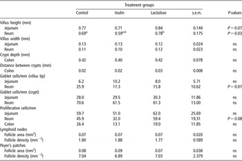
Results of measurements on mucosal architecture, goblet cell numbers, lymph follicles in Peyer’s patches and mesenteric lymphoid nodes (each n = 14 if possible) and on cell proliferation in intestinal sections (n = 6) (all values are demonstrated as mean ± s.e.m.)

ns = not significant.
Significant difference between a and b: P = 0.02 (Tukey-Kramer test).
In jejunum villus length showed an overall tendency to be decreased in inulin treated animals and to be increased in lactulose treated animals when compared to the control group (P = 0.07). A very similar but statistically significant difference could be found for villus length in the ileum (P = 0.03). Tukey–Kramer test revealed significant differences between the two experimental feeding groups (P = 0.02) but not between inulin or lactulose treatment and the control group. No nutritional effects could be seen for villus width in jejunum and ileum or crypt depth and distance between crypts in the colon.
Densities of proliferative epithelial cells tended to be decreased by inulin and to be increased by lactulose feeding in ileum (P = 0.08) but not in jejunum and colon.
Goblet cell densities in jejunum and ileum were determined individually for crypts and villus tips. In villus tips of the ileum a tendency for a decrease of goblet cell density (P = 0.07) could be found for both experimental treatments. No further significant differences could be detected.
Area and density of lymph follicles were determined in slides of mesenteric lymphoid nodes and ileal Peyer’s patches, but no significant differences could be found between the different feeding regimes.
Discussion
Prebiotics are one possible alternative to antimicrobial growth enhancers to improve animal health and performance especially during critical periods such as weaning. Therefore a variety of studies on influences of inulin and oligofructose on animal performance have already been accomplished. Quite a number found no or only little effects, but some reported effects similar to this study with improved daily weight gain and feed efficiency (Kaufhold et al., Reference Kaufhold, Hammon and Blum2000; Flickinger et al., Reference Flickinger, van Loo and Fahey2003; Van Loo, Reference Van Loo2007). Interestingly a significantly higher daily weight gain or an improved feed conversion ratio as was found in the inulin group is commonly associated with a better intestinal nutrient absorption because of longer villi (Wu et al., Reference Wu, Meier and Knabe1996; Awad et al., Reference Awad, Ghareeg and Böhm2008), while in this study small intestinal villi were shortened after inulin feeding. A similar decrease of villus length after addition of inulin to the feed has already been reported in weaning piglets (Pierce et al., Reference Pierce, Sweeney, Brophy, Callan, McCarty and O’Doherty2005). But also increases of villus length in rats after feeding of soluble chicory extract (1% to 5%) or purified inulin (5%) (Kim, Reference Kim2002) were found. Supporting the observed shortening of villi a decrease of MIB1-positive and hence considered proliferating cells was found in ileum of calves fed inulin. A comparable lowering of cell proliferation has been reported before in rats fed inulin and oligofructose at a concentration of 10% of the diet (Femia et al., Reference Femia, Luceri, Dolara, Giannini, Biggeri, Salvadori, Clune, Collins, Paglierani and Caderni2002). Concluding it may have to be considered that shorter villi and a decreased proliferation rate may reduce the amount of energy necessary for maintenance of gut architecture thereby providing more energy for growth and fattening. A decrease of goblet cell density in ileal villus tips, also found in the inulin group, may further lower energy requirements for mucus production. In contrast, an increase of goblet cell densities after feeding inulin and other non starch polysaccharides to rats has been detected before (Kim, Reference Kim2002). However, as a support for this study a significant decrease of goblet cell numbers was found in weaned piglets fed a diet supplemented with 10% carob tree seed meal as a fermentable fiber source (Van Nevel et al., Reference Van Nevel, Decuypere, Dierick and Molly2005).
Although lactulose has also been considered as a prebiotic only comparably little data on animal performance has been available up until now. In contrast, to this results Fleige et al. (Reference Fleige, Preißinger, Meyer and Pfaffl2007) found a significantly increased feed consumption and a tendency for improved daily weight gain for calves fed 3% lactulose and a probiotic bacteria strain with their milk replacer. Comparable to these findings with inulin they also found a decrease of villus length in ileum of said calves. Directly opposed to these effects lactulose treatment in this study led to lower feed intakes and daily weight gains and simultaneously increased villus heights in the small intestine compared to the control group. Possibly the addition of Enterococcus faecium to the milk replacer in the study of Fleige et al. (Reference Fleige, Preißinger, Meyer and Pfaffl2007) had a modulating influence on lactulose derived effects leading to different regulations of appetite and villus length. In accordance to the longer villi lactulose also tended to increase the number of proliferative cells in ileum. Similar to this finding other dietary fibers such as highly fermentable guar gum and pectin are also known to increase the proliferation zone in the caecum crypts of rats (Brunsgaard and Eggum, Reference Brunsgaard and Eggum1995). Interestingly the notable decrease of nutrient ingestion (∼10%) of lactulose fed calves was not enough to antagonize the increase of villus length or proliferation rates as may be expected with respect to studies on fasted animals (Clarke, Reference Clarke1975). But, on the other hand, no reduction in villus height was found in pigs fed with a low-energy diet compared to a diet with twice the energy content (Claus et al., Reference Claus, Günthner and Letzguß2006) which would be more similar to the decrease in energy uptake in the lactulose group than starved animals. An expected improvement of animal performance due to a better intestinal morphology ( Wu et al., Reference Wu, Meier and Knabe1996; Awad et al., Reference Awad, Ghareeg and Böhm2008) may be overruled by the less nutrient ingestion observed in the lactulose group. But similar to inulin lactulose also led to a decrease in goblet cell density in villus tips in ileum.
The differences in effects on animal performance and mucosal architecture between the treatment groups are not easily explained by differences in nutrient consumption alone but could be based on partly different fermentation properties of the two prebiotics. It has been found that pre-caecal fermentability of lactulose in pigs was lower than that of inulin (Branner et al., Reference Branner, Böhmer, Erhardt, Henke and Roth-Maier2004) maybe leading to different sites of action in the calves’ intestines. Additionally both substances differed in stimulatory effects on beneficial bacterial subpopulations and fermentation product profiles (Rycroft et al., Reference Rycroft, Jones, Gibson and Rastall2001). Goblet cell numbers might be regulated by other factors that may be similar between both prebiotics, such as viscosity (Ito et al., Reference Ito, Satsukawa, Arai, Sugiyama, Sonoyama, Kiriyama and Morita2009).
In conclusion this study has shown regulative effects of two commonly used prebiotics on intestinal villus length, proliferation and goblet cell numbers supporting observations already made before in other animals. But the differences in the effects of the two prebiotics on animal performance and intestinal morphology observed in this study raise the question whether prebiotics and their health promoting actions can be generalized. In conclusion the usage of prebiotics would have to be fine tuned for particular purposes raising the need for additional research study.
Acknowledgements
The current research study was supported by grants from the ‘Vereinigung zur Förderung der Milchwissenschaftlichen Forschung’ (Germany). Special thanks go to Milei GmbH for providing the lactulose preparation, to Milkivit, Trouw Nutrition (Burgheim) for the preparation of the milk replacer and to Orafti for the Beneo® ST as well as to the Landesuntersuchungsamt (LUA, Oberschleißheim) for their help with paraffin embedding and to Dr Ales Tichopad for his help with statistics.