Case report
A 17-year-old male patient with past medical history significant for asthma presented to the paediatric cardiology clinic secondary to a syncopal event associated with exercise. At the time of track running, he felt dizzy, began stumbling, and had loss of consciousness. He was unconscious for several minutes and then had a subsequent brief syncopal spell immediately thereafter. Other than feeling dizzy, he denies any other symptoms before or after the event. Physical examination revealed a blood pressure of 120/66 millimetres of mercury and a regular heart rate of 51 beats per minute. His cardiac examination was within normal limits with no murmurs.
Electrocardiography showed only sinus bradycardia. An echocardiogram revealed a left atrial mass measuring 1.1 by 1.4 centimetre in diameter with attachments to the anterior leaflet of the mitral valve and projecting into the left atrium. The mass appeared well circumscribed, pedunculated, and freely mobile, moving to and fro across the mitral valve annulus. It was homogeneous with no echolucent areas seen. There was no mitral valve stenosis or insufficiency, and no other cardiac abnormalities were seen. On the basis of appearance and location, the mass was thought most likely to be a myxoma. No features were seen, suggesting a haemangioma such as a sessile mass with central areas of necrosis or calcifications.
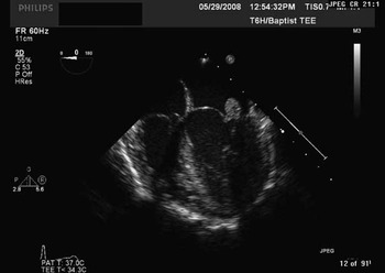
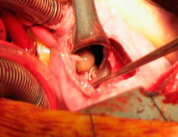
Nine days following the diagnosis, he underwent surgical excision of the mass. Transoesophageal echocardiography was performed to confirm the diagnosis (Fig 1). Intraoperatively, the mass showed a smooth reddish brown surface typical of a myxoma, but arose from the coapting edge of the anterior leaflet of the mitral valve, more consistent with a fibroelastoma (Fig 2). The mass was carefully excised at its base including a small portion of the normal leaflet, and the valve reconstructed with an autologous pericardium. The patient tolerated the procedure well and was discharged to home on post-operative day 4.
Transoesophageal echocardiogram shows the left atrial mass attached to the anterior leaflet of the mitral valve leaflet.

Tumour specimen.

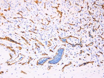
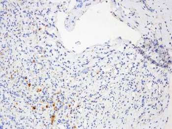
The specimens were evaluated in histochemical sections with CD31 and calretinin stains. The CD31 stain was positive, highlighting the endothelial cells in the haemangioma (Fig 3). The calretinin stain was negative (Fig 4). These histology results support the diagnosis of a benign atrial cardiac haemangioma.
CD31 stain.

Calretinin stain.

It is likely that the initial syncopal event was unrelated to the haemangioma, and this now 18-year-old male patient continues to be asymptomatic. His most recent echocardiogram shows no recurrence of the tumour and mild-to-moderate mitral valve regurgitation with mild left atrial enlargement. He has normal left ventricular size and function. An angiotensin-converting enzyme inhibitor was added to his medical regimen.
Discussion
Cardiac haemangiomas are rare, benign tumours, representing 5% to 10% of benign primary cardiac tumours.Reference Oshima, Hara, Kono, Shibamoto, Mishima and Akita1 These lesions can affect patients of all age groups and are described in all chambers of the heart, including the pericardium.Reference Soberman, Plauth, Winn, Forest, Hatcher and Sink2 Clinical presentation is highly variable depending on location, extension, and size of the tumour. Despite the possibility of some patients being asymptomatic, the reported symptoms include the following: chest pain, congestive cardiac failure, thromboembolic complications, pericarditis, pericardial effusion, haemodynamic instability, and cardiac tamponade.Reference Serri, Schraub, Lafitte and Roudaut3, Reference Solum, Romero, Ledford, Parker, Madani and Coletta4
Two-dimensional echocardiography is the most appropriate screening and imaging modality for the detection of cardiac tumours given the reliability, cost effectiveness, and safety of echocardiography. A complete echocardiogram including evaluation of all chambers of the heart should be obtained to evaluate for multiple tumours that are reported.Reference Acikel, Dogan, Akdemir, Kilic, Yesilay and Cagirci5 Computerised tomography scan and magnetic resonance imaging provide better soft tissue contrast and can yield more information about the invasiveness of the tumour and allow for visualisation of the entire mediastinum for extracardiac extent. Coronary angiography may reveal a characteristic tumour blush as seen with haemangiomas.Reference Pigato, Subramanian and McCaba6 Most often, a definitive diagnosis requires surgical excision with histological examination. In our case, the echocardiogram showed an atypically located haemangioma on the anterior leaflet of the mitral valve. In our review of the literature, this is the first child reported to have a haemangioma with attachments exclusively to the mitral valve. Detection of valvular haemangiomas is extremely rare, most likely because valves are predominantly avascular structures.Reference Yaganti, Patel, Yaganti and Victor7
The natural history of cardiac haemangiomas is extremely variable. Rarely, tumours may completely involute while others proliferate indefinitely.Reference Tabry, Nassar, Rizk, Touma and Dagher8 Patients may remain asymptomatic until death from some other cause or death may result from one of the previously mentioned complications of the tumour. Surgical resection is the treatment of choice when the diagnosis is unknown or for symptomatic patients.Reference Soberman, Plauth, Winn, Forest, Hatcher and Sink2 Complete surgical excision is preferable and possible in most cases as there have been cases of recurrence.Reference Chalet, Mace, Franc, Neveux and Lancelin9 Limited data with small case series have shown successful treatment with radiation therapy for unresectable tumours.Reference Gaspar, Mascarenhas, da Costa, Dias, Afonso and Silvestre10
In summary, we describe a patient with a very rare benign haemangioma of the mitral valve who is currently asymptomatic at 1 year status after resection. We believe that definite diagnosis of atypically located tumours can be made with the most certainty from histopathologic examination.