Introduction
In pulmonary atresia with intact ventricular septum and a suitable anatomy for biventricular repair, stenting the arterial duct, conventional Blalock–Taussig shunt surgery, and long-term prostaglandin infusion therapy are acceptable strategies that allow, if necessary, an adequate source of pulmonary blood flow after balloon pulmonary valvuloplasty.Reference Burkholder and Balaguru 1 These techniques provide a period of time long enough to allow the right ventricle to grow and to decide whether its diastolic function is normalised or elective bidirectional Glenn operation is still needed.Reference Burkholder and Balaguru 1 , Reference Raval, Thakkar and Madan 2 Percutaneous ductal stenting is an arising method that has several advantages including reduced procedure-related complications and hospital stay duration.Reference Santoro, Gaio and Giugno 3 – Reference Schranz, Michel-Behnke and Heyer 6 Although ductal stenting has been studied in a small series in the setting of mixed patients with ductal-dependent pulmonary circulation, rare studies reviewed ductal stenting strategy in a selected group of pulmonary atresia with intact ventricular septum.Reference Mallula, Vaughn, El-Said, Lamberti and Moore 7 – Reference Alwi, Choo, Radzi, Samion, Pau and Hew 9 In this study, we highlight the efficiency of this technique and analyse our short- and medium-term results of ductal stenting in pulmonary atresia with intact ventricular septum (or critical pulmonary stenosis) with a right ventricular anatomy suitable for biventricular repair.
Materials and methods
Patients’ selection
This is a single medium size centre, non-randomised, and retrospective study conducted between January 2007 and March 2017, which was reviewed and approved by the institutional ethical committee. During this period, only 18 patients suffering from pulmonary atresia with intact ventricular septum (or critical pulmonary stenosis) who underwent ductal stenting after balloon pulmonary valvuloplasty at our hospital were identified. Patients’ medical records were reviewed. Demographic, echocardiographic, procedural, and clinical data were collected and analysed. Our patients were divided into two groups according to ductal stenting timing after balloon pulmonary valvuloplasty. Group A consisted of patients in whom ductal stenting was anticipated at the time of balloon pulmonary valvuloplasty. These patients had a diminished right ventricular cavity on echocardiography prior to balloon pulmonary valvuloplasty, expecting the need for an additional pulmonary blood flow. Group B consisted of patients who received ductal stenting secondarily, a few days following balloon pulmonary valvuloplasty. These patients did not tolerate prostaglandin infusion or remained hypoxic (SaO2 <70%), despite the various therapeutic measures in order to assist the right ventricular diastolic function.
Technique
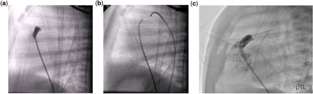
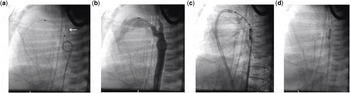
An informed consent was obtained prior to patient enrolment. All procedures were done by the same operator. After diagnosis, all neonates were on a continuous prostaglandin infusion, which was stopped 1 to 2 hours prior to catheterisation in group A patients. The procedure was done in the catheterisation laboratory, under general anaesthesia or deep sedation. Special attention was given to minimise hypothermia. One femoral artery was cannulated with a 4-F short sheath and one contralateral femoral vein is accessed with a 5-F short sheath. Prograde pulmonary blood flow was established by a snare-assisted balloon pulmonary valvuloplasty after certifying that right ventricular-dependent coronary circulation and infundibular atresia were absent. Early in our study, the atretic pulmonary valve was perforated using the stiff tip of a 0.014” coronary guide wire (Fig 1a–b), and later in time, using the floppy tip of a special 0.014” wire conceived for coronary chronic total occlusion with high perforating capacity (Fig 1c).
At this point, ductal stenting was attempted immediately in the same setting in group A patients. On the other hand, patients judged dispensed from ductal stenting returned to the intensive care unit for close surveillance and a trial off of prostaglandin. During the first few days, we were able to determine whether ductal patency was still required by testing the tolerance (oxygen saturation level) at prostaglandin weaning. These repetitive stopping tests of prostaglandin make it possible, on the one hand, to prove ductal dependency and, on the other hand, to identify the time interval necessary for the closure (or the restriction) of the arterial duct. Patients who did not tolerate prostaglandin infusion or remained hypoxic (despite maximal dosages of prostaglandin infusion) were sent to ductal stenting (group B). With this in mind, the prostaglandin infusion was stopped at the right time before the second setting procedure to achieve the maximal arterial duct pulmonary end restriction, which will help to stent. Thus, it was mandatory that the initial angiogram gives adequate information about the ductal anatomy for a subsequent possible ductal stenting.
( a ) Right infundibular angiogram in the lateral view using a 4-F right coronary catheter. The catheter tip is in contact with the atretic pulmonary valve and the stiff tip of the 0.014” guidewire is protruding in the pulmonary trunk. ( b ) Establishment of a snare-assisted arterio-venous circuit. ( c ) Crossing the atretic pulmonary valve with the floppy tip of special coronary 0.014” guidewire (HI-TORQUE CROSS-IT 200XT).

The equivalent vascular accesses were taken as for balloon pulmonary valvuloplasty. Through the femoral vein, a 0.014” guidewire was placed in the descending aorta, across the arterial duct. Bare metal, balloon pre-mounted coronary stents, were selected in all cases and delivered in a prograde approach using either a 5-F right coronary or a multi-purpose guiding catheter. For patients weighing 3.0–4.0 kg, we choose a 3.5-mm or a 4.0-mm diameter stent and for those weighing <3.0 kg, we choose a 3.0-mm diameter stent. The ductal length was measured in lateral view after a guide wire was positioned, since it straightened it and improved the accuracy of length determination. Stent length was chosen so that both pulmonary and aortic ends were covered while avoiding a posterior aortic wall contact. We kept in mind that 5–15% stent shortening might occur upon stent full expansion, especially in the tortuous arterial duct and when stent over-dilatation took place. The nasogastric tube and the endotracheal tube (when the patient was intubated) were useful landmarks. Once the tip of the stent passed across the narrowest part of the arterial duct, repeated angiography was performed and fine adjustments were made to position the stent accurately. Once satisfied with the positioning, the assistant rapidly inflated the balloon while the operator kept the ensemble steady as the stent tends to jump forward during inflation. Once the stent was fully expanded, the balloon was deflated and angiograms were repeated. After a satisfying stent placement, the fully deflated balloon should be carefully retrieved to avoid stent mobilisation. For that, the operator would maintain the intra-stent balloon position while advancing the catheter to recapture it. Then, the unified “balloon in catheter” system was retrieved from the stent while pushing the wire forward. The wire was kept across the arterial duct for about 10 minutes in case of an inadvertent event of acute stent thrombosis or displacement. Prophylactic antibiotic therapy using intravenous cefazolin at 30 mg/kg was given to all patients. Anticoagulation, using intravenous heparin at a dose of 25 units/kg/h, was provided for 24 hours. Aspirin therapy (5 mg/kg/day) was subsequently administered and continued as long as needed. Follow-up after discharge was done by referring paediatric cardiologists on a monthly basis during the first 6 months and then was progressively spaced. It consisted of a physical examination complemented with an echocardiogram and a pulse oximetry measurement. The indications for discontinuation of antiplatelet therapy were the spontaneous stent closure, the exclusive left–right interatrial shunt (if present) on echocardiogram, or the capillary saturation above 95% after an elapsed period of 6 months post-ductal stenting.
Statistics
Discrete variables were summarised as percentages and continuous variables as mean with standard deviation or median with range. Statistical analysis of the categorical variables was conducted using Fisher’s exact test and by t-test and Mann–Whitney U-test for continuous variables. Statistical analyses were computed using the Statistical Package for the Social Sciences (SPSS) Statistics, version 21 for Mac (IBM, Armonk, New York, United States of America), with a p-value <0.05 considered statistically significant. All reported p-values are two sided.
Results
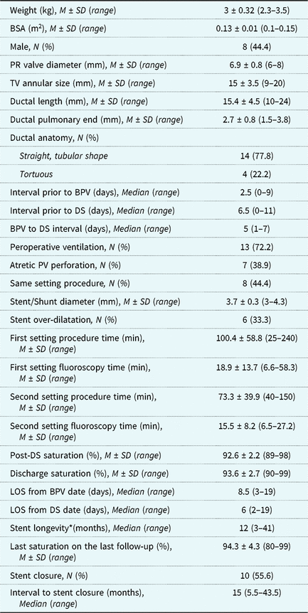
Patient characteristics (Table 1)
Eighteen neonates were identified and of them eight (44.4%) were male. The mean body weight was 3 ± 0.32 kg, with a range from 2.3 to 3.5. Fourteen patients had a straight, tubular-shaped arterial duct. Eight patients underwent ductal stenting in the same setting after balloon pulmonary valvuloplasty (group A). The overall median interval prior to ductal stenting was 6.5 days, with a range from 0 to 11 days.
Demographic and procedural characteristics

M ± SD = Mean ± standard deviation;
BSA = body surface area; PR = pulmonary ring; TV = tricuspid valve; BPV = balloon pulmonary valvuloplasty; DS = ductal stenting; PV = pulmonary valve; LOS = length of stay.
*Stent longevity = period of time from DS to last documented open stent on cardiac ultrasound.
Stent (Tables 1 and 2)
All ducts in group A were considered to be “straight”. In group B, 40% of ducts were deemed tortuous. There were no “vertical” ducts. Coronary stents of similar design, on low-profile balloon dilation catheters, were used in all patients: [8 Veriflex Coronary stents (Boston Scientific, Marlborough, Massachusetts, United States of America), 4 Multilink Vision stents (Abbott Vascular, Abbott Park, Illinois, United States of America), 2 FlexMaster F1 stent (Abbott, Abbott Park, Illinois, United States of America), 1 Integrity Coronary stent (Medtronic Vascular, Minneapolis, Minnesota, United States of America), 1 Omega BMS (Boston Scientific), 1 Skylor™ stent (Invatec, Roncadelle, Italy), and 1 Amazonia CroCo Stent (Minvasys, Paris, France)]. All the patients received one single stent to establish ductal patency. As for procedure incidences, two stent aortic displacements were bailed out with balloon adjustment in group A, without any complication. In another patient, the stent was short and did not cover the 1–2 mm of the arterial duct aortic end. Nevertheless, the repeated angiogram showed satisfactory results and the stent patency was proven on follow-up. Seven neonates (38.9%) underwent transcatheter mechanical perforation of the atretic pulmonary valve, of which four were in group B. Ductal stenting was performed as a staged procedure in 10 patients (55.5%). The median interval from balloon pulmonary valvuloplasty to ductal stenting in group B patients was five days, with a range from 1 to 7 days.
Procedural outcomes (Tables 2 and 3)
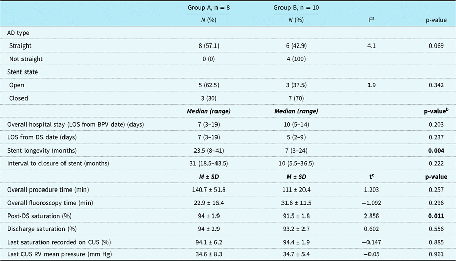
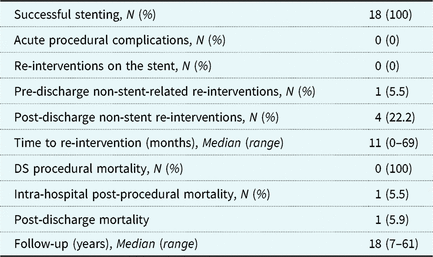
Procedural success was 100% in both groups. The saturation at the end of ductal stenting was 94 ± 1.9% in group A versus 91.5 ± 1.8% in group B (p < 0.05). There was no difference between saturations on hospital discharge day in both groups. Median hospital stay post-ductal stenting was 7 (3–19) days in group A versus 5 (2–9) days in group B (p = 0.237). However, the median overall hospital stay (post-balloon pulmonary valvuloplasty) in group A was 7 (3–19) days versus 10 (4–14) days in group B. No potential complications such as acute thrombosis, spasm of the arterial duct, or distal migration of expanded stent were documented. On follow-up, no re-intervention for stent failure or overflow were noted. During the same hospitalisation, one patient in group A needed an aortic balloon valvuloplasty for severe stenosis four days post-stenting. Post-discharge from the hospital, four re-interventions not related to the ductal stenting procedure were undertaken. One patient in group A had stent re-dilatation, in the occasion of pulmonary re-ballooning 2.5 months after ductal stenting. This same patient needed another pulmonary balloon angioplasty 8.5 months later. One group B patient needed a bidirectional Glenn surgical shunt repair after 15 months, while another group B infant underwent a transcatheter atrial septal defect closure at five years of age. There were no deaths attributable to the stenting procedure. However, one intra-hospital death was noted in group B. It occurred at day 16 of life (8 days after ductal stenting, 14 days after balloon pulmonary valvuloplasty) when the baby developed sepsis followed by multiple organ dysfunctions. Stent patency was proven 2 hours before death. On follow-up, another 15 months aged patient died 24 hours after a modified Glenn surgical procedure that was undertaken for right mid-ventricular stenosis. A patent stent was identified before surgery. The median duration of follow-up for the whole study was 18 (7–61) months. Adequate right ventricular growth with a two-ventricle physiology occurred in all survivors after 12 months. The stent closure was documented in 10 patients (55.6%) after a median of 15 (5.5–43.5) months. In patients with a persisted ductal flow, the median overall longevity of the stent was 12 (3–41) months. However, when comparing the two groups, we noted that the stent lifespan was remarkably higher in group A in comparison to group B (23.5 > 7, p = 0.004), with a higher median interval to closure time.
Groups’ characteristics comparison

AD = arterial duct; LOS = length of stay; BPV = balloon pulmonary valvuloplasty; DS = ductal stenting; CUS = cardiac ultrasound; RV = right ventricle.
a Fisher Test; b Mann–Whitney U-test; c t-test.
Group A = Same setting procedures; Group B = Different setting procedures.
M ± SD = Mean ± standard deviation.
*Stent longevity = period of time from DS to last documented open stent on cardiac ultrasound.
Morbidity and mortality

DS = ductal stenting.
Discussion
Previous reports have suggested that the establishment of pulmonary blood flow from the right ventricle to the main pulmonary artery is the key to achieve biventricular repair in cases of pulmonary atresia with an intact ventricular septum or critical pulmonary stenosis.Reference Alwi, Geetha and Bilkis 10 Balloon pulmonary valvuloplasty is performed for both these conditions and may be more technically challenging in pulmonary atresia with intact ventricular septum, since the valve must be perforated during cardiac catheterisation, either mechanically or with radiofrequency or using laser techniques.Reference Burkholder and Balaguru 1 At the present time, no anatomical criteria can accurately predict the need for systemic to pulmonary artery shunt after balloon pulmonary valvuloplasty.Reference Weber 11 It was also noted that in at least half of pulmonary atresia with intact ventricular septum cases, right ventricular growth may take several weeks to months to be achieved despite successful balloon pulmonary valvuloplasty.Reference Chubb, Pesonen and Sivasubramanian 12 , Reference Xu, Xia, Zhang, Li, Zhou and Yu 13 Thus, it is mandatory, in these cases, to continue prostaglandin therapy or to create a systemic to pulmonary artery shunt because of persisting deep hypoxemia. The systemic to pulmonary artery shunt could be obtained by a surgical approach with its significant morbidity and mortality rate as well as by a long-term prostaglandin infusion with its multiple side effects.Reference Burkholder and Balaguru 1 Moreover, since it has been estimated that most of the infants suffering from pulmonary atresia with intact ventricular septum can be managed by cardiac catheterisation, ductal stenting prevails as a logical non-surgical alternative to maintain ductal patency.Reference Mallula, Vaughn, El-Said, Lamberti and Moore 7 , Reference Mortera, Rissech, Bartrons, Carretero and Prada 8 Notwithstanding its rising popularity, this attractive technique is a challenge for the interventionist as much as for the delicacy of the manoeuvers performed, as for the choice of advanced equipment and particularly the timing of the procedure. Ductal stenting has been successfully used in diseases with duct-dependent pulmonary blood flow.Reference Raval, Thakkar and Madan 2 , Reference Glatz, Petit and Goldstein 4 – Reference Schranz, Michel-Behnke and Heyer 6 . In fact, the best results are obtained in diseases where the arterial duct is short and less tortuous, such as in pulmonary atresia with an intact ventricular septum and critical pulmonary stenosis.Reference Alwi and Mood 14 In addition to the favourable arterial duct anatomy, stent delivery in a prograde venous approach after balloon pulmonary valvuloplasty gives one more advantage for easier positioning. On top of this, ductal stenting is a preferable choice in pulmonary atresia with intact ventricular septum, where ductal patency is needed for a medium period of time in order to decide whether biventricular repair is achieved or subsequent therapy is needed.Reference Mallula, Vaughn, El-Said, Lamberti and Moore 7
Ductal stenting timing remains a real challenge. Most of the ductal stenting interventions were performed later in time after balloon pulmonary valvuloplasty, as a staged procedure. This practice was in some of these cases necessary to prove ductal dependency after balloon pulmonary valvuloplasty and also helped us with timing assessment of ductal constriction after prostaglandin weaning. In fact, obtaining ductal optimal constriction is mandatory for a safe stenting,Reference Alwi 15 , Reference Alwi 16 since it avoids the need for stent over dilatation for its successful anchoring. Consequently, this may also help to minimise problems related to over circulation such as necrotising enterocolitis, prolonged invasive ventilation, and long hospital stay.Reference Udink Ten Cate, Sreeram, Hamza, Agha, Rosenthal and Qureshi 17 On the other hand, ductal stenting when performed, immediately after balloon pulmonary valvuloplasty, turned out to have multiple advantages as well: it avoided repetitive vascular access as well as repetitive transfers to the catheterisation laboratory. Over that, when we compared the two groups, we noticed that group A had a shorter overall hospital stay with a shorter overall time of irradiation. However, longer overall procedural time was noted in some group A cases, since it was necessary to wait for the optimal ductal restriction, after stopping the prostaglandin infusion while monitoring the decrease in saturation by 1% every 10 to 15 minutes.
Critical pulmonary stenosis to pulmonary atresia with intact ventricular septum ratio was unexpectedly high in our study.Reference Lucas 18 In fact, eight of the eleven critical pulmonary stenosis cases had nearly complete atresia with an extremely tiny opening in the pulmonary valve. It is noteworthy that the presence of this tiny valvular opening allowed an easier passage of the guide wire tip, and this was essential to reduce procedural time by avoiding the demanding mechanical perforation technique.
Recently in the literature, it has been discussed that various types of chronic total occlusion coronary stiff wires based on the tip load with variable penetrating characteristics can be used as an alternative to the usage of the stiff tip of a 0.014” coronary guide wire for perforation of atretic pulmonary valve.Reference Bakhru, Marathe and Saxena 19 , Reference Alwi, Budi, Mood, Leong and Samion 20 In the last consecutive three patients with pulmonary atresia with intact ventricular septum encountered in our centre, we believe that using the CROSS-IT 200 XT (Abbott Laboratories, Chicago, Illinois, United States of America) or the PROGRESS 200T (Abbott Laboratories) Hi-Torque guide wires contributed to the effectiveness and the safety of the perforation technique while substantially reducing time procedure (Fig 1c).
Similar to earlier reports,Reference Mallula, Vaughn, El-Said, Lamberti and Moore 7 , Reference McMullan, Permut, Jones, Johnston and Rubio 21 , Reference Santoro, Gaio and Palladino 22 ductal stenting was successful in 100% of our cases. All of our ductal stenting procedures were performed in a transvenous prograde approach to avoid arterial vascular access complications. This approach also promoted guidewire stability when positioned distally in the descending aorta. The manipulation of catheters requires advanced skills to overcome the tortuosity of miniaturised trajectories in newborns. In fact, in one group B patient, we were not able to cross the tortuous and extremely restricted pulmonary ductal end in a prograde fashion. This anatomical challenge was overcome by a retrograde approach and the creation of a snare-assisted arterio-venous circuit for venous stent delivery.
Stent diameter was chosen according to the body weight. A 3.5-mm diameter stent was implanted in 13 patients (72.2%). A 4-mm diameter stent was chosen in four cases (of which three were group A patients) to achieve optimal results. A 3-mm stent was implanted in one group B patient. In six cases (33.3%), the stent was over-dilated larger than its nominal size (mean of 0.2 ± 0.1 mm), using a high-pressure technique, to optimise results and positioning. When stenting an arterial duct that was not optimally restricted, especially in the setting of an extended procedural time with an unstable clinical state of the patient, the over-dilation of the stent was sometimes necessary to bail out its displacement. In two cases encountered in our series, it was noticed that after deflating the balloon the stent slid forward on the wire into the aorta. The stent was repositioned into the pulmonary stenotic end zone by pulling it back on the balloon and pushing its aortic end with the arterial catheter. After that, the balloon was inflated to a larger diameter to ensure the stent anchoring. No acute perforations, dissections, or other serious complications occurred during over-dilatation. Further, over-dilation of the stent did not induce a re-intervention for failure or overload on the mid-term follow-up.
Stent length was directed by the ductal length. In fact, when the patient was stable, measurements were taken using the markers of a 2-cm long balloon introduced into the arterial duct (Fig 2a–d). However, when ductal stenting took place urgently (due to clinical deterioration), especially during the initial procedure, the stent length was chosen on the basis of angiographic measurements. The nasogastric tube was used as a marker of the posterior wall of the aorta.
( a ) After balloon pulmonary valvuloplasty, a 2-cm long balloon catheter is transvenously delivered in the straight arterial duct for accurate sizing. Note the long arrow pointing to the nasogastric tube which is a valuable marker for stent placement. ( b ) Aortography indicating the possible stent length. Note the vertical arrow pointing to the pulmonary end ductal restriction. ( c ) Adequate stent placement. ( d ) Lateral view of the stented duct after guidewire retrieval. Compare the straight stent shape to the “wavy” stent deformation in Figure 3d.

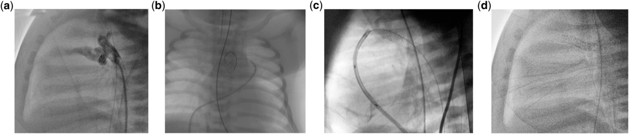
The shape of the arterial duct in this study was determined by echocardiography at baseline and by angiography. Recently, in the setting of ductal-dependent heart lesions, many tortuous arterial ducts were stented in our center. In these cases, as well as in some tortuous arterial duct cases enrolled in our study, we noted that although the rigid part of the 0.014” guide straightened the arterial duct as it passes through, a “wavy” stent deformation was noted at the removal of the guide but did not influence the permeability of the stent as well as its longevity (Fig 3a–d). Taking tortuosity into consideration, an overestimation of the stent length of 5–15% during the initial measurements was required for better results. Furthermore, when positioning the stent, slight pulmonary end protrusion is acceptable by many operators. However, extremely protruded stent may spit in the right ventricle in case of a well-dilated and leaky pulmonary valve. Heart failure may be precipitated by a coexisting tricuspid valve regurgitation. In our series, we didn’t encounter this issue, and on follow-up no patient needed diuretics or any special manoeuvers to reduce the stent flow.
( a ) An aortogram in lateral view showing the tortuous ductal anatomy. ( b ) The floppy wire’s segment is in the duct delineating its tortuosity in an anteroposterior view. ( c ) The ductal tortuosity is straightened by the stiff wire. ( d ) Note the “wavy” stent shape after the wire withdrawal.

While previous studies have shown that ductal stenting efficacy decreases after 4 months due to in-growth of neointimal tissue,Reference Matter, Almarsafawey, Hafez, Attia and Abuelkheir 23 we noticed, in our series, that stent overall longevity was 12 months. No balloon stent angioplasty was required to overcome neointimal proliferation. This was a desirable outcome for our patients since this period of time was sufficient to indicate whether our patients progressed to two ventricles circulation or additional surgical repair was still needed. Only one patient needed a Glenn bidirectional surgery at 15 months of age for right ventricular diastolic dysfunction, but despite the success of the operation, he died of major hyperthermia. On follow-up, while the stent patency became progressively redundant and in particular when the biventricular repair was achieved, we did not rash to percutaneously occlude these persistent shunts in the hope of ultimate spontaneous closure.
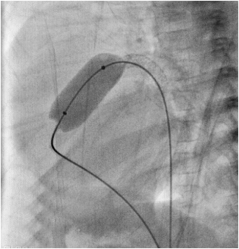
Other studies reported higher rates of stent re-intervention on short- and mid-term follow-ups in order to improve patency or to occlude the redundant unnecessary shunt.Reference Mallula, Vaughn, El-Said, Lamberti and Moore 7 , Reference McMullan, Permut, Jones, Johnston and Rubio 21 We should note that the only re-intervention on the stent encountered in our series did occur on the occasion of pulmonary re-ballooning. In this particular case, since anchoring the guidewire into the descending aorta across the stented arterial duct gives more circuit stability, we thought that stent angioplasty over the pre-existing intra-stent wire would extend its patency (Fig 4).
Balloon valvuloplasty is undertaken 2.5 months after ductal stenting. A TyShak balloon (12 mm to 2 cm) is advanced on a 0.035” guidewire placed across the arterial duct in the descending aorta for a better system stability.

Finally, the catheter management has revolutionised the prognosis in pulmonary atresia with an intact ventricular septum. Nevertheless, the rate of re-interventions for comorbidities is still being noticeable.Reference Mallula, Vaughn, El-Said, Lamberti and Moore 7 In our series, an aortic valvuloplasty was performed in a non-syndromic patient in whom aortic valve stenosis was found, while another infant underwent a transcatheter atrial septal defect closure when he was 5 years old.
Limitations
This is a retrospective study of a small patient series from a single congenital cardiovascular medium size centre. We believe that few ductal stenting procedures, particularly in group A, might be judged avoidable knowing that right ventricular diastolic function may improve in a few weeks, after balloon pulmonary valvuloplasty, with no need for further intervention. Additionally, we feel that the decision to stent the arterial duct, in some group B patients, may have been precipitated due to clinical deterioration.
Definitely, well-designed, prospective cohort studies are needed to compare the outcomes of different alternative therapies in pulmonary atresia with an intact ventricular septum and critical pulmonary stenosis.
Conclusion
With the current technology, ductal stenting is a feasible, safe, and efficient alternative in the therapeutic arsenal of selected pulmonary atresia with intact ventricular septum patients. It avoids surgical shunt as well as long-term prostaglandin infusion while shortening hospitalisation and reducing complication rates and treatment costs. Crucial to the success of the procedure and the medium-term outcome are case selection, choice of appropriate technique, proper skills, and the right timing for ductal stenting. In our series, all stents were successfully deployed without any procedure related to major complication. The minimal 6 months stent longevity provides a period of time long enough to decide whether the right ventricular diastolic function is normalised or if further interventions are needed. The contribution of ductal stenting to promoting right ventricular growth is still to be determined by well-conducted studies.
Author ORCIDs
Raymond N. Haddad 0000-0001-7605-4708
Acknowledgements
None.
Financial Support
The authors have no financial relationships relevant to this article to disclose.
Conflict of Interest
None.
Ethical Standards
This study was reviewed and approved by Saint Joseph University Research Ethics Board.