Methods
We performed a detailed review of PubMed for articles pertaining to hypoplastic left heart syndrome, which produced over 2700 articles that were then narrowed down to 67 articles based on relevance to histology, pathology, valve, myocardium, coronaries, endocardial fibroelastosis, and pathologenesis. We searched from 1940 until the present. We needed to evaluate articles dating back to 1940 because it was during that time period that much of the histological analysis for hypoplastic left heart syndrome was being performed. We also wanted to know the histologic appearance of specimens before any intervention. We included all articles we could locate pertaining to the pathogenesis of hypoplastic left heart syndrome.
Variable anatomy of hypoplastic left heart syndrome
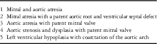
Hypoplastic left heart syndrome is a severe and devastating heart defect that affects ~1 in 5–10,000 children born each year and accounts for up to 25% of all neonatal deaths from CHD.Reference Rosamond, Flegal and Furie 1 Hypoplastic left heart syndrome is characterised by a diverse spectrum of malformations distinguished by underdevelopment of the left ventricle and its components, rendering it unable to support systemic circulation.Reference Barron, Kilby, Davies, Wright, Jones and Brawn 2 , Reference Sedmera, Cook, Shirali and McQuinn 3 The presence of anatomic variations within the classification of hypoplastic left heart syndrome yields a continuum of phenotypic heterogeneity that can be divided into broad subgroups.Reference Sedmera, Cook, Shirali and McQuinn 3 , Reference Sinha, Rusnak, Sommers, Cole, Muster and Paul 4 These variations are dependent upon the presence or absence of the following: an inlet to the left ventricle, a patent outflow tract, a ventricular septal defect, and/or any other associated cardiac defects. Each subtype can be associated with any atrial arrangement, with situs solitus being the most common.Reference Sedmera, Cook, Shirali and McQuinn 3 It is necessary to analyse these subgroups individually because of their differing histological characteristics and the possibility for differing inciting events. Sedmera et al provide an excellent breakdown of hypoplastic left heart syndrome subtypes, which appear in Table 1.
Morphologic subgroups of hypoplastic left heart syndrome.Reference Sedmera, Cook, Shirali and McQuinn 3

First, hearts with combined mitral and aortic atresia present with a thin-walled, slit-like left ventricle.Reference Sinha, Rusnak, Sommers, Cole, Muster and Paul 4 The ascending aorta and arch are extremely hypoplastic, and flow is retrograde.Reference Tweddell and Hoffman 5 Systemic output is ductal dependent. In the setting of combined mitral and aortic atresia, if a ventricular septal defect is present, a larger left ventricular cavity will develop and the wall of the left ventricle will be proportionately thicker. The larger the ventricular septal defect, the more closely the left ventricle will approach normal dimensions.Reference Sedmera, Cook, Shirali and McQuinn 3 The observation that left ventricular dimensions are proportional to the size of the inlet suggests that myocardial development is dependent on adequate blood flow during development. Second, mitral atresia can also occur with a stenotic aortic valve, patent aortic root, and ventricular septal defect. These cases are characterised by a hypoplastic ascending aorta with a widely patent ductus arteriosus.Reference Sinha, Rusnak, Sommers, Cole, Muster and Paul 4 In the setting of a large ventricular septal defect, irrespective of mitral valve dimensions, the aortic valve is rarely atretic, which suggests that valvular development is also dependent to some degree on adequate blood flow during development. Third, hearts with isolated aortic atresia and a patent mitral valve without a ventricular septal defect demonstrate thickening of the left ventricular free wall, ventricular septum, and endocardium. As in combined mitral and aortic atresia, the ascending aorta and arch are hypoplastic, and all systemic output is ductal dependent.Reference Tweddell and Hoffman 5 Fourth, hypoplastic left heart syndrome hearts with aortic valve stenosis and a patent mitral valve maintain antegrade flow through the aortic valve. The degree of ascending aortic and arch hypoplasia is less than observed with aortic atresia.Reference Tweddell and Hoffman 5 Fifth, in general, the degree of aortic arch hypoplasia correlates with the dimensions of the aortic root, but there is one subset of hypoplastic left heart syndrome hearts with severe aortic arch hypoplasia that is out of proportion to the degree of aortic root hypoplasia.Reference Sedmera, Cook, Shirali and McQuinn 3 , Reference Szumilo, Skomra, Swatek and Korobowicz 6 This subtype of hypoplastic left heart syndrome can have multiple combinations of mitral and/or aortic valve abnormalities.
Lev et al described some common qualities between subtypes of hypoplastic left heart syndrome. A detailed analysis of 230 hypoplastic left heart syndrome hearts revealed that specimens had an enlarged heart with an apex forming the right ventricle. In all cases the right atrium was hypertrophied and enlarged, the left atrial appendage was small, and the tricuspid orifice was enlarged and impinged upon the infundibular region. The right ventricle was hypertrophied and enlarged.Reference Bharati and Lev 7 Although right ventricular abnormalities are found in hypoplastic left heart syndrome, many of these abnormalities may be the result of the right ventricle compensating for an increased workload secondary to the primary left ventricular malformation.
Myocardial histopathology of normal hearts versus hypoplastic left heart syndrome hearts
Cardiac muscle cell orientation in non-diseased hearts exhibits a similar pattern in the embryo, foetus, child, and adult.Reference Bulkley, Weisfeldt and Hutchins 8 Normally, the major portion of both ventricular walls and the mid portion of the interventricular septum have an orderly parallel arrangement of cells. Myocardial fibre disarray, defined as a lack of orderly parallel arrangement of myocytes, has been described in hypoplastic left heart syndrome. Myocardial fibre disarray can present in one of the following patterns: fibres branching at sharp angles to one another, groups of fibres cut longitudinally interspersed with fibres cut transversely, or fibres forming concentric whorls. These patterns may be found alone or in combination within a given heart.Reference Bulkley, D’Amico and Taylor 9 Myocardial fibre disarray is not unique to hypoplastic left heart syndrome but can be found to some degree in normal hearts, and more extensively in hearts with conditions such as hypertrophic cardiomyopathy,Reference Bulkley, D’Amico and Taylor 9 pulmonary atresia,Reference Bulkley, Weisfeldt and Hutchins 8 and tetralogy of Fallot.Reference Jones, Ferrans, Morrow and Roberts 10
Histopathology of left ventricle in hypoplastic left heart syndrome
Certain subtypes of hypoplastic left heart syndrome present with organised myocardial architecture at birth. Autopsy specimens of hearts with combined mitral and aortic atresia collected before the availability of effective surgical palliation demonstrate a normally arranged cell pattern without evidence of myofibre disarray in the left ventricular myocardium and rudimentary septum.Reference Bulkley, Weisfeldt and Hutchins 8 In the event of mitral atresia with a large ventricular septal defect and a fully developed left ventricle chamber, myocardial fibre orientation is normal.Reference Bulkley, Weisfeldt and Hutchins 8 A common finding in both of these cases is that they likely do not have increased left ventricular cavity haemodynamic pressure burden during development. The normal myocardial architecture in these cases also suggests that the aetiology of these cases is likely valvular.
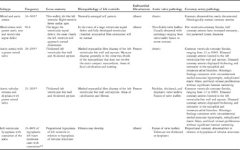
Hypoplastic left heart syndrome specimens that are exposed to increased left ventricular haemodynamic pressure in utero (patent inflow and obstructed outflow) display myocyte disarray as newborns. Hearts with aortic atresia and a patent mitral valve demonstrate marked myocardial fibre disarray of both the ventricular septum and the left ventricular free wall. O’Connor et al observed that the distribution of disarray is generally in the inner two-thirds of the myocardium and does not involve the outer compact myocardium or right ventricle.Reference O’Connor, Cash, Cottrill, Johnson and Noonan 11 Areas of focal calcification or scarring may also involve the subendocardial region of the left ventricle, interventricular septum, and papillary muscles.Reference O’Connor, Cash, Cottrill, Johnson and Noonan 11 Patients with hypoplastic left heart syndrome and coarctation of the aorta will develop myocardial pathology that correlates with valvular pathology. Postnatally, this subgroup can develop hypertrophy and fibrosis of the left ventricle.Reference Sedmera, Cook, Shirali and McQuinn 3 Characteristics of each subgroup are summarised in Table 2.
Characteristics of hypoplastic left heart syndrome subtypes in newborns.

* Frequencies documented before Norwood procedureReference Mahle, Spray, Wernovsky, Gaynor and Clark 12 , Reference Tweddell, Hoffman and Mussatto 13
** Frequencies unrelated to Norwood procedureReference Szumilo, Skomra, Swatek and Korobowicz 6 , Reference Hawkins, Clark and Doty 14 , Reference Von Rueden, Knight, Moller and Ewards 15
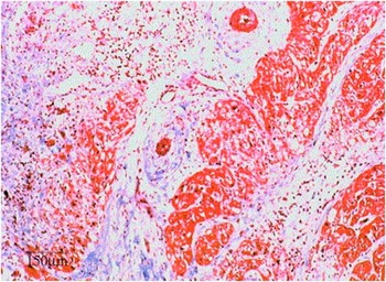
A recent histologic analysis of the myocardium of hypoplastic left heart syndrome hearts found large areas of randomly oriented, disorganised bundles of myocytes with variable myocyte size found between bundles in all left ventricle and right ventricle samples (Fig 1).Reference Bohlmeyer, Helmke and Ge 16 Although subgroup classifications were not documented in this report, all cases presented with myocyte disarray.Reference Bohlmeyer, Helmke and Ge 16 Previously reported histological studies observed organised myocardium in the subgroups with combined aortic artesia/mitral atresia or mitral atresia with a ventricular septal defect.Reference Bulkley, Weisfeldt and Hutchins 17 The obvious difference between the study groups was the age of the patients and the advancements in palliative techniques. In early publications, the patients’ mean lifespan was typically 1 week or less versus the more modern study group in which the lifespan was 6 days to 10 months.Reference Bohlmeyer, Helmke and Ge 16 The more modern patients were also exposed to inotropic support. This observation suggests that increased exposure to abnormal blood flow in the neonate will lead to myocardial disorganisation and scar formation in some hearts that perhaps had a histologically normal myocardium at birth. The differing myocyte architecture observed between subtypes of hypoplastic left heart syndrome during development highlights the potential for differing inciting events between subtypes. The cases of hypoplastic left heart syndrome with abnormal myocyte architecture at birth may have a primary myocardial pathogenesis that alters blood flow patterns and results in abnormal development of the immature valve structures. Recent studies have highlighted the possibility that some cases of hypoplastic left heart syndrome may result from abnormal cardiomyocyte proliferation during development.Reference deAlmeida, McQuinn and Sedmera 18 , Reference Grossfeld 19
Hypoplastic left heart syndrome. Disorganised bundles of myocytes, large areas of fibrosis, and abnormal vascular formations (left ventricle from 3-month-old hypoplastic left heart syndrome patient, trichrome stain, original magnification 25×). Courtesy of Bohlmeyer.Reference Bohlmeyer, Helmke and Ge 16

Endocardial fibroelastosis
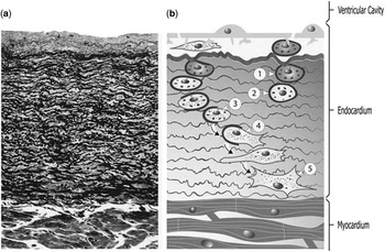
In an eloquent review, LurieReference Lurie 20 describes endocardial fibroelastosis as a reaction of the endocardium, not a disease state. Grossly, the reaction is identified as a pearly or opaque white appearance of the endocardium, especially of the ventricles.Reference Lurie 20 The normal endocardium is transparent and only around 10 μm in thickness.Reference Neustein, Lurie and Fugita 21 Endocardial fibroelastosis is defined by thickening of the endocardium by layers of collagenous and elastic fibres to >20 μm.Reference Lurie 20 LurieReference Lurie 20 described the endocardial fibroelastosis reaction as a chronologic sequence of smooth muscle hyperplasia followed by their transformation and translocation from the inner, sub-endothelial layers to the outer, juxtamyocardial layers (Fig 2). In the foetus, the fibroelastic lining is highly cellular, containing numerous smooth muscle cells and fibroblasts. Antepartum, the cellular characteristics disappear, leaving layers of collagen and elastic fibres.Reference Sedmera, Cook, Shirali and McQuinn 3 The endocardial fibroelastosis reaction is most active during foetal life and during periods of active growth.
( a ) Histologic stained section of endocardium (3420), showing dense fibres of collagen and elastin typical of endocardial fibroelastosis. Reproduced from Am J Pathol 1972; 66: 483–496, Figure 3; with permission from The American Society for Investigative Pathology and kind cooperation of Grover M. Hutchins, MD.13 ( b ) In contrast is the parallel diagram derived from electron microscopy showing cellular activity that generates the fibroelastosis. 1. Dark smooth muscle cell; 2. light smooth muscle cell; 3. light smooth muscle cell with loss of basement membrane; 4. leiomyoid cell; and 5. fibroblast. Adapted from the Archives of Pathology Laboratory Medicine 1979; 103: 218, Figure 8; Copyright 1979 American Medical Association. All rights reserved. Courtesy of Lurie.Reference Lurie 20

Left ventricular endocardial fibroelastosis in hypoplastic left heart syndrome is often found in the setting of a patent mitral valve and severely stenotic or atretic aortic valve. Hearts that develop the endocardial fibroelastosis reaction are always under stress, either from pressure overload due to mechanical obstruction or from volume overload due to cardiac muscle disease.Reference Lurie 20 , Reference Black-Schaffer 22 However, not every heart under stress will develop endocardial fibroelastosis.Reference Sinha, Rusnak, Sommers, Cole, Muster and Paul 4 , Reference Lurie 20 In addition, endocardial fibroelastosis is not seen within the left ventricle in the setting of combined mitral and aortic atresia when blood flow is absent from the left ventricleReference Noonan and Nadas 23 but may affect the left atrium and mitral valve in the presence of mitral valve obstruction.Reference Noonan and Nadas 23
There is strong evidence that prenatal infection could account for some cases of left ventricular hypoplasia with endocardial fibroelastosis.Reference Ni, Bowles and Kim 24 Coxsackie virus and mumps virus were recognised as aetiologic agents of endocardial fibroelastosis in the 1960s and 1970s.Reference Lurie 20 These findings were further verified with more recent work identifying mumps genome in the majority of hearts with endocardial fibroelastosis that were preserved from an era when mumps was a widespread infection.Reference Lurie 20 The incidence of endocardial fibroelastosis sharply declined with the administration of the mumps vaccine.Reference Lurie 20
Infants with foetal ventricular hypertrophy lack the endocardial fibroelastosis reactionReference Lurie 20 even though they are exposed to similar mechanical stressors seen with certain subtypes of hypoplastic left heart syndrome (patent inflow with obstructed outflow) that may present with endocardial fibroelastosis. Animal studies have been performed creating a severe left ventricular outflow tract obstruction in first-trimester sheep. All foetuses had elevated end-diastolic pressure, but none developed endocardial fibroelastosis.Reference Eghtesady, Michelfelder, Altaye, Ballard, Hirsh and Beekman 25 This finding suggests that mechanical stressors alone do not always cause endocardial fibroelastosis. Lurie hypothesised that in the conditions that increase contractility, such as hypertrophic cardiomyopathy, the endocardium may inhibit the endocardial fibroelastosis reaction. It is also possible that in genetically susceptible patients contributions from both mechanical and immunologic factors are necessary for pathology. In hypoplastic left heart syndrome, a two-hit phenomenon may contribute to the expression and/or severity of the endocardial fibroelastosis reaction.
In the past, endocardial fibroelastosis was thought to be a primary disease that could potentially lead to left ventricular hypoplasia, termed the “contracted type” of primary endocardial fibroelastosis.Reference Lurie 20 The current understanding is that endocardial fibroelastosis does not cause hypoplastic left heart syndrome,Reference Lurie 20 but its presence and severity worsen the prognosis.Reference McElhinney, Vogel and Benson 26 McElhinney et alReference McElhinney, Vogel and Benson 26 recently reported that the severity of endocardial fibroelastosis, as determined by prenatal echocardiography in patients with aortic stenosis and evolving hypoplastic left heart syndrome, had an association with postnatal outcome following in utero balloon valvuloplasty. Patients with more severe endocardial fibroelastosis had a lower probability of postnatal biventricular outcome. They also reported that from pre-intervention to late gestation the time-indexed change in left ventricular end-diastolic volume was significantly greater in foetuses with mild endocardial fibroelastosis compared with those with severe endocardial fibroelastosis.Reference McElhinney, Vogel and Benson 26 Although our understanding of endocardial fibroelastosis has improved greatly over the last half century, many questions remain regarding the fascinating relationship between hypoplastic left heart syndrome and endocardial fibroelastosis.
Congenital coronary artery fistulas
We now shift focus to the coronary artery histopathology found in hypoplastic left heart syndrome and its contribution to the pathogenesis of the syndrome. We will first evaluate coronary artery fistulas, followed by macro coronary artery disease or malformations, and will finally evaluate the microvasculature observed in hypoplastic left heart syndrome. A congenital coronary artery fistula is an abnormal direct communication between any part of the coronary system and a cardiac chamber or great vessel, having bypassed the myocardial capillary bed.Reference Mavroudis, Backer, Rocchini, Muster and Gevitz 27 Microscopic evaluation of hypoplastic left heart syndrome specimens with ventriculo-coronary connections demonstrate that sinusoids from the endocardial surface may extend into the myocardium.Reference O’Connor, Cash, Cottrill, Johnson and Noonan 11 Blake categorised these ventriculo-coronary connections into three subtypes:
-
∙ Arterio-luminal subtype – connects the left ventricular chamber directly to the vertically penetrating branches of the coronary arteries.
-
∙ Arterio-sinusoidal subtype – connects the left ventricular chamber indirectly to smaller ramifications of the coronary arteries.
-
∙ Arterio-capillary subtype – connects the left ventricular chamber indirectly to a rich network of thin-walled, capillary-sized vessels.
O’Connor et alReference O’Connor, Cash, Cottrill, Johnson and Noonan 11 found a variable combination of ventriculo-coronary connections in all cases of hypoplastic left heart syndrome with patent inflow and obstructed outflow. The largest number of connections was found in subgroups with either stenosis or complete closure of the foramen ovale. No ventriculo-coronary connections were identified in the subgroup with mitral and aortic atresia.Reference O’Connor, Cash, Cottrill, Johnson and Noonan 11 Baffa et alReference Baffa, Chen, Guttenberg, Norwood and Weinberg 28 found ventriculo-coronary connections in 27 of 89 specimens with a patent mitral valve combined with aortic atresia and in 2 out of 52 specimens with mitral and aortic atresia. Most of the connections in hypoplastic left heart syndrome are the arterio-sinusoidal subtype rather than the direct arterio-luminal subtype.Reference Baffa, Chen, Guttenberg, Norwood and Weinberg 28 , Reference Sauer, Gittenberger-de Groot, Geishauser, Babic and Buhlmeyer 29 The arterio-sinusoidal subtype frequently coexists with endocardial fibroelastosis of the left ventricle.Reference Baffa, Chen, Guttenberg, Norwood and Weinberg 28 The regional distribution of myofibre disarray supports the concept that vascularisation parallels myocardial organisation in the developing heart.Reference O’Connor, Stahr, Cottrill, Todd and Noonan 30 It has been postulated that elevated left ventricular intracavitary pressure incites the persistence of embryonic microvascularity, which leads to the creation of complex arterio-luminal connections to allow for the egress of blood.Reference Beckman, Moller and Edwards 31
Coronary artery pathology
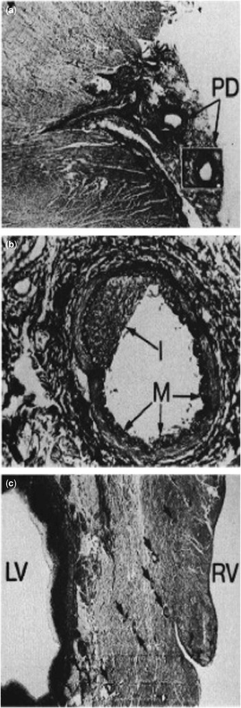
Physiologic coronary perfusion occurs in a cyclical pattern, with the major contribution during diastole. In the setting of an anatomically obstructed left ventricle, the coronary perfusion occurs during systole, as demonstrated by selective aortic root angiography in prior studies of hypoplastic left heart syndrome.Reference Freedom, Culham, Moes and Harrington 32 The high-pressure blood flow during ventricular systole may lead to wall thickening and tortuosity in the epicardial coronary arteries and their intramyocardial branchesReference O’Connor, Cash, Cottrill, Johnson and Noonan 11 , Reference Sauer, Gittenberger-de Groot, Geishauser, Babic and Buhlmeyer 29 (Fig 3). O’Connor et al observed that diseased vessels were limited to the left ventricular free wall and septum. The histological findings consisted of circumferential medial muscular hypertrophy, reduplicated elastic fibres, and focal intimal fibrous proliferation without significant luminal narrowing.Reference O’Connor, Cash, Cottrill, Johnson and Noonan 11 Sauer et alReference Sauer, Gittenberger-de Groot, Geishauser, Babic and Buhlmeyer 29 found that specimens with mitral and aortic atresia did not demonstrate histological coronary artery abnormalities.
Coronary artery wall thickening observed in cases of hypoplastic left heart syndrome with patent inflow and obstructed outflow. ( a ) Photomicrograph of a branching posterior descending artery in the epicardial groove. Magnification 14×. ( b ) Vessel outlined in ( a ) shows a prominent musculoelastic zone (M). Eccentric intimal thickening (I) is present but does not appear to significantly narrow the calibre of the lumen. Magnification 120×. ( c ) Low-power view of interventricular septum with prominent muscularised arteries (arrows). Magnification 10×. All elastic stain. Courtesy of O’Connor.Reference O’Connor, Cash, Cottrill, Johnson and Noonan 11 LV=left ventricle; PD=patent ductus; RV=right ventricle.

Coronary artery histopathologic findings are most prominent in arteries that communicate directly with the lumen (arterio-luminal subtype) and less prominently in those that communicate indirectly with the lumen (arterio-sinusoidal and arterio-capillary subtypes).Reference O’Connor, Cash, Cottrill, Johnson and Noonan 11 Baffa et alReference Baffa, Chen, Guttenberg, Norwood and Weinberg 28 noted increased tortuosity of the left coronary artery in mitral stenosis/aortic atresia specimens, but observed on histological examination that the ratio of coronary wall thickness relative to lumen diameter in the left anterior descending artery and circumflex artery revealed no significant differences from control hearts. Lloyd et alReference Lloyd, Evans and Marvin 33 also observed that the luminal diameters of the coronary arteries and ostia in hypoplastic left heart syndrome were not different from control specimens.
Anomalous coronary artery origins and coronary artery hypoplasia have rarely been reported in hypoplastic left heart syndrome.Reference DeRose, Corda, Dische, Eleazar and Mosca 34 Infrequently, case reports have described an anomalous origin of the left coronaryReference Malec, Mroczek, Pajak, Zajac, Kolcz and Januszewska 35 – Reference Nosal, Omeje and Poruban 38 or circumflexReference Bartram, Grunenfelder and Van Praagh 39 , Reference Villa, Brancaccio, Carotti, Francalanci and Di Donato 40 artery from the right pulmonary artery in the setting of hypoplastic left heart syndrome. Ito el alReference Ito, Niino and Ishikawa 41 reported a case of hypoplastic left heart syndrome with a single coronary artery originating from the pulmonary artery. Saroli et alReference Saroli, Gelehrter, Gomez-Fifer, van der Velde, Bove and Ensing 37 reported three cases of hypoplastic left heart syndrome with superior origin of the left coronary artery. The origin of the right coronary artery from the descending thoracic aorta has been documented in one case of hypoplastic left heart syndrome.Reference Cheatham, Ruyle, McManus and Gammel 42 DeRose et alReference DeRose, Corda, Dische, Eleazar and Mosca 34 described a case of hypoplasia of the left anterior descending artery that resulted in marked left ventricular ischaemia during a Norwood procedure, ultimately leading to the patient’s demise.
Abnormal coronary artery fistulas have also been described. Raghib et alReference Raghib, Bloemendaal, Kanjuh and Edwards 43 described a case of hypoplastic left heart syndrome due to aortic atresia with a fistula present between the left circumflex coronary artery and the coronary sinus. A recent case report documented a left ventricle–left coronary artery–pulmonary artery fistula associated with combined mitral and aortic atresia and a ventricular septal defect.Reference Paladini, Caruso, Sglavo, Lapadula, Parisi and Martinelli 44 The authors concluded that the coronary fistula was the primary cause of hypoplastic left heart syndrome in this case because of the existence of a ventricular septal defect that nullified the intraventricular pressure gradient, which is typical of hypoplastic ventricles and thought to be responsible for the secondary development of fistulas in other cases.Reference Paladini, Caruso, Sglavo, Lapadula, Parisi and Martinelli 44
In an autopsy report of 122 patients who died after a Norwood procedure, Bartram et alReference Bartram, Grunenfelder and Van Praagh 39 found impairment of coronary artery perfusion to be the most frequent cause of death (33 patients, 27%). However, the cause of stenosis in the vast majority of these patients (31 of 33) was secondary to either intraluminal stenosis at the anastomosis or external kinking of the graft.Reference DeRose, Corda, Dische, Eleazar and Mosca 34 , Reference Bartram, Grunenfelder and Van Praagh 39 The poor coronary perfusion in these cases was therefore the result of the surgical technique and was not the underlying coronary disease. Surgical ligation of an aberrant left circumflex coronary artery from the right pulmonary artery occurred in one case, resulting in biventricular infarction.Reference Bartram, Grunenfelder and Van Praagh 39 Generalised coronary artery hypoplasia was also found in one patient.Reference Bartram, Grunenfelder and Van Praagh 39 Despite the rare occurrence of coronary anomalies in hypoplastic left heart syndrome, detailed coronary artery assessment is recommended as part of the routine echocardiographic evaluation of hypoplastic left heart syndrome before surgical intervention.Reference Saroli, Gelehrter, Gomez-Fifer, van der Velde, Bove and Ensing 37 The coronary arteries in hypoplastic left heart syndrome may become thickened or tortuous in cases with high intraluminal pressure, but have a preserved lumen, and with rare exception are likely not primary to the pathogenesis of disease.
Microvasculature
Focussing on the microvasculature of hypoplastic left heart syndrome, Salih et alReference Salih, Sheppard and Ho 45 described an interesting finding that unoperated hearts with hypoplastic left heart syndrome have a higher mean and maximal diffusion distance from any arbitrary point to the nearest capillary than do normal age-matched control hearts. No differences were noted between left and right ventricles or between subtypes. The authors believe the reduction in capillarisation may be an inherent abnormality of hypoplastic left heart syndrome that may have implications for ventricular development. Rakusan et alReference Rakusan, Flanagan, Geva, Southern and Van Praagh 46 noted that congenital aortic stenosis and coarctation of the aorta are characterised by an increase in capillary supply proportional to myocyte volume, maintaining capillary density similar to control hearts. They observed that pressure-overload left ventricular hypertrophy in children demonstrates proportional capillary angiogenesis, whereas in adults hypertrophy appears to be associated with failure of compensatory angiogenesis.
Could the pathogenesis of hypoplastic left heart syndrome stem in part from premature failure of compensatory angiogenesis? Jacobs suggested that the decreased capillarisation observed by Salih et al might not be an unalterable inherent abnormality of hearts with hypoplastic left heart syndrome, but rather a snapshot of the supply–demand mismatch encountered with unaltered hypoplastic left heart syndrome anatomy. As stated previously, perfusion of the myocardium in unoperated hearts with hypoplastic left heart syndrome occurs primarily in systole, rather than in diastole, as there is considerable diastolic runoff into the pulmonary circulation. Coronary perfusion is further limited by excessive myocardial wall tension.Reference Jacobs 47 Elimination of the diastolic runoff, by means of closure of the systemic to pulmonary artery shunt at the time of the second-stage surgery, results in the restoration of more normal diastolic coronary perfusion as well as reduction in the volume of work of the ventricle.Reference Jacobs 47 JacobsReference Jacobs 47 proposes that this higher diastolic blood pressure may result in improved capillarisation of the myocardium early in life. Further research is necessary to confirm or refute this theory, but it is clear that there are primary myocardial differences in capillary density in hypoplastic left heart syndrome compared with other left-sided CHDs, which further suggests that alterations in the myocardial development of hypoplastic left heart syndrome could contribute to the observed phenotype.
Inappropriate expression of platelet-endothelial cell adhesion molecule-1, also known as CD31, has been identified in heart samples with hypoplastic left heart syndromeReference Bohlmeyer, Helmke and Ge 16 and may give some insight into myocardial differences in capillary density, or capillary angiogenesis, observed in hypoplastic left heart syndrome. CD31 is a member of the cell adhesion molecule family, which has a primary role in the regulation of tissue morphogenesis.Reference Bohlmeyer, Helmke and Ge 16 Before a recent study, cardiac myocytes have never been shown to express CD31 at any stage of development or in any disease state.Reference Bohlmeyer, Helmke and Ge 16 The authors suggest that inappropriate expression of CD31 or a related gene under the same regulatory control may be responsible for the disorganisation of hypoplastic left heart syndrome cardiac myocytes and potentially the higher-order cardiac structural abnormalities associated with this disease.Reference Bohlmeyer, Helmke and Ge 16
CD31 has also been associated with cell migration and cancer angiogenesis.Reference DeLisser, Christofidou-Solomidou and Strieter 48 Recent work suggests that CD31 has a novel role in arteriogenesis and collateral remodelling.Reference Chen, Rubin and Tzima 49 CD31 has also been identified as the first molecule that determines pre-existing collateral diameter.Reference Chen, Rubin and Tzima 49 These finding highlight the possibility that abnormal CD31 expression might be an attempt by the myocardium to induce angiogenesis secondary to the decreased capillarisation seen in hypoplastic left heart syndrome. Alternatively, the atypical capillarisation in hypoplastic left heart syndrome could be the product of abnormal CD31 expression within the myocardium.
Pathogenesis of hypoplastic left heart syndrome
The aetiologic mechanisms leading to hypoplastic left heart syndrome are largely unknown. About one-fourth of hypoplastic left heart syndrome cases occur in the context of recognised genetic disorders including, but not limited to, Turner, Jacobsen, Noonan, and Holt–Oram syndromes. However, studies involving non-syndromic family members have suggested that heritability is complex. No single disease-causing pathway has yet been identified.Reference Grossfeld, Ye and Harvey 50 , Reference Grossfeld 51 Clinical observations indicate that obstruction of blood flow through the left ventricle in an otherwise normal four-chambered foetal heart, caused by either aortic and/or mitral stenosis, leads to the development of left ventricular hypoplasia.Reference Allan, Sharland and Tynan 52 Prenatal diagnosis of hypoplastic left heart syndrome can be made as early as 14 weeks’ gestation,Reference Carvalho 53 but numerous cases of hypoplastic left heart syndrome have presented with normal or even dilated left ventricular cavities on routine foetal ultrasound at >19 weeks’ gestation.Reference Allan, Sharland and Tynan 52 , Reference Anderson and Brown 54 The unifying aetiologic explanation is that the growth and development of vascular structures are dependent to some degree on the relative quantity and quality of blood flow during development.Reference Sedmera, Cook, Shirali and McQuinn 3 Recent studies have shown that hypoplastic left heart syndrome myocytes are well differentiated, but have prolonged expression of foetal or “heart failure” genes.Reference Bohlmeyer, Helmke and Ge 16 These findings suggest that intrauterine insult to the foetus may occur after embryogenesis and highlights the possibility that foetal development of hypoplastic left heart syndrome may occur later in gestation.Reference Makikallio, McElhinney and Levine 55 , Reference Tworetzky, Wilkins-Haug and Jennings 56 Recent hypotheses also question whether immunologic,Reference Eghtesady 57 infectious,Reference Sedmera, Cook, Shirali and McQuinn 3 , Reference Oyer, Ongcapin, Ni, Bowles and Towbin 58 or autoimmune insults to genetically susceptible hosts may contribute to left ventricular hypoplasia from either direct myocardial injury or secondary to reduced left ventricular blood flow through damaged valves.
Genetic factors that alter valve development have been proposed as the aetiology in hypoplastic left heart syndrome.Reference Hor, Border, Cripe, Benson and Hinton 59 , Reference Hinton, Martin, Tabangin, Mazwi, Cripe and Benson 60 Mutations in the signalling and transcription regulator, NOTCH1, have caused early aortic valve defects in animal models.Reference Tweddell and Hoffman 5 Genes involved in downstream NOTCH signalling and cardiac gene expression have been implicated in familial forms of hypoplastic left heart syndrome.Reference Dai, Cserjesi, Markham and Molkentin 61 A recent study showed a shared genetic linkage with hypoplastic left heart syndrome and bicuspid aortic valve.Reference Hinton, Martin, Rame-Gowda, Tabangin, Cripe and Benson 62 Bicuspid aortic valve is very common, affecting 1% of the general population, and is a known risk factor for aortic valve disease. Is it possible that a small subset of patients with most restrictive bicuspid aortic valves progress to hypoplastic left heart syndrome? Isolated aortic valve atresia and congenital aortic stenosis do not always progress to hypoplastic left heart syndrome, suggesting that aortic valve defects and hypoplastic left heart syndrome have distinct genetic susceptibilities.Reference Grossfeld, Ye and Harvey 50 Genetic factors are clearly present, but epigenetic modifiers and/or environmental influences might be necessary for the phenotypic expression of hypoplastic left heart syndrome.Reference Grossfeld, Ye and Harvey 50
The complex heritability of hypoplastic left heart syndrome suggests the potential for environmental contributions to the observed phenotype. Maternal upper respiratory infection during the first trimester has been documented as a significant risk factor for hypoplastic left heart syndrome.Reference Tikkanen and Heinonen 63 Population-based studies in the Baltimore–Washington region have identified environmental risk factors for hypoplastic left heart syndrome, including maternal exposure to organic solvents.Reference Kuehl and Loffredo 64 A recent study showed a seasonal pattern in the presentation of hypoplastic left heart syndrome with preponderance in summer months in contrast to the random pattern observed in other left-sided heart diseases.Reference Eghtesady, Brar and Hall 65 These findings support a role for an environmental component in the aetiology of hypoplastic left heart syndrome.
An immune-mediated mechanism for the pathogenesis of hypoplastic left heart syndrome has been proposed where antibodies cross the placenta and create disease in genetically susceptible hosts.Reference Eghtesady 57 In rheumatic heart disease, antibodies have already been shown to “cross-react” with human valvular and myocardial antigens through a mechanism known as molecular mimicry.Reference Cunningham 66 Our lab recently demonstrated that transplacental transfer of maternal anti-cardiac myosin autoantibodies leads to structural congenital cardiac defects in affected progeny that included diminished left ventricular cavity dimensions.Reference Cole, Yutzey and Brar 67 Foetuses that developed hypoplastic left heart syndrome-like phenotype had elevated serum titres of anti-β adrenergic receptor as well as increased protein kinase A activity, suggesting a potential mechanism for the observed pathological changes.Reference Cole, Yutzey and Brar 67
There is evidence that prenatal viral infection could account for some cases of left ventricular hypoplasia with endocardial fibroelastosis.Reference Sedmera, Cook, Shirali and McQuinn 3 , Reference Ni, Bowles and Kim 24 , Reference Oyer, Ongcapin, Ni, Bowles and Towbin 58 Viral insult is a well-accepted cause of cardiomyopathy. Studies have been reported that viral RNA is present in myocardial samples from foetal hearts with aortic valvular stenosis.Reference Sedmera, Cook, Shirali and McQuinn 3
Alternative mechanisms for the pathogenesis of hypoplastic left heart syndrome have been suggested in which abnormal cardiac myocyte proliferation may be the primary defect. In this case, left ventricular hypoplasia could result from abnormal cellular signal transduction, which is critical for normal myocyte division and left ventricular growth.Reference Grossfeld 19 Conversely, the thickening of the left ventricular myocardium in subtypes of hypoplastic left heart syndrome with a patent inflow tract and obstructed outflow tract is thought to result from cardiomyocyte hyperplasia during development.Reference Sedmera, Cook, Shirali and McQuinn 3 , Reference Cole, Yutzey and Brar 67 The abnormal thickening of the left ventricular myocardium could lead to reduced left ventricular cavity dimensions, and valvular abnormalities as a result of altered haemodynamics. Many primary myocardial abnormalities have been identified that could contribute to the pathogenesis of hypoplastic left heart syndrome.
Conclusion
Because of the variability in subtypes and presentation of hypoplastic left heart syndrome, a common aetiology for all subtypes is unlikely. Genetic and environmental factors are likely working in concert to create the spectrum of phenotype observed. Cases of hypoplastic left heart syndrome with mitral atresia and aortic atresia or with mitral atresia, a ventricular septal defect, and an intact aortic root have been shown to have normal myocardial architecture at birth and normal coronary vasculature, which suggests that these subtypes are likely due to primary valvular pathogenesis. Future research efforts in these subtypes should look for genetic and environmental causes of valvular agenesis. Subtypes of hypoplastic left heart syndrome with patent inflow and obstructed outflow present with disarray of the myocyte architecture and thickening of the myocardium. These hearts are also more prone to vascular abnormalities, such as coronary artery fistulas. As stated previously, the regional distribution of myofibre disarray supports the concept that vascularisation parallels myocardial organisation in the developing heart. Future studies should evaluate for primary myocardial disorders or dis-regulation of cardiomyocyte proliferation during development for these subtypes. In addition, some phenotypes of hypoplastic left heart syndrome, most likely the subtypes with an atretic valve and thickened ventricle, may require a combined insult to both the myocardium and valve structures. Futhermore, continued investigation into the reduction in capillarisation in hypoplastic left heart syndrome and the potential contribution of CD31 in this abnormality is warranted. If the pathogenesis of this disease is uncovered, the possibility for more effective treatment or perhaps even the prevention of certain subtypes of hypoplastic left heart syndrome may one day become reality.
Acknowledgement
The authors would like to thank Anoop Brar PhD and R. Scott Baker for their contribution in editing this review.
Financial Support
Financial support was provided by NIH R21-HL104391, NIH F32-HL103054 and Saving Tiny Hearts Society.
Conflicts of Interest
The authors have no relationships with industry or financial associations that might pose a conflict of interest with the submitted article.