Background
COA accounts for 5–7% of CHD, with an incidence of 0.3–0.4/1000 live births. Reference Rosenthal1,Reference Kuehl, Loffredo and Ferencz2
Although it results from abnormalities in the development of the embryonic fourth and sixth aortic arches, COA is quite diverse in severity and presentation. The underlying mechanisms are not fully understood, but two concepts have been proposed: the ductal tissue theory and reduced-flow theory. Reference Hamdan3
L.M. Bonnet first categorised CoA into an infantile form and an adult form. Reference Jenkins4 A commonly accepted classification is based on stenosis location: pre-ductal, isthmic, and post-ductal. Reference Secchi, Iozzelli, Papini, Aliprandi, Di Leo and Sardanelli5
Imaging of COA could be done by different imaging modalities. Echocardiography has paramount importance for evaluation of the thoracic aorta and the functional assessment of COA. Reference Evangelista, Flachskampf and Erbel6 Multidetector CT is commonly used for the evaluation of thoracic arch morphology and abnormalities with good results. Reference Turkvatan, Buyukbayraktar, Olcer and Cumhur7–Reference Finlay, Johnson and Forbes9 MRI offers a functional assessment of coarctation without high ionising radiation dose, making it suitable for long-term follow-up. Reference Shepherd, Abbas and McParland10
Surgical repair has been the standard treatment for coarctation of the aorta (CoA). However, endovascular interventions have emerged as less invasive alternatives, particularly for older patients. Despite advancements in treatment, long-term complications like hypertension remain a concern. Reference Dikmen, Ozcinar and Eyileten11
Hypertension is the most important complication of COA. It is associated with stroke, coronary artery disease and renal failure in the long run. Reference Buonacera, Stancanelli and Malatino12–Reference Hamrahian and Falkner14 The mid-term outcome regarding residual hypertension in COA is similar between stented patients and those who underwent surgical repair. Reference Bouet, Bonello and El Tahlawi15
Although some studies suggested that anatomical variations in the aortic arch are associated with increased incidence of CoA and stroke, Reference Rotundu, Nedelcu and Tepordei16,Reference Dodge-Khatami, Ott, Bernardo and Berger17 up till now, there is scarce data regarding the effect of the relative inter-branch distance of the aortic arch on the short- and long-term prognosis after COA repair.
We hypothesise that there is a strong association between the relative anatomical distance between aortic arch branches and recoarctation, suggesting that this anatomical parameter may play a role in the development of recoarctation.
Here, we aim to explore the association between aortic arch anatomy, particularly inter-branch distances, and the short- and long-term outcomes of surgical coarctation repair.
Methods
This is a retrospective study enrolling patients with surgically corrected COA in 2 different centres during the period between November 2007 and March 2016.
We followed the patients prospectively during the recruitment and for at least 2 postoperative years or till the development of one of the following endpoints (recoarctation “defined as a re-elevation of peak-to-peak systolic pressure gradient over 20 mm Hg across the site of the previous COA repair”, Persistent/Re-elevation pressure gradient, or Persistent/Re-elevation of blood pressure necessitating reintervention).
Inclusion criteria
We included all patients under the age of 8 years of both sexes with surgically corrected COA during the indicated period.
Exclusion criteria
We excluded patients who
-
1. died during or after surgery.
-
2. underwent percutaneous intervention for COA.
-
3. were lost during follow-up or
-
4. lacked echocardiography or multidetector CT data.
We reviewed patient records, including clinical data, echocardiograms, multidetector CT scans, and surgical reports.
Patient and clinical data
The medical records were reviewed for
-
Demographic data: age at operation, sex, weight, and associated genetic or hereditary syndromes.
-
Clinical data: resting blood pressure, upper limb/lower limb pressure gradient,
-
Intraoperative data: type of surgical repair,
-
Postoperative data: arm/leg systolic pressure gradient before discharge, blood pressure, complications, and mortality.
-
The follow-up data including clinical data and the need for redo or stenting.
All these data were reviewed and recorded.
Echocardiographic data were analysed from echocardiographic records, including:
-
Pre-operative traditional 2D with the anatomical description of the aortic arch, aortic valve anomalies, presence of hypoplastic transverse aorta and associated lesions, dimensions, and Doppler arch pressure gradient and flow.
CoA was diagnosed based on the presence of a significant pressure gradient across the narrowed segment of the aorta, (typically located in the aortic arch or descending aorta) ≥ 20 mmHg with diastolic flow by continuous Doppler, the presence of flow turbulence across the coarctated segment by colour Doppler and low amplitude spectral Doppler of the abdominal aorta in subcostal view. Reference Doshi and Chikkabyrappa18
-
Postoperative echocardiography was reviewed for peak pressure gradient across the repaired arch using continuous-wave Doppler and for the recurrence and/or persistence of forward flow in both the thoracic descending and the abdominal aorta using spectral Doppler. The dimensions, arch anatomy, and arch flow were also reviewed.
-
Follow up on the same measurements.
Pre-operative multi-detector CT
CT was performed using a multidetector CT scanner (Philips Ingenuity Core128, Philips Healthcare, the Netherlands).
Image processing and measurements
Images were post-processed on a separate workstation (IntelliSpace Portal V5, Philips Healthcare). Oblique multiplanar 3D maximal intensity projection reconstructed planes were created till getting the best image, displaying the root of all aortic arch branches in the same plane. The curved distance from the midpoint of the innominate artery root to the midpoint of the left common carotid artery was measured along the greater curvature of the aortic arch. The straight distance from the midpoint of the root of the first arch branch to the last arch branch (LSCA) was also measured, as shown in Figure 1.
Measurement of inter-branch distances in MDCT images.

We measured the inter-branch distances in millimetres in all patients, regardless of age. As there is no standardised reference range for these measurements, we could not use Z-scores to normalise the data.
We related such (LCCA-IA) distance to the total (LSCA- IA) distance to get the distance ratio. We classified the patients according to the distance ratio of their aortic arches into 2 groups: Group I, whose distance (LCCA- IA) is short and less than 50% of the total distance (LSCA- IA), and Group II, whose such distance is long and ≥ 50% of the total distance (LSCA- IA).
Other features of COA in the multidetector CT were recorded, such as the inclusion of LSCA in the COA segment and the presence of hypoplastic arch. The hypoplastic arch was defined if the diameter of the transverse arch was less than 50% of that of the ascending and/or descending aorta. Reference Huuskonen, Hui and Runeckles19
Results
The study included 73 patients (38 males and 35 females) with their ages ranging between 10 days and 8 years, mean ± SD of age equals 10.4 ± 32.3 months.
The mean duration of follow-up was 6.3 ± 2.8 years.
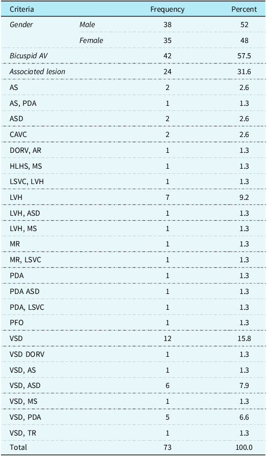
The characteristics of the included patients are presented in table 1
The characteristics of the recruited patients

Type of surgical repair
Anastomosis end-to-end with interposition graft was done in 64 patients (84.2%), Crafoord (end-to-end) in 6 patients (7.9%), and aortoplasties with a patch in 4 patients (5.2%). Subclavian flap was needed in 4 patients (5.2%), and excision of the ductal zone was done in 4 patients (5.2%).
The majority of surgical repairs (87%) were performed through a left posterolateral thoracotomy. The most common surgical technique employed was end-to-end anastomosis.
In cases where the coarctation is extensive or the aortic arch is hypoplastic, patch aortoplasty was applied. This technique involves widening the narrowed segment of the aorta using a patch of synthetic material or autologous pericardium.
None of the subjects had a highly curved arch.
The distances between the left common carotid artery (LCCA) and the innominate artery (LCCA-IA), and between the left-common carotid artery and the left subclavian artery (LSCA-IA) were measured for each patient. The LCCA-IA distance ranged from 0 to 22.3 mm, with a mean ± SD of 13.8 ± 7.5 mm.
The distance ratio (LCCA- IA) / (LSCA-IA) has a range of Zero to 89%.
Group I involved 20 patients (27.4%), while Group II contained 53 patients (72.6%).
There was no significant difference between both groups regarding age, sex, and age at operation.
The descriptive anatomy of the aortic arch is presented in figure 2 for all enrolled populations.
The descriptive anatomy of the aortic arch.

The outcome of the patients during the follow-up period is presented in figure 3
The prognosis of the operated patients during the follow-up period.

There was a significant difference between both groups regarding recoarctation, re-elevation of blood pressure, and re-elevation of pressure gradient (P = 0.0001, 0.011, and 0.014, respectively), with more occurrences in those patients with small (LCCA- IA) distance.
There was a positive correlation between the distance ratio (LCCA- IA) / (LSCA-IA) and pressure gradient across the repaired segment (P = 0.0001) (Table 2).
Correlations between the distance (LCC-left innom art) and each of reCOA, persistent/Re-elevation PG, and persistent/Re-elevation of BP

** Correlation is significant at the 0.01 level (2-tailed).
There was also a positive correlation between the inclusion of left subclavian artery in the COA segment and both the re-elevation of pressure gradient across the repaired segment and the re-elevation of blood pressure (Table 3).
Correlations between the presence of LSCA included in the lesion and all worse prognostic determinants

** Correlation is significant at the 0.01 level (2-tailed).
We found no association between the presence of hypoplastic arch and recoarctation.
By performing logistic regression analysis to determine independent predictors of recoarctation, we found that only the anatomical distance ratio (LCCA- IA) / (LSCA-IA) can independently predict recoarctation in the long term.
Figure 4 presents a case with a common origin of both left IA and LCCA (bicarotid arch), who presented with recoarctation 2 years after COA repair.
Presents a case with common origin of both left IA and LCCA (bovine arch) who presented with re-elevation of of BP 2 years after COA repair.

Subgroup analysis
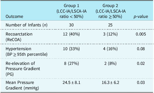
We conducted a subgroup analysis for infants who underwent extended end-to-end anastomosis. A total of 55 infants were included. Using the same anatomical ratio (LCCA-IA / LSCA-IA) as described in our main study, we divided the cohort into two groups:
-
Group 1: LCCA-IA to LSCA-IA ratio < 50% (shorter inter-branch distances).
-
Group 2: LCCA-IA to LSCA-IA ratio ≥ 50% (longer inter-branch distances).
Our findings revealed that Group 1 had a significantly higher incidence of recoarctation compared to Group 2 (P = 0.005). Additionally, Group 1 showed a significantly higher incidence of pressure gradient re-elevation (P = 0.02), with a higher mean pressure gradient overall (P = 0.03). However, the difference in hypertension rates between the two groups was not statistically significant (P = 0.08) (Table 4).
Subgroup analysis for the infants underwent extended end-to-end anastomosis repair

Discussion
In our cohort, we address, for the first time, the impact of different anatomical aspects, particularly the relative inter-branch distances, on the outcome of surgically repaired CoA. Our study found that a shorter LCCA-IA distance was associated with a higher incidence of recoarctation, persistent hypertension, and re-elevation of blood pressure. Conversely, a longer LCCA-LSCA distance was also associated with more adverse outcomes. There is considerable interaction between the anatomical structure of the aortic arch and the expected arch anomalies. Reference Dodge-Khatami, Ott, Bernardo and Berger17 One theory (anatomical theory) suggests that CoA may result from tissue from the ductus arteriosus invading the descending aorta distal to the aortic isthmus, leading to narrowing. However, this theory cannot explain cases of coarctation occurring outside the aortic isthmus. Reference Hamdan3
Another theory (haemodynamic theory) attributes the development of COA to altered fetal haemodynamics, specifically reduced blood flow through the aortic arch and isthmus. Reference Buyens, Gyselaers and Coumans20
We hypothesised that the displacement of left-common carotid artery towards innominate artery might cause stretching of the transverse arch; therefore, the constrictive effect of the invading ductal tissue may produce recoarctation and consequently re-elevation of blood pressure. The same effect could occur because the left subclavian artery is more inferiorly displaced and therefore the (LSCA-IA) distance increases.
The determinants of COA prognosis after surgery are still an area of research. While previous studies have demonstrated the impact of the anatomical shape and diameters of the arch on the prognosis regarding recoarctation, Reference Turek, Conway and Cavanaugh21,Reference Ashcraft, Jones and Border22 persistent HTN, or re-elevation of blood pressure, Reference Ou, Celermajer and Raisky23 we studied the relative branch anatomy and the impact of their inter-relationship on the prognosis after COA repair.
Meyers et al studied the distance between arch branches and sinotubular junction in both normal & bicarotid arch in patients with COA indexed to BSA; however, they did not provide clinical data about the impact of these measurements on the outcome such as recoarctation or HTN. Reference Meyer, Turek and Froud24
Additionally, some authors have noted the difficulty in consistently classifying the various morphologies of the aortic arch, which may limit the clinical utility of these classifications. Reference Lashley, Curtin, Malcolm, Clark and Freeman25
Arch geometry has been previously reported as a risk factor for residual HTN. Reference Ou, Bonnet and Auriacombe26,Reference Iriart, Laïk and Cremer27
Iriart et al found that gothic arch could contribute to persistent HTN after COA stenting despite the absence of residual COA. This may be due to shortened or absent transverse aortic arch that could alter the function of baroreceptors. Reference Iriart, Laïk and Cremer27 This geometry could also affect arterial distensibility and increase central aortic stiffness. Reference Ou, Celermajer and Raisky23
Besides, the aortic angle in a gothic arch would lead to early and large systolic wave reflection. The resultant vascular remodelling in the aortic wall with increased wall stress and decrease compliance may finally lead to high central aortic pressure. Reference London28
Patients with a laterally displaced LSCA and a short aortic isthmus may develop a Gothic arch configuration after repair, leading to acute angulation of the aorta. Reference Ou, Mousseaux and Celermajer29
Indeed, the extended aortic arch anastomosis increases the size of the segment between the LCCA and the LSCA, but does not usually enlarge the segment between the innominate and the LCCA, and may, therefore, lead to a gothic geometry of the aorta, Reference Siewers, Ettedgui, Pahl, Tallman and Delnido30 this may in part support our findings.
A previous study proved a negative correlation between the distance LCCA- LSCA and the duration before recoarctation with early reintervention for recoarctation in patients with long LCCA-LSCA segment. Reference El Tahlawi and Tharwat31
The common brachiocephalic trunk is another geometry that might affect the result of repaired COA. It is commonly referred to as a ‘‘bicarotid or bovine arch’’ where the left-common carotid artery arises more proximally on the arch, so as to originate immediately close to or as a branch of the IA.
A previous study by Reinshagen et al. highlighted the relatively high prevalence of bicarotic trunk in patients with coarctation of the aorta. This anatomical variation may influence the hemodynamics of the aortic arch and the risk of complications after surgical repair. Reference Reinshagen, Vodiskar, Mühler, Hövels-Gürich and Vazquez-Jimenez32
Turek et al Reference Turek, Conway and Cavanaugh21 found that there were significantly higher recoarctation rates in the cohort with bovine arch (bicarotid) anatomy (P < 0.03). These results may be in concordant with ours, as we found that the possibility for recoarctation and persistence of HTN increases when LCCA is close to left IA.
The extended end-to-end anastomosis technique, commonly used for the surgical repair of coarctation of the aorta (COA), is designed to address a range of anatomical challenges, particularly in infants. Reference Vasile, Laforest, Bulescu, Jalal, Thambo and Iriart33 However, its effectiveness may be influenced by certain anatomical variations, such as a shorter distance between the LCCA and the IA. Reference Backer, Mavroudis, Zias, Amin and Weigel34 In cases where this inter-branch distance is reduced, the surgical repair may be more challenging, as the aortic arch may be less flexible, leading to less optimal surgical outcomes. A shorter LCCA-IA distance could contribute to a more acute angulation or a less favourable geometry of the aortic arch after the anastomosis, potentially predisposing patients to complications like recoarctation or persistent pressure gradients.
Turek et al. suggested that a bovine arch configuration limits the extent of possible aortic arch reconstruction due to a shortened transverse aortic arch, making extended end-to-end anastomosis technically challenging because of the insufficient clampable arch segment. Reference Turek, Conway and Cavanaugh21
In the same context, a systematic review concerning the COA repair in neonates, the authors linked Gothic arch anatomy to postoperative hypertension and bicarotid arch to restenosis and increased mortality in neonatal coarctation repair. Reference Margarint, Youssef and Robu35
However, Lashley et al Reference Lashley, Curtin, Malcolm, Clark and Freeman25 studied the arch geometry in an adult population that had undergone surgical correction for COA, taking into account the A/T index (ratio of height/transverse diameter, which is higher in theory when the arch is gothic). They did not find a link with residual HTN (at rest and during exercise).
Similarly, in the study of Liu et al Reference Liu, Jones and Cheung36 using ambulatory blood pressure monitoring in patients with corrected COA, they could not correlate adverse arch geometry with any adverse cardiovascular outcomes.
In addition, some investigators concluded that bovine arch anatomy did not influence recoarctation. However, the cohort prevalence of bovine arch anatomy in these studies was only 3 to 5%. Reference McElhinney, Yang and Hogarty37,Reference Burch, Cowley and Holubkov38
In a recent study that may support our hypothesis, Huang et al found that the relative inter-branch anatomy may influence aortic dissection, as the distance from the left-common carotid artery to the left subclavian artery was significantly stretched and greater in the patient group with acute Type A aortic dissection compared with the control group (P < 0.05). Reference Huang, Liu, Wu, Lai and Cai39
In our study, we found no association between the presence of hypoplastic arch and recoarctation.
A recent study conducted by Lehnert et al reported no association between hypoplasia of the aortic arch and the type of surgical repair on one side and the risk of recoarctation in young infants on the other side. Reference Lehnert, Villemain, Gaudin, Méot, Raisky and Bonnet40
Previous studies have reported conflicting findings regarding the association between aortic arch hypoplasia and the risk of recoarctation. While McElhinney Reference McElhinney, Yang and Hogarty37 found that an absolute transverse arch diameter less than 3.5 mm was an independent predictor of earlier recoarctation necessitating reintervention in infants less than 3 months old. In contrast, however, Kaushal Reference Kaushal, Backer and Patel41 failed to find any association between transverse arch hypoplasia and recoarctation in their cohort of infants and children. Similarly, Truong et al Reference Truong, Tani, Minich, Burch, Bardsley and Menon42 and Callahan et al Reference Callahan, Saudek, Creighton, Kuhn, Mitchell and Tweddell43 reported no association between the occurrence of recoarctation and arch Z-score in patients who underwent successful arch repair.
Bicuspid aortic valve (BAV) hasn’t also any association with recoarctation in our study. This result was in accordance with Lehnert et al study, Reference Lehnert, Villemain, Gaudin, Méot, Raisky and Bonnet40 which could not associate between BAV and the risk of recoarctation.
The subgroup analysis of infants undergoing extended end-to-end anastomosis revealed significant differences in adverse outcomes based on the anatomical ratio of LCCA-IA to LSCA-IA (Group 1). Infants with a shorter ratio demonstrated a notably higher incidence of recoarctation. Additionally, Group 1 had a higher mean pressure gradient and a greater incidence of re-elevation of pressure gradient, suggesting that a smaller LCCA-IA/LSCA-IA ratio may be associated with more challenging aortic arch anatomy and worse long-term outcomes. Although the difference in hypertension rates did not reach statistical significance, the trend towards higher hypertension in Group I further supports the notion that anatomical features could influence post-surgical vascular function. These findings suggest that the LCCA-IA to LSCA-IA ratio may serve as an important predictor of surgical outcomes in infants, particularly in predicting recoarctation and pressure gradients, and could help refine pre-operative assessments and surgical planning.
We hypothesise that specific anatomical features, such as a shorter LCCA-IA distance, may contribute to difficulties in achieving optimal aortic arch geometry, which could predispose patients to adverse outcomes. The surgical technique itself, while tailored to the patient’s specific anatomy, might not fully correct these inherent anatomical challenges, leading to residual pressure gradients or recoarctation, as demonstrated in Group 1 of our subgroup analysis.
In our cohort, age at surgery did not significantly impact the outcome. However, other studies have suggested that older age may be associated with a higher risk of residual hypertension after COA stenting, potentially due to age-related changes in aortic compliance. Reference Iriart, Laïk and Cremer27
Multidetector CT is a valuable tool for three-dimensional assessment of the aortic root and arch anatomy. Reference Sakrana, Nasr, Ashamallah, Abuelatta, Naeim and El Tahlawi44 In our cohort, we used maximum intensity projection image to draw the measurements. While maximum intensity projection images are widely used in clinical practice due to their ease of interpretation and automated measurement capabilities, they can be subject to limitations, particularly when the aortic arch curves significantly out of the plane of the image. This can lead to inaccuracies in measuring inter-vascular distances. Maximum intensity projection projections can potentially distort spatial relationships and may introduce variability in measurement, especially when the vessel plane deviates from the imaging plane. Curved plane reformatted images, on the other hand, allow for visualisation of the aortic arch along its centreline, potentially providing a more accurate assessment of these distances. However, they may require more complex image processing techniques and manual measurements. Reference Bartolozzi, Neri and Caramella45
More precise techniques—such as confirming measurements on thin-slice reconstructions or using dedicated oblique planes for each segment (IA–LCCA and LCCA–LSCA)—can reduce variability. Alternatively, employing a single oblique or curved multiplanar reformation that includes the origin of all three arch branches could enhance anatomical fidelity.
While the LSCA–IA distance was measured as a straight line, which may introduce error in markedly curved (“gothic”) aortic arches, we did not find any overtly gothic arches in our study cohort, thereby minimising the likelihood of this source of bias affecting our results. Nonetheless, we acknowledge that in future studies, a more accurate approach would be to measure both segments along the aortic centreline or to compare IA–LCCA to the total curved distance (IA–LCCA + LCCA–LSCA), which would better account for variations in arch curvature.
In our study, we primarily utilised a left posterolateral thoracotomy approach, which is a well-established technique for coarctation repair. The specific surgical technique employed was tailored to the individual patient’s anatomy and clinical condition. Tulzer et al concluded that, For patients with severe aortic arch hypoplasia (z-score < −4.59), a median sternotomy approach with cardiopulmonary bypass is recommended to minimise the risk of postoperative complications, such as recurrent coarctation. Reference Tulzer, Mair, Kreuzer and Tulzer46
Limitations
The most important limitations of the current manuscript are the restriction to surgical repair without including transcatheter management, the consideration of all surgical repair techniques, the retrospective nature of the primary data, and the relatively small sample size.
Additionally, while we performed measurements using maximum intensity projection images due to their practicality and capacity to visualise the arch branches in one projection, this method may be suboptimal in highly curved or tortuous aortic anatomies. In such cases, there is a potential for projectional distortion and increased measurement variability. Ideally, inter-branch distances should be confirmed on thin-slice datasets using either two dedicated oblique planes aligned to each branch segment or a single optimised curved plane that includes all three origin points.
Conclusion
Aortic arch geometry and relative inter-branch anatomy influence the long-term outcome after surgical repair of COA. There is a strong association between the anatomical distance LCCA-Left innominate artery and recoarctation, persistent/re-elevation pressure gradient, and persistent/re-elevation of blood pressure. The anatomical distance ratio (LCCA-Left IA )/ (LSCA-Left IA) is the only anatomical factor that can independently predict recoarctation in the long term.
Recommendations
Careful attention should be paid to the anatomy of aortic arch in COA patients before repair. Close follow-up should be directed to the repaired COA with high (LCCA-Left IA )/ (LSCA-Left IA) ratio. Further studies should be done to evaluate the best technique for each anatomical variety.
Availability of data and materials
The datasets used and/or analysed during the current study are available from the corresponding author on reasonable request.
Acknowledgements
Not applicable.
Author contributions
M Eltahlawi: collected cases, wrote the first draft, submitted the manuscript; M Tharwat: helped in measurements and results; M Safwat: helped in collecting cases; M Alanwar: participated in surgical data and collecting cases; Amal Sakrana: revised images acquisitions, helped in measurements, helped in drafting the manuscript; A Fraisse: critical and final revision of the manuscript.
Financial support
The authors received no funding for conducting this study.
Competing interests
The authors declare that they have no competing interests.
Ethical standard
Written informed consent from patients or relatives and approval of Ethical Committee were taken.
All methods were carried out in accordance with relevant guidelines and regulations.
Consent for publication
Not applicable.