Introduction
Vascular pulsatile tinnitus is a perceived and not infrequently heard sound synchronous with the heartbeat, without any external stimulus. It can be attributed to both arterial and venous causes. It is generated by turbulent blood flow through stenotic or irregular vascular structures in and around the petrous bone.Reference Sonmez, Basekim, Ozturk, Gungor and Kizilkaya1 A sigmoid sinus diverticulum is increasingly identified as a treatable cause of pulsatile tinnitus.
A sigmoid sinus diverticulum is usually defined as a well-circumscribed sac, in which the sigmoid sinus focally protrudes into the adjacent mastoid area.Reference Wang, Zeng, Liu, Liang, Xian and Wang2, Reference Otto, Hudgins, Abdelkafy and Mattox3 The exact mechanism of how a sinus diverticulum develops remains unclear, but it is likely that pulsatile tinnitus occurs as a result of turbulent blood flow in the diverticulum transmitted through bone to the cochlea. Studies have suggested that pulsatile tinnitus can resolve after remodelling of venous blood flow, such that the diverticulum is excluded from the circulation.
Materials and methods
In those felt to have a symptomatic and technically accessible sigmoid sinus diverticulum, an extensive cortical mastoidectomy is performed via a post-auricular incision. Using a selection of cutting and diamond burrs, the bone is removed from and around the sigmoid sinus in its entirety, superiorly and inferior to the diverticulum. This involves retro-labyrinthine and occipital dural plate exposure, to allow for compression of the sigmoid sinus, enabling proximal control in the event of a breach of the sinus or its diverticulum. The diverticulum can additionally be identified with the aid of intra-operative Doppler ultrasonography. Bone is removed circumferentially in a 360-degree fashion around the diverticulum and its neck.
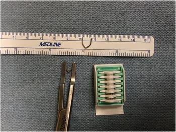
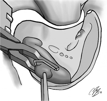
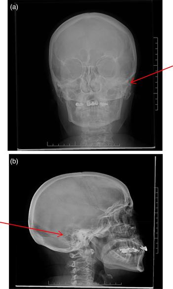
A large Weck Horizon clip (Teleflex Medical, Durham, North Carolina, USA) (Figure 1) is placed around the neck of the diverticulum, isolating the diverticulum from the sinus (Figure 2). The absence of turbulent flow within the diverticulum sac can be confirmed by Doppler ultrasonography at the end of the procedure. Confirmation of the clip(s) position can be achieved by standard lateral and anteroposterior skull X-rays (Figure 3).
Large Weck Horizon clip, used to ligate a sigmoid sinus diverticulum.

Illustration showing clipping of a left-sided sigmoid sinus diverticulum using a Weck Horizon clip.

Post-operative anteroposterior (a) and lateral (b) skull X-rays confirm the clip position (arrows).

Results
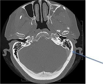
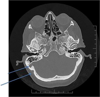
This procedure has been performed in two patients. There was an audible bruit identified behind the ear in both cases. Figures 4 and 5 show the pre-operative computed tomography (CT) scans of the patients. Clipping of the diverticulum was performed in these patients. Intra-operative Doppler ultrasonography was used for one patient. At six months’ follow up, both patients reported complete resolution of their pulsatile tinnitus with no complications.
Axial temporal bone computed tomography scan demonstrates a 6 × 4 mm venous out-pouching, projecting from transverse–sigmoid junction through the defect, into the left posterior mastoid air cells (arrow).

Axial temporal bone computed tomography scan shows a prominent bony canal (5 mm) extending from the right sigmoid sinus to the mastoid air cells (short arrow), and an enlarged occipitomastoid suture (long arrow).

Discussion
The premise of treating pulsatile tinnitus secondary to a sigmoid sinus diverticulum is to isolate the diverticulum from the sigmoid, to prevent turbulent blood flow within. This can be performed via transmastoid surgery.Reference Otto, Hudgins, Abdelkafy and Mattox3–Reference Harvey, Hertzano, Kelman and Eisenman5 Others have reported using an endovascular approach in order to coil the sac to promote thrombosis within, or to stent across the neck of the diverticulum to direct blood flow away.Reference Paramasivam, Furtado, Shigamatsu and Smouha6–Reference Shastri, Chaudhary, Pandey, Telian and Gemmete8 Various surgical techniques have been described, such as compression of the diverticulum using autologous material or reconstruction of the sinus wall.
Otto et al. described using either temporalis muscle and fascia or bone wax placed extraluminally to invaginate the sac.Reference Otto, Hudgins, Abdelkafy and Mattox3 Eisenman et al. performed the same approach, but used a soft-tissue graft of temporalis fascia or Neuro AlloDerm® placed between the dura and the posterior fossa bony plate, to reconstruct the soft-tissue sinus wall.Reference Eisenman4
Gologorsky et al. also reported open surgical treatment of the diverticulum.Reference Gologorsky, Meyer, Post, Winn, Patel and Bederson9 After coagulation and shrinkage of a diverticulum, they used a series of self-tying U clips along the sinus wall defect, to support the wall and prevent recurrence. We believe that the clipping of an aneurysm is a safe and effective alternative method of eliminating the diverticulum from circulation and improving pulsatile tinnitus.
Sinus thrombosis leading to increased intracranial pressure remains a potential risk with both transmastoid surgery and an endovascular approach; the risk may be greater if the diverticulum emanates from the dominant sinus.Reference Otto, Hudgins, Abdelkafy and Mattox3, Reference Eisenman4, Reference Paramasivam, Furtado, Shigamatsu and Smouha6, Reference Amans, Stout, Doed, Higashida, Hetts and Cook7 As some studies have observed a correlation between a sigmoid sinus diverticulum with pulsatile tinnitus and idiopathic intracranial hypertension by imaging, it would be prudent to consider the existence of both, especially in a symptomatic patient.Reference Liu, Dong, Wang, Han, Zhao and Lv10 For those pulsatile tinnitus patients with both idiopathic intracranial hypertension and a sigmoid sinus diverticulum, not only are surgical and endovascular treatments required for the sigmoid sinus diverticulum, but treatment of idiopathic intracranial hypertension is also necessary to improve the clinical outcome of other symptoms and to avoid recurrence.
Theoretically, the clipping of a sigmoid sinus diverticulum might minimise the risk of propagation of thrombogenic material, and lower the risk of thrombosis leading to subsequent intracranial hypertension from central venous obstruction in an involved dominant sigmoid sinus. However, long-term follow up and flow studies (magnetic resonance angiography or CT angiography) are needed to confirm this.
Conclusion
Transmastoid ligation of the neck of the sigmoid sinus diverticulum, using a Weck Horizon clip, is a safe and effective method of treating vascular pulsatile tinnitus.
Competing interests
None declared