National mastitis surveillance studies provide information on the current distribution of mastitis pathogens in a country. This information can be used for advising veterinarians and farmers and for tailoring mastitis prevention programmes towards the pathogen distribution. The prevalence of intramammary infection (IMI) was determined in The Netherlands in 1973, 1975, 1980 and in 1985 (Vecht et al. Reference Vecht, Wisselink and Defize1989). In the 1970s, Streptococcus agalactiae was the predominant mastitis pathogen, while in the 1980s Staphylococcus aureus became the most frequently isolated pathogen. After 1985, no comparable studies were carried out in The Netherlands. In Finland and Norway, however, large mastitis prevalence surveys were conducted recently (Pitkälä et al. Reference Pitkälä, Haveri, Pyörälä, Myllys and Honkanen-Buzalski2004; Østerås et al. Reference Østerås, Sølverød and Reksen2006). The results of the 2001 Finnish study were compared with studies performed in 1995 and 1988 (Myllys et al. Reference Myllys, Asplund, Brofeldt, Hirvelä-Koski, Honkanen-Buzalski, Junttila, Kulkas, Myllykangas, Niskanen, Saloniemi, Sandholm and Saranpää1998). The decrease in bulk milk SCC (BMSCC) from 1988 to 1995 was accompanied by a significant shift in pathogens. Although the average Finnish BMSCC remained stable from 1995 to 2001, distribution of pathogens changed considerably (Pitkälä et al. Reference Pitkälä, Haveri, Pyörälä, Myllys and Honkanen-Buzalski2004). Since 1985, average BMSCC in The Netherlands has decreased from 331 000 to 227 000 cells/ml in 2003. A decrease in prevalence of subclinical mastitis and a shift in pathogens involved in IMI can therefore be expected.
The objectives of this study were therefore: 1) to determine the overall and pathogen-specific prevalence of IMI in Dutch dairy herds and 2) to compare the distribution with earlier studies in The Netherlands. The implications of this for mastitis prevention programmes will be discussed.
Materials and Methods
Herds and cows
Similarly to earlier Dutch studies on prevalence of IMI (Vecht et al. Reference Vecht, Wisselink and Defize1989) herds were randomly selected stratified over the 12 Dutch provinces. The number of selected herds within the 12 provinces was based on the actual number of herds in each province. To be selected, herds had at least 40 lactating cows and participated in the Dutch milk recording system. To select a representative sample of the population with respect to the prevalence of major pathogens [given that approximately 25% of cows have a high SCC (HSCC); an accepted error of 5% and a CI of 95%] at least 400 culture-positive quarter milk samples with major pathogens from cows with HSCC were needed. The estimation was that 20% of HSCC samples would be culture-positive; therefore a total of 2000 milk samples from these cows was needed. Additionally, a subset of approximately 25% of cows with a low SCC (LSCC) was sampled to complete the description of the herds. To select a representative sample of the population we conservatively calculated that with a herd size of 40 lactating cows approximately 50 herds were needed. A total of 61 herds was selected and 49 agreed to participate in the study. Cows that were dried off, had clinical mastitis, were culled or treated with antibiotics in the meantime, were excluded from sampling. Samples were collected between April 2003 and September 2004, similarly to earlier Dutch studies (Vecht et al. Reference Vecht, Wisselink and Defize1989). All herds were visited by trained personnel of the GD Animal Health Service (GD) in Deventer, The Netherlands.
Milk samples were collected within 1 week after arrival of the results of DHI from all quarters of a selection of cows, based on cow SCC (CSCC) of the last milk recording. Two groups of cows were selected: 1) a HSCC group that consisted of all cows with a SCC ⩾250 000 cells/ml and all heifers with a SCC ⩾150 000 cells/ml, based on the currently used cut-off levels in The Netherlands used by the Dutch DHI, and 2) a LSCC group that consisted of approximately 25% randomly chosen cows and heifers with a SCC <250 000 cells/ml and <150 000 cells/ml, respectively. A cow was defined as being culture-positive if at least one quarter milk sample was culture-positive.
Bulk milk SCC (BMSCC) was measured once, approximately 2 weeks before the sampling of the individual cows. Farms were divided into three BMSCC categories for this last analysis (low, <150 000 cells/ml; medium, 150 000–250 000 cells/ml; and high, ⩾250 000 cells/ml).
Sampling
The selected cows were sampled during regular milking times. Pre-milking udder preparation with a cotton or paper towel was carried out by the farmer. Single quarter foremilk samples were collected aseptically according to the NMC protocol by trained personnel of the GD. Teat ends were disinfected with a cotton swab soaked in methylated alcohol, and after the third stripping, milk was collected. Milk samples were transported on ice to the GD for bacteriology and determination of SCC.
Laboratory analyses
The samples were split and SCC was determined using a Fossomatic cell counter (Foss Electric, Hillerød, Denmark). Bacteriological culturing was carried out according to NMC protocols (Harmon et al. Reference Harmon, Eberhart, Jasper, Langlois and Wilson1990). An inoculum of 0·01 ml of milk was spread on a 6% sheep blood agar plate and on Edward's medium (Biotrading, The Netherlands). Edward's medium was used for selective isolation of streptococci combined with morphology, haemolytic properties, CAMP reaction and hydrolysis of aesculin. Streptococci were grouped according to the Lancefield groups B, C, D and G. Str. dysgalactiae and Str. agalactiae were identified using Lancefield media (Oxoid, Basingstoke, UK) with Str. dysgalactiae defined as positive for group C and Str. agalactiae as positive for group B. Str. uberis were further identified by a negative reaction with Lancefield group D antisera and no growth in Tryptose Phosphate Broth with 6·5% NaCl (Oxoid). All plates were incubated at 37°C and examined after 24 h and 48 h. Isolates of staphylococci showing α, α and β, or β haemolysis were presumptively identified as Staph. aureus. Coagulase-negative staphylococci were distinguished from Staph. aureus using Slidex Staph Plus (bioMérieux, France) agglutination test. Sensitivity to β-lactam antimicrobials such as penicillin was tested on all isolated strains of Staph. aureus with nitrocephin (Oxoid, SR0112C, Haarlem, The Netherlands). Strains that were β-lactamase-positive were considered to be resistant to penicillin. Escherichia coli found on the blood agar plate were confirmed with BBL Crystal ID system. Identification of Bacillus spp. was based on morphology of the colonies on blood agar plates and on the morphology of the cells as large endospore-forming rods after Gram staining. Identification to species level was not carried out. Identification of Corynebacterium bovis was based on morphology of the colonies on blood agar plates.
A milk sample was considered culture-positive when ⩾100 cfu/ml of a major pathogen (i.e. Staph. aureus, Str. uberis, Str. agalactiae, Str. dysgalactiae and Esch. coli) or ⩾500 cfu/ml of a minor pathogen (i.e. CNS, Bacillus spp. and C. bovis) were cultured. Milk samples that were culture-positive for more than two bacterial species were considered to be contaminated.
Statistical analyses
Differences in quarter SCC between the HSCC group and the LSCC group were analysed with the two-sample t test after natural logarithmic transformation of SCC. Differences in age and DIM between the HSCC group and the LSCC group were analysed with the median test. Differences in prevalence of pathogen-specific IMI between the two SCC groups, among parities, and among BMSCC categories were evaluated using chi-square analysis on contingency tables (Rothman & Greenland, Reference Rothman and Greenland1998). These statistical analyses were performed using Statistix 8.0 (Analytical Software, Tallahassee FL, USA). Statistical significance was defined at P<0·05.
Pathogen-specific prevalence of IMI and 95% CI were calculated on quarter and cow-level for the HSCC and LSCC group separately. To determine herd-level and national prevalence, the number of sampled HSCC and LSCC animals were adjusted for the total number of HSCC and LSCC animals per herd. The national prevalence was calculated by summing the herd results. Additionally, a correction was made for the fact that cows within a herd were more correlated to each other than random cows by entering the herd number as repeated measurement (random effect) in the logistic regression procedure in STATA (Version 9, 2005). Pathogen-specific IMI prevalence was compared among parities; prevalences were calculated per parity in this same data-set by means of a regression analysis. In these analyses, culture results were included as dependent variables, parity was included as independent variable and a correction was made for the repeated measurements within a herd.
Additionally, the national pathogen-specific quarter IMI prevalences that were determined in this study were compared with the prevalences found in the studies of Vecht et al. (Reference Vecht, Wisselink and Defize1989). The distribution in time of the pathogen-specific prevalence of IMI was tested using logistic regression in STATA with positive or negative quarter as dependent variable and year as independent variable. A frequency weight was added to the model to correct for the number of positive quarters sampled and number of negative quarters sampled in each of the studies. In this analysis the correction for clustering within herds was excluded. In this way our results could be compared with those of Vecht et al. (Reference Vecht, Wisselink and Defize1989), using the same methods of analysis used by those authors.
Results
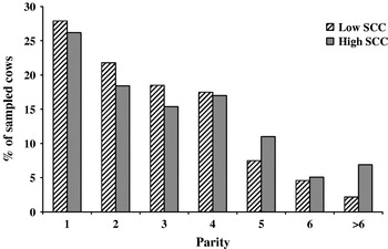
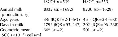
In the 49 herds, 22·2% (95% CI, 20·7–23·7%) of 2994 lactating cows had HSCC at the last milk test-day before sampling (Table 1). Quarter milk samples were collected from 553 of these 665 HSCC cows. The remaining 112 HSCC cows were either dry at the moment of sampling, had clinical mastitis, or were culled between the last milk test-day and the sampling date. Additionally, quarter milk samples were collected from 519 of 2329 LSCC cows. HSCC cows were older than LSCC cows (Table 1). In the LSCC group, relatively more cows had a parity <4, while in the HSCC group relatively more cows had parity >4 (Fig. 1).
Parity distribution of the 519 sampled low somatic cell count (at last milk test-day before sampling: heifers SCC <150 000 and cows SCC <250 000 cells/ml) and 553 sampled high somatic cell count cows.

Characteristics of cows with a low somatic cell count (LSCC) and cows with a high somatic cell count (HSCC) included in the study
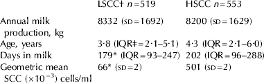
† LSCC at last milk test-day before sampling: heifers with SCC <150 000 and cows SCC <250 000 cells/ml
‡ Interquartile Range
* Different (P<0·05) from HSCC
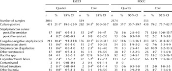
Bacterial growth occurred in 37·7% of milk samples from HSCC cows compared with 21·1% in milk samples from LSCC cows (Table 2; P<0·0001). Coagulase-negative staphylococci (CNS) were the most frequently isolated group of bacteria in both the HSCC and the LSCC group, 14·6 and 10·4% of quarters, respectively, while at cow-level the prevalence was 38·5 and 30·1%, respectively. All major and minor pathogens were more frequently isolated in the HSCC group than in the LSCC group. Prevalence of contaminated samples was 0·05%.
Quarter and cow-level distribution of mastitis pathogens from cows with a low somatic cell count (LSCC) and from cows with a high somatic cell count (HSCC)

† LSCC at last milk test-day before sampling: heifers with SCC <150 000 and cows SCC <250 000 cells/ml
a Different (P<0·05) from HSCC quarters
b Different (P<0·05) from HSCC cows
SCC was not different between major pathogen-positive quarters of HSCC and LSCC cows. However, SCC of culture-negative (97 000 v. 38 000 cells/ml), CNS-positive (163 000 v. 60 000 cells/ml), Bacillus-positive (64 000 v. 31 000 cells/ml) and quarters positive with other streptococci (109 000 v. 21 000 cells/ml) was higher in HSCC cows than in LSCC cows (P<0·05).
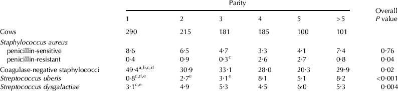
Pathogen distribution differed considerably among parity groups (Table 3). Prevalence of CNS IMI was highest in first parity heifers (P<0·05). Prevalence of Str. uberis, Str. dysgalactiae and penicillin-resistant Staph. aureus IMI increased with increasing parity (P<0·05). The association was, however, not completely linear. The prevalence of penicillin-sensitive Staph. aureus IMI was not associated with parity.
Cow-level prevalence of the most important mastitis pathogens per parity
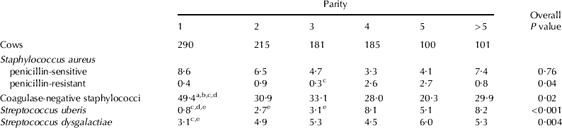
a Different (P<0·05) from parity 2
b Different (P<0·05) from parity 3
c Different (P<0·05) from parity 4
d Different (P<0·05) from parity 5
e Different (P<0·05) from parity >5
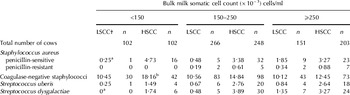
CNS were found in all herds, while penicillin-sensitive Staph. aureus was isolated in 73·5% of the herds. Penicillin-resistant Staph. aureus was found in 9 herds (18%), Str. uberis and Str. dysgalactiae were cultured in samples of >50% of the herds in this study, while Str. agalactiae was not found in any of the milk samples cultured.
A lower proportion of HSCC cows was culture-positive for CNS in the high-BMSCC group (13 farms) compared with the low-BMSCC group (19 farms) (Table 4). The proportion of LSCC cows that was Str. dysgalactiae or penicillin-sensitive Staph. aureus-positive was higher in high- than in low-BMSCC herds.
Prevalence of the most important mastitis pathogens in milk samples from low (LSCC) and high somatic cell count (HSCC) cows in three bulk milk somatic cell count categories

† LSCC at last milk test-day before sampling: heifers with SCC <150 000 and cows SCC <250 000 cells/ml
a Different (P<0·05) from BMSCC ⩾250 000 cells/ml (LSCC)
b Different (P<0·05) from BMSCC ⩾250 000 cells/ml (HSCC)
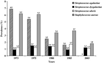
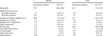
The overall national quarter and cow-level prevalence of pathogen-specific IMI corrected for clustering within herds is presented in Table 5. Overall, quarter-level prevalence of Str. agalactiae and Staph. aureus IMI was lower in 2003 than in 1973, 0 v. 6·9% and 1·8 v. 6·2%, respectively (Fig. 2; P<0·0001). Quarter-level prevalence of Str. uberis and Str. dysgalactiae IMI were more stable at 1·1–1·7 and 0·9–1·5%, respectively.
Dutch national quarter-level prevalence of intramammary infection with the most important major mastitis pathogens from 1973 to 2003.

National quarter and cow-level prevalence of mastitis pathogens

Discussion
Over the years, several national mastitis surveys were performed in The Netherlands. The first one was conducted in 1973, and surveys were repeated in 1975, 1980 and in 1985 (Vecht et al. Reference Vecht, Wisselink and Defize1989). The selection strategy used was a random stratified sampling designed to achieve a representative distribution across The Netherlands for herd size and location. Together, the results of these studies offer a unique opportunity to study the prevalence of IMI in dairy cows in The Netherlands over a prolonged period of time. The studies conducted in UK, Finnish and Norwegian herds (Pitkälä et al. Reference Pitkälä, Haveri, Pyörälä, Myllys and Honkanen-Buzalski2004; Østerås et al. Reference Østerås, Sølverød and Reksen2006; Bradley et al. Reference Bradley, Leach, Breen, Green and Green2007) were also true national surveillance studies. In all these studies, herds were randomly selected from the national database and in that respect were comparable to our study. The herds included in a study in New York and Pennsylvania (Wilson et al. Reference Wilson, Gonzalez and Das1997) were not randomly selected, with many herds with mastitis problems included. In a study conducted in Germany in Brandenburg (Tenhagen et al. Reference Tenhagen, Koster, Wallmann and Heuwieser2006) farms were also not randomly selected. Thus, neither the USA study nor the study conducted in Germany is comparable to our study.
The milk samples taken in our study were collected by trained personnel of the GD. This procedure may explain the low number of contaminated milk samples (0·05%) which was comparable to the study of Myllys et al. (Reference Myllys, Asplund, Brofeldt, Hirvelä-Koski, Honkanen-Buzalski, Junttila, Kulkas, Myllykangas, Niskanen, Saloniemi, Sandholm and Saranpää1998). In other studies (Makovec & Ruegg, Reference Makovec and Ruegg2003; Pitkälä et al. Reference Pitkälä, Haveri, Pyörälä, Myllys and Honkanen-Buzalski2004; Østerås et al. Reference Østerås, Sølverød and Reksen2006) in which milk samples were collected by farmers, dairy advisors and veterinarians, respectively 15·3, 4·2 and 6·9% of the samples were contaminated. In the study of Bradley et al. (Reference Bradley, Leach, Breen, Green and Green2007), however, 0·2% of the samples collected by farmers were contaminated. Contamination of samples can be avoided by proper sampling techniques.
In our study, all quarters of lactating cows with an individual SCC ⩾250 000 cells/ml, and all quarters of heifers with an individual SCC ⩾150 000 cells/ml were sampled, as were a subset (approximately 25%) of the lactating cows and heifers with SCC below these thresholds. Ideally all lactating animals in the participating herds are sampled as was done in the earlier Dutch studies (Vecht et al. Reference Vecht, Wisselink and Defize1989) and in some other international studies (Gonzalez et al. Reference Gonzalez, Jasper, Farver, Bushnell and Franti1988; Pitkälä et al. Reference Pitkälä, Haveri, Pyörälä, Myllys and Honkanen-Buzalski2004). However, by selecting cows we were able to reduce costs of the survey, as was done in other studies in Norway and in England and Wales (Østerås et al. Reference Østerås, Sølverød and Reksen2006; Bradley et al. Reference Bradley, Leach, Breen, Green and Green2007). We chose a stratified sampling approach over a random selection independent of SCC of cows. In this study, we have determined the overall and pathogen-specific prevalence of IMI, while in another study the possible risk factors for IMI were reported (Sampimon et al. Reference Sampimon, Barkema, Berends, Sol and Lam2008).
CNS were the most frequently isolated species followed by Bacillus spp. and by Staph. aureus and Str. dysgalactiae of the major pathogens. In the study of Vecht et al. (Reference Vecht, Wisselink and Defize1989) CNS was not individually mentioned. In that study CNS were included in the group of ‘other bacteria’. CNS were the most frequently isolated group of organisms isolated in almost all recent mastitis surveys (Pitkälä et al. Reference Pitkälä, Haveri, Pyörälä, Myllys and Honkanen-Buzalski2004; Bradley et al. Reference Bradley, Leach, Breen, Green and Green2007). It is not clear, however, whether IMI with CNS always result in inflammation. Challenge studies would be necessary to find that out. In Norway (Østerås et al. Reference Østerås, Sølverød and Reksen2006) not CNS but Staph. aureus was the most frequently isolated pathogen. Prevalence of CNS IMI was remarkably lower in Norway than in Finland, 3·3% compared with 16·6%. In the Finnish study and in our study, a sample was considered CNS-positive when ⩾500 cfu/ml was found, while in the Norwegian study a sample was considered positive when ⩾4000 cfu/ml was found. Therefore, the differences in prevalence of CNS IMI between these studies can be partly explained by differences in definition of a positive culture for CNS. International agreement on the definition of IMI would make studies more comparable. When comparing results of clinical trials and other studies, selection criteria for herds and animals, sampling techniques and laboratory procedures may be different (Barkema et al. Reference Barkema, Schukken and Zadoks2006). The definition of a positive sample is just one of them.
Based on earlier work in The Netherlands, we expected to find a higher prevalence of Esch. coli IMI (Lam et al. Reference Lam, Lipman, Schukken, Gaastra and Brand1996). However, only two Esch. coli positive samples were found. Because of this low number, they were categorized as ‘other bacteria’. Esch. coli IMI mainly occur during the dry and in early lactation (Bradley & Green, Reference Bradley and Green2000). Our study, however, was a prevalence study, which was not specifically aimed at these periods and cows were sampled in all stages of lactation. In our study, the number of culture-negative samples was higher in the LSCC group than in the HSCC group.
Prevalence of Bacillus spp. was, at 4·2%, relatively high. Bacillus spp. was found with equal frequency in the HSCC and LSCC groups (Table 2). Prevalence was higher than the 0·8–1·9% found in other studies (Wilson et al. Reference Wilson, Gonzalez and Das1997; Bradley et al. Reference Bradley, Leach, Breen, Green and Green2007) but it was comparable with results of samples submitted to our GD laboratory. In 2003 and 2004 the prevalence of Bacillus spp. was 3·3 and 3·5%, respectively in more than 50 000 milk samples sent to the GD laboratory on a yearly base of cows with a high SCC. It is not likely that the high prevalence of Bacillus spp. is due to contamination. In both, prevalence study and routine, we found pure cultures of Bacillus spp. Additionally, contamination of samples with Bacillus spp. is not likely to have played an important role in this study because our sampling procedures were very strict, as is illustrated by the very low prevalence of contaminated samples. Finally, the effect of Bacillus spp. on udder health seems to be low: in this study 68% of the samples containing Bacillus spp. had SCC <100 000 cells/ml.
Staph. aureus was the most frequently isolated major pathogen in quarters of LSCC cows (1%). Streptococci were also found in quarters of LSCC cows. These findings agree with Schukken et al. (Reference Schukken, Wilson, Welcome, Tikofsky and Gonzalez2003). They also found a low number of quarters with a major pathogen IMI with quarter SCC between 96 000 and 250 000 cells/ml. This means cows with SCC below the cut off values can have IMI with major pathogens. Monitoring cow SCC on a regular basis will give early insight into development of these major pathogen IMI. However, as was found in our study, cows with HSCC are more likely to harbour major pathogens.
Mean quarter SCC in quarters with a major pathogen IMI did not differ between the HSCC and LSCC groups. Culture-negative quarters of LSCC cows had on average a lower SCC than culture-negative quarters of HSCC cows. One explanation for this phenomenon is that milk samples of subclinically infected cows can be cultured false-negative as a result of the shedding pattern of Staph. aureus (Sears et al. Reference Sears, Smith, English, Herer and Gonzalez1990) or other pathogens. Another explanation may be that quarters of HSCC cows have been challenged more often by major pathogens than quarters of LSCC cows.
Distribution of mastitis pathogens varied considerably among parities. CNS were significantly more frequently isolated in heifers than in older cows, as was found by Tenhagen et al. (Reference Tenhagen, Koster, Wallmann and Heuwieser2006). Prevalence of Str. uberis, Str. dysgalactiae and penicillin-resistant Staph. aureus IMI was (although the association was not linear) higher in cows with higher parity, comparable to what was found in the German study (Tenhagen et al. Reference Tenhagen, Koster, Wallmann and Heuwieser2006). Penicillin-resistant Staph. aureus was also more prevalent in older cows, as was found by Sol et al. (Reference Sol2002). In that study older cows had a lower cure rate after treatment of subclinical mastitis cases with antibiotics at drying-off and during lactation, as well as in clinical cases. In another Dutch study, Zadoks et al. (Reference Zadoks, Allore, Barkema, Sampimon, Wellenberg, Grohn and Schukken2001) found a higher incidence of new Staph. aureus and Str. uberis IMI in third and higher parity cows than in first and second parity cows. The higher prevalence of Staph. aureus and Str. uberis IMI in cows with higher parity may therefore be a result of both, a higher incidence of new IMI and selection of IMI that are less sensitive to antimicrobial treatment. Because cure rates of IMI decreases with duration, to increase treatment success, early detection of IMI with Staph. aureus and Str. uberis is important (Barkema et al. Reference Barkema, Schukken and Zadoks2006).
Distribution of mastitis pathogens varied among herds. CNS were present in all herds, as was the case in the study conducted in Germany (Tenhagen et al. Reference Tenhagen, Koster, Wallmann and Heuwieser2006). In our study, CNS were more frequently isolated from cows with HSCC on farms with a low BMSCC than on farms with a high BMSCC. Harmon & Langlois (Reference Harmon and Langlois1995) mentioned that CNS were the most prevalent organisms isolated from milk samples in herds using effective mastitis control procedures as reflected in the reduction of major pathogens in the herd. Instead of CNS, Staph. aureus and Str. dysgalactiae were more frequently isolated in milk samples of LSCC cows on farms with a high BMSCC. Farms with a high prevalence of mastitis pathogens can have cows with SCC below the cut-off values but still have quarters containing major pathogen IMI. Similarly to what was found in other studies, quarters with a low SCC can have major pathogens present (Harmon, Reference Harmon1994). In contrast with penicillin-sensitive Staph. aureus, penicillin-resistant Staph. aureus was not found on the 19 farms with a BMSCC <150 000 cells/ml. It may be hypothesized that penicillin-resistant Staph. aureus is associated with high BMSCC.
Prevalence of IMI with the contagious pathogens Staph. aureus and Str. agalactiae has decreased considerably in the last 30 years. This decrease in The Netherlands is similar to what is found in other countries (Pitkälä et al. Reference Pitkälä, Haveri, Pyörälä, Myllys and Honkanen-Buzalski2004; Bradley et al. Reference Bradley, Leach, Breen, Green and Green2007) and occurred at the same time as a decrease in BMSCC, mainly due to the introduction of a penalty-system for high BMSCC (Nightingale et al. Reference Nightingale, Dhuyvetter, Mitchell and Schukken2008). In another Dutch study (Barkema et al. Reference Barkema, Schukken, Lam, Beiboer, Benedictus and Brand1998) adoption of the 5-point contagious mastitis prevention and control plan was significantly better in low-BMSCC herds compared with herds having a high BMSCC. The decrease in BMSCC is therefore most likely the result of an increased adoption of the 5-point contagious mastitis prevention and control programme (Neave et al. Reference Neave, Dodd, Kingwill and Westgarth1969). Farmers became more aware of the necessity to treat or cull cows to avoid BMSCC penalties (Sol, Reference Sol2002). This may also be the explanation for the absence of Str. agalactiae in our study, and the absence of penicillin-resistant Staph. aureus in the low-BMSCC group. The same explanation was also used in Finland to explain the decrease of IMI prevalence from 38% in 1995 to 31% in 2001 (Pitkälä et al. Reference Pitkälä, Haveri, Pyörälä, Myllys and Honkanen-Buzalski2004). Sol (Reference Sol2002) also found a decreased prevalence of Staph. aureus and Str. agalactiae IMI and an increase of Str. uberis IMI between 1975 and 2000 in milk samples submitted to the GD. However, milk samples submitted to the GD were mainly samples of cows with an elevated SCC; therefore these results are less comparable with the survey of Vecht et al. (Reference Vecht, Wisselink and Defize1989) and our current study.
Apparently, the changed circumstances did not have a similar effect on the prevalence of Str. uberis and Str. dysgalactiae IMI. No obvious explanation is available for the Str. dysgalactiae observations. The epidemiology of this pathogen, based on field studies behaves intermediate between contagious and environmental. In the study of Neave et al. (Reference Neave, Dodd, Kingwill and Westgarth1969) the 5-point contagious mastitis programme worked well for Str. agalactiae, less well for Str. uberis and was intermediate for Str. dysgalactiae. Str. uberis behaves in many situations more like an environmental pathogen being associated with nutrition and housing and in other situations as a contagious pathogen (Barkema et al. Reference Barkema, Schukken, Lam, Beiboer, Wilmink, Benedictus and Brand1999; Zadoks et al. Reference Zadoks, Gillespie, Barkema, Sampimon, Oliver and Schukken2003). These streptococci seem to be less sensitive to changes in udder health management made over the last few decades. More research is needed to provide tools to control IMI with Str. dysgalactiae and Str. uberis. Although these pathogens may share some risk factors. The incidence of Str. uberis IMI and clinical mastitis is highest when cows are on pasture, while the incidence of Str. dysgalactiae IMI and clinical mastitis is highest in winter, as was described in Norway and Canada (Østerås et al. Reference Østerås, Sølverød and Reksen2006; Olde Riekerink et al. Reference Olde Riekerink, Barkema and Stryhn2007). When it is known which factors increase the incidence of IMI with these pathogens, the control programme for Str. uberis and Str. dysgalactiae IMI may need to be adjusted. In addition to these streptococci, the importance of CNS in mastitis control programmes also needs further investigation.
Conclusions
This was the first true random prevalence study on IMI in The Netherlands since the survey of 1985. CNS were the most frequently isolated pathogen in our study. The importance of CNS in mastitis control programmes needs further investigation. Prevalence of Staph. aureus and Str. agalactiae IMI is much lower now than it was in the 1970s and 1980s. The combined prevalence of Str. uberis and Str. dysgalactiae IMI, however, seems to have hardly decreased during this period. These non-agalactiae streptococci seem to have become relatively more important in The Netherlands.
Regular sampling of quarter milk samples of HSCC cows in a dairy herd is necessary to evaluate the distribution of pathogens on a herd level. Additionally it is very informative to conduct national mastitis prevalence studies on a regular basis because, also at this level, the distribution of pathogens changes over time. Mastitis control programmes can be adjusted accordingly.
This study is part of the five year mastitis programme of the Dutch Udder Health Centre and was financially supported by the Dutch Dairy Board. The authors express their appreciation to Dr A Rothkamp for technical laboratory assistance during this project.