DEVELOPMENT OF WHITE MATTER IN HUMANS – HISTOLOGICAL DATA
Myelinated axons are a principal constituent of the white matter of the nervous systems of almost all vertebrates and are also found in some invertebrate taxa (Schweigreiter et al., Reference Schweigreiter, Roots, Bandtlow and Gould2006; Hartline and Colman, Reference Hartline and Colman2007). The process of myelination may be more or less extended in time and its detailed spatiotemporal characteristics show considerable variability between species (Nadon et al., Reference Nadon, Crotzer and Stewart1995; Hamano et al., Reference Hamano, Takeya, Iwasaki, Nakayama, Ohto and Okada1998; Nunez et al., Reference Nunez, Nelson, Pych, Kim and Juraska2000).
A classical method to study myelination is to use some type of myelin-specific staining of histological sections, e.g. Weigert hematoxylin or Fast Blue staining (Kiernan, Reference Kiernan2007). Since the pioneering work of Paul Flechsig (Reference Flechsig1920), such techniques have been employed in a number of human postmortem studies. These studies have established several basic characteristics of the myelination process (Kinney et al., Reference Kinney, Brody, Kloman and Gilles1988). The most general observations are that while myelination is rapid during the first years of infancy, human myelination is a process that takes place over many years, and that the timing of myelination differs widely between brain regions. These regional differences are seen both for the onset time, the rate and the termination of myelination (Lucas Keene and Hewer, Reference Lucas Keene and Hewer1931; Yakovlev and Lecours, Reference Yakovlev, Lecours and Minkowski1967; Gilles et al., Reference Gilles, Shankle, Dooling, Gilles, Leviton and Dooling1983; Brody et al., Reference Brody, Kinney, Kloman and Gilles1987; Kinney et al., Reference Kinney, Brody, Kloman and Gilles1988). Studies on larger samples indicate that there are also substantial individual differences in the timing of myelination in the same brain region (Gilles et al., Reference Gilles, Shankle, Dooling, Gilles, Leviton and Dooling1983). Concerning the specific temporal order of myelination of different brain regions, a few principles have been observed. While several exceptions can be found, there is an overall tendency for caudal regions of the central nervous system to myelinate earlier than rostral regions. Several spinal cord and brain stem structures thus finish their myelination during the first year, or even prenatally (Yakovlev and Lecours, Reference Yakovlev, Lecours and Minkowski1967; Brody et al., Reference Brody, Kinney, Kloman and Gilles1987), while, for example, the association cortex of the forebrain appears to continue its myelination into adulthood (Yakovlev and Lecours, Reference Yakovlev, Lecours and Minkowski1967; Benes, Reference Benes1989). Projection pathways typically myelinate before association pathways, and proximal regions tend to myelinate before distal regions (Kinney et al., Reference Kinney, Brody, Kloman and Gilles1988). Within the telencephalon myelination proceeds from the region of central sulcus towards the poles (Kinney et al., Reference Kinney, Brody, Kloman and Gilles1988). This general picture of the human myelination process is consistent with neurochemical and immunohistochemical studies, where the sequential appearance of specific myelin-associated lipids and proteins during myelination is characterized (see e.g. Kinney et al., Reference Kinney, Karthigasan, Borenshteyn, Flax and Kirschner1994; Haynes et al., Reference Haynes, Borenstein, Desilva, Folkerth, Liu and Volpe2005).
Importantly, studies of the microstructure of white matter have shown that the development of myelination is accompanied by a growth in thickness of the axons themselves. For example in the pyramidal tract a dramatic increase in the diameter of the thickest axons occurs during the period from birth to early adolescence (Verhaart, Reference Verhaart1950; Nathan and Smith, Reference Nathan and Smith1955; Eyre et al., Reference Eyre, Miller and Ramesh1991). One important consequence of this is that the conduction velocity increases so that the conduction delay is kept approximately constant in spite of body growth (Eyre et al., Reference Eyre, Miller and Ramesh1991). During development, the growth of the myelin sheath and the growth of the axon are carefully coordinated, to produce a nerve fiber where the ratio between axonal diameter and outer diameter of the myelin sheath (g-ratio) is optimal for maximal conduction velocity (Smith and Koles, Reference Smith and Koles1970; Friede and Bischhausen, Reference Friede and Bischhausen1982; Michailov et al., Reference Michailov, Sereda, Brinkmann, Fischer, Haug and Birchmeier2004).
NEUROIMAGING STUDIES OF HUMAN WHITE MATTER DEVELOPMENT
Histological studies thus show that white matter development, including myelination and axonal growth, continues into adulthood (Yakovlev and Lecours, Reference Yakovlev, Lecours and Minkowski1967; Benes, Reference Benes1989), but the majority of these studies have focussed on the prenatal and infant period. In the last decade, research on white matter development has been revolutionized by the appearance of a number of magnetic resonance (MR) imaging techniques that allow investigations on healthy individuals in vivo and the use of longitudinal designs. Several different such techniques have been employed for the study of white matter. The first developmental MR studies provided evaluations of changes in signal intensities in conventional T1- and T2-weighted MR images as a function of age (Barkovich et al., Reference Barkovich, Kjos, Jackson and Norman1988; Martin et al., Reference Martin, Kikinis, Zuerrer, Boesch, Briner and Kewitz1988; van der Knaap and Valk, Reference van der Knaap and Valk1990; for a review see Paus et al., Reference Paus, Collins, Evans, Leonard, Pike and Zijdenbos2001). In line with the postmortem literature, these studies demonstrated that T1 and T2 relaxation times in white matter change dramatically during the first 2–3 years of life, presumably reflecting the rapid myelination during infancy. After this age a pattern of MR intensities that qualitatively resembles the adult stage is seen.
MR studies of late myelination have benefited from the development of more sensitive, quantitative methods to analyze conventional MR images as well as entirely new MR techniques. These developments include, first, studies of regional and global white matter volume or density. Such studies can employ either manual delineation of anatomical structures or procedures involving automated segmentation of white and gray matter, such as voxel-based morphometry (Good et al., Reference Good, Johnsrude, Ashburner, Henson, Friston and Frackowiak2001). This work has confirmed the protracted nature of human white matter development. During the first years of infancy, increases in white matter volume are slow compared to increases in gray matter volume (Knickmeyer et al., Reference Knickmeyer, Gouttard, Kang, Evans, Wilber and Smith2008). Giedd et al. (Reference Giedd, Blumenthal, Jeffries, Castellanos, Liu and Zijdenbos1999) found a linear increase of white matter volume with age in all lobes throughout the studied age range (4–22 years), in study on a large sample (n = 145) using a mixed cross-sectional and longitudinal design. These results are in line with cross-sectional studies on the development of total white matter volume (Pfefferbaum et al., Reference Pfefferbaum, Mathalon, Sullivan, Rawles, Zipursky and Lim1994; Reiss et al., Reference Reiss, Abrams, Singer, Ross and Denckla1996; Iwasaki et al., Reference Iwasaki, Hamano, Okada, Horigome, Nakayama and Takeya1997) and white matter density in corticospinal and frontotemporal pathways (Paus et al., Reference Paus, Zijdenbos, Worsley, Collins, Blumenthal and Giedd1999). The increase in white matter volume during adolescence is seen in both sexes but is steeper in males (Perrin et al., Reference Perrin, Leonard, Perron, Pike, Pitiot and Richer2009). Studies on older adults indicate no substantial decrease in total white matter volume before age 60 (Good et al., Reference Good, Johnsrude, Ashburner, Henson, Friston and Frackowiak2001).
Another MR technique of relevance for white matter studies is magnetization transfer imaging (MTI), which relies on interaction between proton magnetization in macromolecular structures (e.g. myelin) and in free water (Wolff and Balaban, Reference Wolff and Balaban1989; Henkelman et al., Reference Henkelman, Stanisz and Graham2001). A common MTI measure is the magnetization transfer ratio (MTR). MTR is higher in white matter than in gray matter and modeling of data from MTI of the optic nerve indicates that it largely reflects the degree of myelination of the white matter (Stanisz et al., Reference Stanisz, Kecojevic, Bronskill and Henkelman1999). A few studies have reported increasing MTR values in human white matter during childhood and adolescence, with the most rapid increases taking place during infancy (Engelbrecht et al., Reference Engelbrecht, Rassek, Preiss, Wald and Modder1998; van Buchem et al., Reference van Buchem, Steens, Vrooman, Zwinderman, McGowan and Rassek2001).
Finally, the development of white matter can be studied with diffusion tensor imaging (DTI). DTI is an MR technique that measures the local magnitude and direction of water diffusion (Le Bihan, Reference Le Bihan2003). The presence of barriers such as axonal membranes and myelin sheaths will make the diffusion anisotropic, in the case of white matter more rapid along the nerve fibers than perpendicular to them. DTI has proven particularly sensitive to the more subtle developmental changes in white matter occurring after infancy (Lobel et al., Reference Lobel, Sedlacik, Gullmar, Kaiser, Reichenbach and Mentzel2009). Different DTI parameters can be used to analyze white matter microstructure. Among the more commonly used are the apparent diffusion coefficient (ADC), which is a measure of overall diffusion, and the fractional anisotropy (FA), which reflects the degree of anisotropy. Importantly, DTI studies have shown that both ADC and FA continue to change with age also during adolescence and early adulthood. FA and other measures of anisotropy increase with age in widespread white matter regions (Ben Bashat et al., Reference Ben Bashat, Ben Sira, Graif, Pianka, Hendler and Cohen2002; Engelbrecht et al., Reference Engelbrecht, Scherer, Rassek, Witsack and Modder2002; McGraw et al., Reference McGraw, Liang and Provenzale2002; Zhai et al., Reference Zhai, Lin, Wilber, Gerig and Gilmore2003; Barnea-Goraly et al., Reference Barnea-Goraly, Menon, Eckert, Tamm, Bammer and Karchemskiy2005; Lobel et al., Reference Lobel, Sedlacik, Gullmar, Kaiser, Reichenbach and Mentzel2009). ADC, in contrast, decreases with age (Engelbrecht et al., Reference Engelbrecht, Scherer, Rassek, Witsack and Modder2002; Zhai et al., Reference Zhai, Lin, Wilber, Gerig and Gilmore2003; Lobel et al., Reference Lobel, Sedlacik, Gullmar, Kaiser, Reichenbach and Mentzel2009). The increase in FA is presumably dependent on a number of factors apart from increased myelination, e.g. changes in axonal diameters, packing density and distribution of axonal macromolecules (Beaulieu, Reference Beaulieu2002; Lobel et al., Reference Lobel, Sedlacik, Gullmar, Kaiser, Reichenbach and Mentzel2009). The reduction in ADC with age probably reflects an overall decrease in white matter water content, which in turn may be related to myelination (Lobel et al., Reference Lobel, Sedlacik, Gullmar, Kaiser, Reichenbach and Mentzel2009).
CORRELATIONS BETWEEN WHITE MATTER DEVELOPMENT AND FUNCTION
The developmental changes in white matter structure discussed above parallel the development of function (Wozniak and Lim, Reference Wozniak and Lim2006; Giedd, Reference Giedd2008; Marsh et al., Reference Marsh, Gerber and Peterson2008). DTI, with its high sensitivity for differences in white matter microstructure, has been particularly useful for elucidating relationships between function and structure. Prefrontal increases in FA and white matter density parallel the development of higher cognitive functions such as general intelligence, working memory, attention and inhibition (Olesen et al., Reference Olesen, Nagy, Westerberg and Klingberg2003; Barnea-Goraly et al., Reference Barnea-Goraly, Menon, Eckert, Tamm, Bammer and Karchemskiy2005; Schmithorst et al., Reference Schmithorst, Wilke, Dardzinski and Holland2005). Increases in FA in the corpus callosum may reflect the development of cognitive functioning as well as sensorimotor control (Barnea-Goraly et al., Reference Barnea-Goraly, Menon, Eckert, Tamm, Bammer and Karchemskiy2005; Hasan et al., Reference Hasan, Karnali, Kramer, Papnicolaou, Fletcher and Ewing-Cobbs2008) and have recently been shown to be correlated with bimanual coordination ability (Muetzel et al., Reference Muetzel, Collins, Mueller, Schissel, Lim and Luciana2008). The gradual development of the pyramidal tracts and arcuate fasciculus parallels the development of manual fine motor skills and language (Paus et al., Reference Paus, Zijdenbos, Worsley, Collins, Blumenthal and Giedd1999).
Relations between individual differences in behavior and white matter structure are also found independently of age. General intelligence is related to higher FA (Schmithorst et al., Reference Schmithorst, Wilke, Dardzinski and Holland2005) as well as larger volume (Haier et al., Reference Haier, Jung, Yeo, Head and Alkire2004; Ullén et al., Reference Ullén, Forsman, Blom, Karabanov and Madison2008) in widespread white matter regions, including fronto-parietal association bundles. We have recently found that prefrontal white matter volume is positively related both to cognitive ability and millisecond stability in a simple, automatic timing task (Ullén et al., Reference Ullén, Forsman, Blom, Karabanov and Madison2008). A possible link between white matter structure and cognitive performance may thus be that white matter connectivity influences millisecond accuracy of neuronal activity, which in turn affects higher cognitive functions (Ullén et al., Reference Ullén, Forsman, Blom, Karabanov and Madison2008; Madison et al., Reference Madison, Forsman, Blom, Karabanov and Ullén2009). Other studies have demonstrated positive relations between FA in different white matter regions and performance in a wide range of tasks. Among demonstrated relations are: temporo-parietal FA and reading ability (Klingberg et al., Reference Klingberg, Hedehus, Temple, Salz, Gabrieli and Moseley2000; Niogi and McCandliss, Reference Niogi and McCandliss2005); FA in the anterior corona radiata and working memory capacity (Niogi and McCandliss, Reference Niogi and McCandliss2005); FA in visuospatial attention pathways and reaction time (Tuch et al., Reference Tuch, Salat, Wisco, Zaleta, Hevelone and Rosas2005); FA in the corpus callosum and bimanual coordination (Johansen-Berg et al., Reference Johansen-Berg, Della-Maggiore, Behrens, Smith and Paus2007; Bonzano et al., Reference Bonzano, Tacchino, Roccatagliata, Abbruzzese, Mancardi and Bove2008; Muetzel et al., Reference Muetzel, Collins, Mueller, Schissel, Lim and Luciana2008); and FA in the longitudinal fascicles and memory retrieval (Fuentemilla et al., Reference Fuentemilla, Camara, Munte, Kramer, Cunillera and Marco-Pallares2009). FA in parietal and frontal pathways was recently shown to be negatively related to visual evoked potential latency (Stufflebeam et al., Reference Stufflebeam, Witzel, Mikulski, Hamalainen, Temereanca and Barton2008).
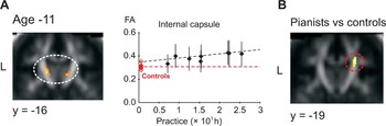
Perhaps most relevant for the question of experience-driven white matter plasticity in humans are studies demonstrating relations between long-term training and white matter structure. Most of this work has been performed on young adult musicians (typically aged 18–45 years) where amount of practicing was estimated retrospectively. A number of studies have found that the corpus callosum is larger in musicians (Schlaug et al., Reference Schlaug, Jäncke, Huang, Staiger and Steinmetz1995; Öztürk et al., Reference Öztürk, Tascioglu, Aktekin, Kurtoglu and Erden2002; Lee et al., Reference Lee, Chen and Schlaug2003). This effect appears to be most pronounced in musicians that started musical training early (<7 years) indicating a practicing effect (Schlaug et al., Reference Schlaug, Jäncke, Huang, Staiger and Steinmetz1995). We found positive relations between FA and training in widespread white matter regions in professional concert pianists (Bengtsson et al., Reference Bengtsson, Nagy, Forsman, Forssberg, Skare and Ullén2005). The correlations with childhood practicing were particularly extensive and strong and included the pyramidal tract, which also had higher FA in the pianists than in a control group of non-musicians (Fig. 1). The finding of higher FA in the pyramidal tract of pianists was recently replicated by Han et al. (Reference Han, Yang, Lv, Zhu, He and Tang2009). Surprisingly, another recent study (Imfeld et al., Reference Imfeld, Oechslin, Meyer, Loenneker and Jancke2009) reported lower pyramidal tract FA values in musicians than in non-musicians. This finding is puzzling given other studies on musicians, as well as the large literature demonstrating increases in FA during development and positive relations between FA and performance (see above). One notable difference to our own study and the study by Han et al. is that Imfeld and coworkers included performers of a large variety of keyboard, string, woodwind and brass instruments in their musician group. Pianists perform a large amount of individual finger movements with both hands during practicing, but for many wind instruments the frequency of finger movements may not exceed that in other professions where, for example, computers are used extensively.
Long-term training affects white matter structure. Correlations between number of hours practiced in childhood and white matter anisotropy (FA) in the right internal capsule (A) in a group of concert pianists. The clusters are overlaid on the mean FA image from all participants. In the diagrams, points and vertical lines represent mean FA values and standard deviations for all voxels in the cluster in one individual. (B) Cluster of voxels in the right internal capsule with significantly higher FA values in the pianists than in a non-musician control group. The mean FA values for each control participant in this cluster are illustrated with red points in the diagram in (A). Modified from Bengtsson et al. (Reference Bengtsson, Nagy, Forsman, Forssberg, Skare and Ullén2005).

IS ACTIVITY-MODULATED MYELINATION A PLASTIC PROCESS IN HUMANS?
Studies on different animal model systems have established that the neural activity in maturing axons influences the myelination process (for reviews, see e.g. Markham and Greenough, Reference Markham and Greenough2004; Fields, Reference Fields2008). As summarized above, a large literature shows that human white matter development is a slow process and that white matter structure has strong relations to functional brain properties, behavior and training. These findings make it plausible that neural activity driven white matter plasticity plays a role also in the human brain.
Many intriguing questions remain regarding the nature and functional roles of white matter plasticity. As mentioned, normal human white matter development involves both myelination and changes in the structure of the axons themselves, including increases in axonal diameters. The ratio between axon diameter and outer myelin sheath diameter (g-ratio) is carefully regulated to have a mean of around 0.65, which is the theoretical optimum for conduction velocity (Smith and Koles, Reference Smith and Koles1970). However, the correlation between axonal thickness and myelin sheath thickness is far from perfect (Leenen et al., Reference Leenen, Meek, Posthuma and Nieuwenhuys1985), which naturally raises the question of the possible functional role of that portion of the variance in myelin sheath thickness that is unexplained by axonal thickness. When discussing myelination plasticity it is thus, from both a functional and mechanistic point of view, probably important to distinguish between what one could call indirect myelination plasticity, i.e. a concomitant change in size of both axon and myelin sheath with preserved g-ratio, and direct myelination plasticity, where the thickness of the myelin sheath would be regulated independently of the axon diameter. It seems highly plausible that indirect myelination plasticity occurs in the human nervous system. Axonal thicknesses increase during development (Verhaart, Reference Verhaart1950; Nathan and Smith, Reference Nathan and Smith1955; Eyre et al., Reference Eyre, Miller and Ramesh1991) and animal data indicate that new myelin wraps are added onto the axons as the animal grows (Berthold et al., Reference Berthold, Nilsson and Rydmark1983; Fields, Reference Fields2008). The functional consequence of indirect myelination plasticity would simply be to maintain a high axonal conduction velocity as the axon grows. Direct myelination plasticity is a more speculative but theoretically interesting possibility. It would imply that the regulatory mechanisms controlling myelin sheath thickness are adjusted in a systematic and functionally meaningful way, e.g. by a change in set-point, so that the g-ratio is altered. This in turn could have effects on both conduction speed and other electrical properties of the axons, such as its refractory period. Fields (Reference Fields2008) has suggested that such regulation of axonal properties by oligodendrocytes could serve, for example, to fine tune conduction velocities in order to influence spike timing and synchronization of neural firing.
In summary, there is strong indirect support for that white matter plasticity occurs in late myelinating axons in humans. Further work will be needed to understand the mechanisms and functional importance of such plasticity and how it changes during the life span.
Statement of interest
None.