DNA is constantly exposed to intracellular damaging events that arise as intermediates in normal endogenous processes, such as DNA replication and meiosis, and to extracellular agents, including oxygen radicals or ionising radiation. Several pathways have evolved to recognise and repair damaged DNA to preserve genomic integrity. Defects in these pathways may allow replication errors, loss or rearrangement of DNA, mutagenesis, carcinogenesis and cell death. During development of T and B cells, three specific mechanisms require repair to targeted DNA damage that has been introduced during development. This review will detail the molecular mechanisms, clinical presentation and treatment of human primary immunodeficiency disorders associated with defects in normal DNA-damage recognition and repair.
DNA-damage sensing and repair
Maintenance of genomic integrity is crucial to prevent cell death or tumour development. Chromosomal damage occurs secondary to exogenous damage from ionising or ultraviolet radiation, chemicals, or from byproducts of normal endogenous cellular physiological processes, such as generation of free radicals and errors occurring during meiosis, which include nucleotide loss and stalling of replication forks. Additionally, specialised cellular developmental programmes might generate DNA damage, such as that occurring during lymphocyte receptor development or generation of antibody diversity. Consequently, numerous interconnected molecular mechanisms have evolved to prevent accumulation of mutations to maintain chromosomal structural integrity. Different repair pathways are required for the different types of DNA damage that can occur; although many use proteins and enzymes specific for that particular pathway, a number of proteins have multiple roles, and function in combination to help sense or repair different forms of DNA damage. Bloom's syndrome protein, an evolutionarily conserved RecQ helicase, has an important role in unwinding secondary DNA structures that inhibit replication fork progression in homologous recombination. Interaction with proteins responsible for resolution of DNA crosslinking that are mutated in Fanconi's anaemia has been demonstrated, as have interactions with ataxia-telangiectasia mutated (ATM) protein (which is involved in DNA-damage sensing, cell cycle checkpoints and DNA repair) and with MLH1, a protein involved in DNA-mismatch repair.
DNA double-strand breaks (DSBs) are a serious form of DNA damage, which can generate harmful mutations and proliferation of damaged cells. Such damage elicits breakage sensing, signal transduction and effector function, leading to cell-cycle-checkpoint arrest and/or apoptosis, and can influence DNA repair. Recruitment of repair proteins to damaged sites involves those binding to the DNA break, and those binding to the surrounding chromatin, and occurs in a highly ordered sequence. The MRE11–RAD50–nibrin (MRN) complex is the initial sensor of DSB damage. Damaged DNA ends are tethered by the MRN complex, which aids the localised activation of ATM protein, and the central component of the signal transduction pathway responding to DSBs. Following ATM activation, several DNA-repair and cell-cycle-checkpoint proteins, including H2AX, MDC1 and nibrin (also known as NBS1 and NBN), are activated, leading to cell cycle arrest and DNA repair. H2AX is phosphorylated to form γ-H2AX, which activates the cascade of repair-protein relocalisation. MDC1 stabilises the MRN complex at the site of the break to the adjacent chromatin, and coordinates assembly of other checkpoint and repair proteins, including the tumour suppressor p53-binding protein 1 (53BP1 or TP53BP1) and the E3 ubiquitin ligase RNF168, to the surrounding chromatin. Nibrin acts downstream of ATM by recruiting targets for ATM-mediated phosphorylation.
Nonhomologous-end-joining repair pathway
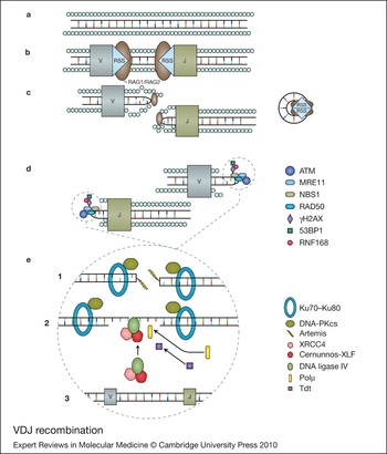
In response to DSBs, cells have developed two general types of repair: homologous recombination (HR) and nonhomologous end joining (NHEJ). HR is generally limited to the late S phase and G2 phase of the cell cycle in mammalian cells, and uses information from a homologous template to accurately repair breaks when sister chromatids present readily available templates. NHEJ is the main DNA-repair pathway that mediates the joining of broken regions of DNA that lack extensive homology, and is the principle mechanism used in vertebrate cells during the G1 phase of the cell cycle. Seven mammalian factors have now been identified as crucial NHEJ components. The DNA-binding subunits Ku70 (XRCC6) and Ku80 (XRCC5) together with the DNA-dependent protein kinase catalytic subunit (DNA-PKcs or PRKDC), form the DNA-PK holoenzyme, which is involved early in the recognition of DSBs. Activated DNA-PK holoenzyme recruits other NHEJ proteins including artemis (DCLRE1C), XRCC4, DNA ligase 4 (LIG4) as well as DNA polymerase µ (Polµ or POLM) to the site of DNA damage (Fig. 1). After phosphorylation by DNA-PKcs, the endonuclease artemis resolves complex DNA ends, such as the heterologous-loop and stem-loop DNA structures, which contain single-stranded DNA adjacent to double-stranded DNA. Polµ is associated with both the Ku heterodimer, and DNA ligase IV (LIG4) and might have a role in gap filling during NHEJ, but is not critical for ligation. LIG4, XRCC4 and cernunnos-XRCC4-like factor (XLF or NHEJ1) are also required for the ligation reaction that rejoins the DSBs.
VDJ recombination. DNA is uncoiled at transcription factories within the cell, where the associated recombination and repair proteins colocalise. (a, b) The lymphoid-specific recombinase-activating gene 1 and 2 (RAG1 and RAG2) proteins recognise and bind the recombination signal sequences (RSS) that flank the VDJ gene segments, and introduce site-specific DNA double-strand breaks (DSBs) through their endonuclease action. (c) The phosphorylated blunt signal ends and the covalently sealed hairpin intermediate of the coding end are held together by the RAG complex. (d) The MRE11–RAD50–NBS1 (MRN) complex binds the broken DNA ends and activates the ATM (ataxia-telangiectasia mutated) protein, which initiates cell cycle arrest and attraction of the repair proteins γH2AX, 53BP1, RNF168 and others, to stabilise the damaged chromatin. (e) (Step 1) The Ku70–Ku80 heterodimer binds the coding ends and recruits DNA-PKcs and artemis, which is required to open the hairpin intermediates. The covalently sealed hairpin intermediate is randomly nicked by the DNA-Pkcs–Artemis complex, which generates a single stranded break with 3′ or 5′ overhangs. (Step 2) XRCC4, DNA ligase IV (LIG4) and cernunnos-XLF (C-XLF) coassociate and are recruited to the ends. The signal ends are directly ligated by the XRCC4–LIG4–C-XLF complex. The opened hairpin intermediate is modified by polymerases, exonucleases and the lymphoid-specific terminal deoxynucleotidyl transferase (TdT), before (Step 3) repair and ligation by the XRCC4–LIG4–C-XLF complex.

DNA-mismatch repair
Mismatch repair (MMR) corrects DNA mismatches generated during DNA replication, preventing mutations from becoming permanent in dividing cells. Nucleotide misincorporation generates DNA base–base mismatches during DNA synthesis at a variable rate, depending on many factors, including which specific DNA polymerases are present. The DNA MMR pathway is highly conserved, and specific primarily for base–base mismatches and insertion–deletion mispairs generated during DNA replication and recombination. A number of human MMR proteins have been identified based on homology to Escherichia coli MMR proteins. Human MSH2 forms a heterodimer with MSH6 or MSH3 to form MutSα or MutSβ respectively, both of which are ATPases with a crucial role in mismatch recognition and initiation of repair. MutSα preferentially recognises base-to-base mismatches and small nucleotide insertion–deletion mispairs of one or two nucleotides, whereas MutSβ preferentially recognises larger insertion–deletion mispairs. Four human MutL homologs (MLH1, MLH3, PMS1, PMS2) have been identified. MLH1 forms a heterodimer with PMS2, PMS1 or MLH3 to form MutLα, MutLβ or MutLγ, respectively. Mutlα is required for mismatch repair by resolving four-stranded DNA structures (DNA Holliday junctions), which form during HR in meiosis. Heterodimers of MSH5 and MSH4 are postulated to form a ‘sliding clamp’ on DNA, and serve as scaffolding for the recombination machinery, including the DNA-repair proteins MLH1 and PMS2. MutLγ has a role in meiosis, but no specific biological role has been identified for MutLβ. MutLα regulates termination of mismatch-provoked excision. Other enzymes involved in mismatch repair include proliferating cellular nuclear antigen (PCNA), DNA polymerase δ (Polδ or POLD1) and DNA ligase I (LIG1). PCNA interacts with MSH2 and MLH1 and is thought to have roles in the initiation and DNA-resynthesis steps of mismatch repair. PCNA also interacts with MSH6 and MSH3 and might help to localise MutSα and MutSβ to mispairs in newly replicated DNA. DNA resynthesis and ligation is performed by LIG1.
Role of DNA-repair proteins in adaptive immunity
DNA-repair proteins and generation of lymphocyte antigen receptors
Effective immunity requires recognition of foreign antigens, requiring the generation of ≈1018 genetically diverse cells, each with a unique receptor that recognises a unique antigen–MHC combination. In jawed vertebrates, these receptors are created by breaking, randomly resorting and joining DNA sequences coding for the antigen-capture region of the receptor, by adapting the ubiquitous DNA-repair mechanisms that maintain genome stability. Recombination is a site-specific event that occurs at the T-cell receptor (TCR) α-, β-, γ- and δ-chain loci, and the B-cell receptor (BCR) immunoglobulin heavy (IgH), and immunoglobulin κ or λ light (IgL) chain loci. Recombination occurs between component variable (V), junction (J), and for TCRβ, TCRδ and BCR IgH loci, diversity (D) gene segments, with the fused VJ or VDJ coding sequence subsequently joined to a constant region segment through RNA splicing. Two recombination-activating gene proteins (RAG1 and RAG2) introduce site-specific DSBs at conserved noncoding DNA sequences adjacent to the points at which recombination occurs, known as recombination signal sequence (RSS) sites, either side of the segments to be rearranged, during the G1 phase of the cell cycle.
After the introduction of DSBs at the coding-sequence–RSS junction, two types of DNA ends arise: coding sequence ends that reconstitute the Ig and TCR genes are generated as hairpin intermediates, and noncoding signal ends containing the motifs targeting site-specific cleavage, which are generated as blunt double-stranded DNA ends. ATM is not required for cleavage of coding joint hairpin intermediates (Ref. Reference Riballo1), but helps to stabilise DNA ends in the RAG postsynaptic cleavage complex, facilitating NHEJ repair of VDJ-recombination-associated breaks (Refs Reference Bredemeyer2, Reference Huang3, Reference Helmink4). ATM, nibrin, γH2AX and 53BPI are associated with RAG-induced DSBs in developing lymphocytes and localise to DSBs and the chromatin region surrounding the recombining loci (Refs Reference Perkins5, Reference Chen6, Reference Celeste7, Reference Stracker8, Reference Difilippantonio9). ATM might contribute to the efficiency of VDJ recombination by activating cell-cycle-checkpoint proteins, which enables monitoring of recombination intermediates. In the absence of ATM, lymphocytes with RAG-induced DSBs might enter the S phase of the cell cycle, which leads to a reduction in productive VDJ recombination and an increased number of abnormal translocations involving Ig and TCR loci (Ref. Reference Kobayashi10).
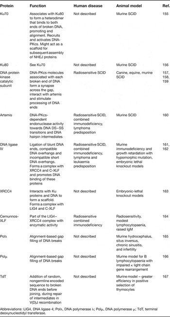
DSBs are repaired using the NHEJ repair pathway (Table 1). The Ku70–Ku80 heterodimer binds DNA ends present at RAG1- or RAG2-generated coding ends and recruits DNA-PKcs, which phosphorylate and activate artemis endonuclease activity to process the coding sequence hairpin intermediates. Following cleavage, coding and signal ends are directly ligated by the XRCC4–LIG4–C-XLF complex (Ref. Reference Lieber11) (Fig. 1). Rejoining of signal ends does not require DNA-PKcs or artemis. Furthermore, VDJ recombination is not completely abolished if any of the seven NHEJ proteins are impaired, because an alternative end-joining pathway exists, in which the frequent use of microhomology and excessive deletions are characteristic. RAG proteins have an essential role in the joining phase of V(D)J recombination, but allow a small degree of alternative NHEJ activity (Ref. Reference Corneo12).
Proteins in the nonhomologous end-joining DSB-repair pathway

Abbreviations: LIG4, DNA ligase 4; Polλ, DNA polymerase λ; Polμ, DNA polymerase μ; TdT, terminal deoxynucleotidyl transferase.
T- and B-cell-receptor recombination occurs in the thymus and bone marrow, respectively. Early lymphocyte progenitors undergo successive stages of lineage commitment, generating a functional lymphocyte receptor repertoire. Between critical developmental stages of VDJ rearrangement of the T-cell β- and α-chain, and B-cell IgH and IgL chain, the lymphocyte precursors undergo intense proliferation. During this phase, cells experience the normal replicative stress of proliferating cells, and in doing so, accumulate abnormal replication intermediates, normally resolved by Bloom syndrome protein (BLM) (Refs Reference Babbe13, Reference Babbe14).
DNA-repair proteins and immunoglobulin class-switch recombination
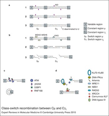
Maturation of the antibody repertoire is required to optimise antibody responses with high antigen affinity. Antibody maturation occurs in the germinal centres of secondary lymphoid organs in response to antigen- and T-cell-driven activation; B cells proliferate vigorously, dividing every 6–8 h and accumulating abnormal replication intermediates acquired during replicative stress, which are resolved by Bloom syndrome protein. Class-switch recombination (CSR) is a somatic DNA-arrangement process, which results in a switch in the IgH constant region of the BCR, expressed from the region encoded by Cµ, to a downstream constant region such as that encoded by Cα, Cγ or Cη. Switch (S) regions lie in the J-C intron. DNA recombination occurs between Sµ and the S region upstream of the C region for the particular antibody isotype, resulting in deletion of the intervening DNA. This results in the production of antibodies of different isotypes (IgG, IgA and IgE) with the same V(D)J specificity and therefore the same antigen affinity (Ref. Reference Iwasato15). Activation-induced cytidine deaminase (AID), a B-cell-specific enzyme that is crucial for CSR, induces DSBs to initiate CSR (Refs Reference Muramatsu16, Reference Revy17). AID selectively deaminates cytosine residues to uracil in the switch and variable regions (Ref. Reference Bransteitter18). Uracil DNA glycosylase (UNG) removes uracil, producing an abasic site, which is cleaved by one of the base excision–repair enzymes to create a DNA single-strand break (SSB) (Refs Reference Rada19, Reference Guikema20). The MMR proteins MSH2–MSH6 recognise U at U:G mismatched bases, and create a further SSB (Ref. Reference Xue, Rada and Neuberger21). If a particular U is on the complementary strand to a previous SSB, a DSB results, enabling CSR to occur (Ref. Reference Wilson22) (Fig. 2). PMS2, acting as a heterodimer with MHL1, converts AID- and UNG-induced SSBs into the DSBs required for CSR by stabilising the recombination intermediate (Refs Reference Schrader, Vardo and Stavnezer23, Reference Péron24). MSH5 is also involved in CSR, and might have a specific role in facilitating CSR between Sμ and Sα (Ref. Reference Sekine25).
Class-switch recombination between Cμ and Cγ1. (a) Activation-induced cytidine deaminase (AID) selectively targets cytosine bases in the Cμ and Cγ1 switch regions of the IgH constant region in the B cell receptor gene (Step 1). Cytosine residues are deaminated to uracil (Steps 2 and 3). (b) Uracil DNA glycosylase (UNG) removes uracil, producing an abasic site, which is cleaved by one of the base excision-repair enzymes (Step 1) to create a DNA single-strand break (Step 2). The mismatch-repair proteins MSH2–MSH6 also recognise U at U:G mismatched bases, and create a further DNA single-strand break, resulting in a DNA double-strand break (DSB) (Step 3). (c) DSB repair is achieved mainly through nonhomologous end joining. ATM (ataxia-telangiectasia mutated) is required for the repair and might recruit or activate 53BP1, γH2AX and RNF168, which are likely to be involved at the synapsis. (d) ATM might also phosphorylate artemis, which is required to repair a subset of DSBs, and organises the damaged DNA ends for subsequent repair steps, or arrests cell cycle progression until the repair is complete. Nibrin/NBS1, MRE11 and RAD50 are phosphorylated by ATM and might be required to activate ATM in CSR. MRE11, nibrin, Ku70–Ku80, DNA-PKcs, XRCC4 and DNA Ligase IV are likely to be involved in the final repair, as is cernunnos-XLF.

A significant role for Bloom syndrome protein is unlikely during CSR (Ref. Reference Babbe26), although it does interact with MLH1 and MSH6 (Refs Reference Schrader, Vardo and Stavnezer23, Reference Pedrazzi27, Reference Pedrazzi28). CSR DSB repair is achieved mainly through NHEJ during the G1 phase of the cell cycle (Ref. Reference Schrader29), although an alternative end-joining mechanism is used when there is impairment of NHEJ (Ref. Reference Yan30). ATM is required for efficient CSR, although the exact function remains unclear. ATM is also associated with MMR factors, including MSH2, MSH6, MLH1 and PMS2 (Ref. Reference Matsuoka31). Additionally, nibrin, MRE11, γH2AX and 53BP1, foci of which can be detected at the DSB switch region during CSR, are phosphorylated by ATM (Refs Reference Berkovich, Monnat and Kastan32, Reference Burma33, Reference Kobayashi34, Reference Ward35). One role of ATM might be to recruit or activate these factors, and organise the damaged DNA ends for subsequent repair steps, or arrest cell cycle progression until the repair is complete. ATM might have a more direct role in the end-processing step through phosphorylation of MRE11 or artemis – two proteins that participate in NHEJ. Artemis is downstream in the ATM signalling pathway for repair of a subset of radiation-induced DSBs, but is dispensable for ATM-dependent cell-cycle-checkpoint arrest (Ref. Reference Riballo1), and also appears dispensable for processing of DSBs for efficient CSR (Ref. Reference Rooney36), although it might be required for repair of the CSR-related chromosomal breaks at the Ig locus (Ref. Reference Franco37) and for the resolution of a subset of breaks generated during CSR (Ref. Reference Rivera-Munoz38). The altered pattern of CSR junctions in artemis-deficient patients also suggests that this protein is required in the predominant NHEJ pathway during CSR (Ref. Reference Du39). Deficiency in MRE11, nibrin, Ku70–Ku80, DNA-PKcs or LIG4 appears to alter the balance between the predominantly used NHEJ and alternative end-joining mechanism in CSR DSB repair, suggesting that these proteins are also involved directly or indirectly in CSR (Ref. Reference Pan-Hammarstrom40).
Little is known about the proteins or mechanism involved in the alternative end-joining mechanism. Poly (ADP-ribose) polymerase 1 (PARP1) is involved in many cellular responses, including base excision repair (BER) and possibly HR (Ref. Reference Helleday, Bryant and Schultz41). It might be involved in the alternative end-joining pathway, with XRCC1 and DNA ligase III (LIG3) (Refs Reference Audebert, Salles and Calsou42, Reference Wang43, Reference Robert, Dantzer and Reina-San-Martin44). LIG1 and LIG3 are required for microhomology-mediated end joining but it is unknown which of these enzymes is involved in alternative end-joining during CSR (Ref. Reference Liang45).
DNA-repair proteins and somatic hypermutation
Although CSR and somatic hypermutation (SHM) occur together in germinal centres, neither is a prerequisite for the other because IgM can be mutated in the absence of any such feature in IgG or IgA isotypes (Ref. Reference Kaartinen46). SHM introduces random mutations into the BCR variable region, resulting in minor conformational changes, which enables positive selection of B cells that carry a BCR with high antigen affinity (Ref. Reference Storb47). SHM is initiated by AID, by RNA editing of variable region C to U residues (Ref. Reference Shivarov48). The MMR proteins MSH2–MSH6 recognise AID-induced U/G residues, and recruit the exonuclease EXO1 and DNA polymerase η (POLH) leading to G:C to T:A transversions (Ref. Reference Schanz49). Whilst NHEJ is not used in SHM, the MRN complex is involved in DNA cleavage at AID-induced abasic sites during SHM (Ref. Reference Larson50), and nibrin has a role in regulating strand-biased repair (Ref. Reference Du39). Bloom syndrome protein does not seem to be involved in SHM (Ref. Reference Sack51).
Primary immunodeficiency syndromes associated with genetic defects in DNA-repair genes
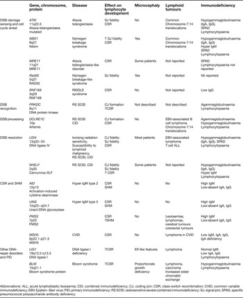
A growing number of genetic defects in the DNA DSB and MMR pathways have been identified in humans with primary immunodeficiency (PID), providing insight into the clinical phenotype of genomic instability on the immune system (Table 2).
Proteins associated with DSB sensing, DNA-repair defects and human primary immunodeficiency

Abbreviations: ALL, acute lymphoblastic leukaemia; CID, combined immunodeficiency; CJ, coding join; CSR, class-switch recombination; CVID, common variable immunodeficiency; EBV, Epstein–Barr virus; PID, primary immunodeficiency; RS SCID, radiosensitive severe combined immunodeficiency; SJ, signal join; SPAD, specific pneumococcal polysaccharide antibody deficiency.
DSB-initiation defects
Recombination-activating gene deficiency
Although not strictly part of the DNA-repair pathway, RAG1 and RAG2 initiate DSBs, which are required for VDJ recombination to develop TCRs and BCRs (Ref. Reference Schwarz52). RAG1 and RAG2 are lymphoid-specific endonucleases, which bind DNA at RSS–coding-sequence junctions. Nonsense mutations in RAG1 or RAG2 give rise to a T-B-NK+ severe combined immunodeficiency (SCID) phenotype, with absent immunoglobulins. Patients usually present in early infancy with persistent viral respiratory or gut infection and growth failure, or Pneumocystis pneumonitis, as for other forms of SCID. Missense mutations give rise to ‘leaky’ SCID, or Omenn syndrome (Ref. Reference Villa53). Infants may present with lymphadenopathy, hepatosplenomegaly, erythroderma, alopecia, agammaglobulinaemia apart from a raised IgE, T lymphocytosis, and absent B cells with accompanying respiratory and gastrointestinal symptoms and failure to thrive (Refs Reference Omenn54, Reference Villa55). T cells are activated and show a restricted TCRVB repertoire (Ref. Reference Rieux-Laucat56). Biopsies of affected skin demonstrate a histopathological pattern that is consistent with graft-versus-host disease, although T cells are autologous. Additional clinical phenotypes include normal immunoglobulin levels, specific antibody responses to infectious agents or vaccine antigens, production of autoantibodies, a predominance of γδ T cells and development of Epstein–Barr virus (EBV)-associated lymphoproliferation in some patients (Refs Reference Ehl57, Reference de Villartay58). T and B lymphocytopaenia, hypogammaglobulinaemia, recurrent viral infection, EBV-associated lymphoma, and extensive granulomatous lesions associated with compound heterozygous mutations in RAG1 and RAG2 are also documented (Ref. Reference Schuetz59).
DSB-damage-sensing and cell-cycle-arrest defects
Ataxia telangiectasia
Ataxia telangiectasia (A–T) is a rare systemic autosomal recessive disorder caused by mutations in ATM (Ref. Reference Chun and Gatti60), manifest by progressive cerebellar ataxia, oculocutaneous telangiectasia, gonadal sterility, postnatal growth retardation and a high incidence of predominantly lymphoid tumours. Patients normally present with cerebellar ataxia before telangiectasia appear. Recurrent sinopulmonary infection can be a presenting feature and might be associated with raised IgM and low levels or absent IgG (Ref. Reference Noordzij61). Sinopulmonary infection combined with recurrent aspiration, can lead to chronic lung disease (Ref. Reference Lefton-Greif62). The incidence of infections is variable and correlates with the presence of two null mutations in ATM (Ref. Reference Staples63). Immunological responses to bacterial antigens, particularly to polysaccharide antigen, are generally reduced (Ref. Reference Sanal64). Lymphocytic interstitial pneumonitis has also been described (Ref. Reference Tangsinmankong65). Median survival is 22 years (Ref. Reference Crawford66). In addition, thymic output is reduced and there is a restricted TCRVB repertoire, indicated by oligoclonal expansions (Ref. Reference Giovannetti67). Chromosomal inversions and translocations, particularly chromosome 7:14 translocations are seen in A-T. B cell repertoire is also restricted and skewed by diffuse oligoclonal expansions with normal VDJ joints. B cells from A-T patients have an intrinsic defect in maturation from IgM to other classes, because of a defect in CSR from Cμ to the most distant loci, reflecting the requirement of ATM for efficient recombination between immunoglobulin switch regions (Ref. Reference Reina-San-Martin68).
Nijmegen breakage syndrome
Nijmegen breakage syndrome (NBS) is associated with a characteristic facial appearance: receding forehead, receding mandible and prominent midface (‘bird-like’ facies) (Ref. Reference Weemaes69). Additional features include epicanthic folds, large ears and sparse hair with microcephaly and mild mental retardation. Patients are prone to sinopulmonary infection and are susceptible to B-cell-lineage lymphomas. Cellular immunity is consistently impaired in NBS patients; lymphcytopaenia is common, with reduced proportions of CD3 and CD4 T cells (Ref. Reference Digweed and Sperling70). Agammaglobulinaemia is reported in about a third of NBS patients, whereas in others the humoral immune deficiency is more variable. Deficiencies of IgA or IgG4, alone or in combination, are common (Ref. Reference Gregorek71). About 10% of patients have normal immunoglobulins. The immunodeficiency might result from reduced fidelity of VDJ recombination, because nibrin is involved with ATM in inducing cell cycle arrest during this process (Ref. Reference Xu72). Frequency of VDJ recombination in NBS patients is normal, however, with normal IgH rearrangement. The deficiency of serum IgG and IgA with normal or raised IgM is probably due to impaired CSR (Refs Reference Kracker73, Reference Reina-San-Martin74).
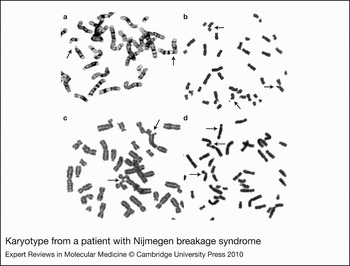
Chromosomal inversions and translocations, particularly chromosome 7:14 translocations are characteristic of NBS (Fig. 3). NBS cells are sensitive to DNA-crosslinking agents such as mitomycin C and diepoxybutane (DEB), in addition to IR. Patients with certain NBS1 mutations have features similar to those seen in Fanconi's anaemia (Refs Reference Nakanishi75, Reference Gennery76), although immunodeficiency is more pronounced, presumably because of the role of nibrin in VDJ recombination and CSR.
Karyotype from a patient with Nijmegen breakage syndrome. (a) Chromosome t(7;14) rearrangement (arrows). (b) chromosomal breakage following exposure to 50 centiGray ionising radiation (arrows). (c) Multiradial formation (arrows) after culture for 72 hours following exposure to mitomycin C at 0.32 µg/ml for 60 minutes. (d) Chromosome breakage (arrows) following lymphocyte culture with diepoxybutane (DEB) for 72 hours (Reproduced with permission from the Paediatric HSCT Unit, Newcastle General Hospital).

Ataxia telangiectasia-like disorder
Ataxia telangiectasia-like disorder caused by mutations in MRE11A is extremely rare, with only 19 patients reported worldwide (Refs Reference Stewart77, Reference Delia78, Reference Fernet79, Reference Khan80, Reference Uchisaka81). Clinical features are similar to those in patients with ataxia telangiectasia; however, progressive cerebellar ataxia is of later onset and slower progression than in patients with ataxia telangiectasia (Ref. Reference Taylor, Groom and Byrd82). Telangiectasia are absent. Immunoglobulin levels are normal, although deficiency in antigen-specific antibodies has been reported, particularly to pneumococcal polysaccharide antigen (Ref. Reference Lahdesmaki83). Lymphoid tumours have not been reported, although two siblings with a novel mutation developed poorly differentiated lung adenocarcinoma (Ref. Reference Uchisaka81). Some patients are microcephalic, and although intelligence is generally normal, psychomotor retardation has been reported (Ref. Reference Uchisaka81). MRE11A encodes a protein that associates in the MRN complex, and patients have features that overlap with both ataxia telangiectasia and NBS. The absence of reported recurrent pulmonary infection due to immunodeficiency might reflect clinical variability as seen in ataxia telangiectasia, or the type of mutation present, the majority of which are homozygous missense mutations. Given the role of MRE11 in CSR, it would appear logical that a degree of hypogammaglobulinaemia is likely in some patients. Defective CSR has been reported, with reduced switching from Sμ to Sα, and an increased usage of microhomology at switch junctions (Ref. Reference Lahdesmaki83).
RAD50 deficiency
One patient has been described with NBS-like features, in whom compound heterozygous mutations in RAD50, one of the components of the MRN complex, were found (Ref. Reference Barbi84). The clinical features comprised intrauterine growth retardation with microcephaly, poor postnatal growth and ‘bird-like’ facies. Speech delay was also noted; ongoing follow-up has demonstrated moderate psychomotor retardation, with mild spasticity and a non-progressive ataxic gait. She developed multiple cutaneous pigmented naevi and hypopigmented areas. There was no history of excessive infections. Lymphocyte numbers, proliferations to mitogens and immunoglobulin levels were normal. However, chromosomal instability with 7:14 translocations was noted and there was radiosensitivity (Ref. Reference Barbi84). At last follow-up, aged 23 years, she had not developed myelodysplasia or lymphoid malignancy. In this individual, one mutation created a premature stop codon, the other led to an abnormally large polypeptide (Ref. Reference Waltes85). No information is available on the use of microhomology at V-J junctions. One report implicates RAD50 in fidelity of end joining of VDJ signal-join substrates (Ref. Reference Donahue86). It is of interest that the phenotype of RAD50 deficiency more closely resembles that of NBS than ataxia telangiectasia, unlike MRE11 deficiency. Although immunodeficiency was not reported in this patient, given the function of RAD50 in the MRN complex in TCR and BCR formation and CSR, it seems likely that immunodeficiency will be a feature of the expanded phenotype in other patients.
Radiosensitivity, immunodeficiency, dysmorphic features and learning difficulties (RIDDLE) syndrome
One Caucasian patient has been described to date with radiosensitivity, immunodeficiency, dysmorphic features and learning difficulties (RIDDLE syndrome) (Ref. Reference Stewart87). He presented with mild facial dysmorphism, short stature, learning difficulties and mild motor abnormalities. There were no oculocutaneous telangiectasia. By 1 year of age, he had low serum IgG levels, but normal IgM and IgA. T- and B-cell numbers were normal. From the age of 3 years, he was treated with replacement immunoglobulin and has remained well. Biallelic mutations in RNF168, coding for a ubiquitin ligase, have subsequently been reported (Ref. Reference Stewart88). B cells from the patient demonstrated increased use of microhomology across the Sμ-Sα and Sα-Sγ3 switch regions, with a reduced frequency of mutations and insertions; findings that are similar, although less severe, to those found in LIG4 deficiency, and suggestive of abnormal CSR. SHM was normal. Fibroblasts exhibited moderately increased sensitivity to ionising radiation, and failed to localise 53BP1 to damaged chromatin. RNF168 has a role in organising chromatin to facilitate long-range NHEJ, which is essential for CSR, but not VDJ recombination, probably explaining the normal cellular immunity observed (Refs Reference Difilippantonio89, Reference Manis90, Reference Ward91).
DSB-recognition defects
DNA-PK deficiency
One Turkish patient, of consanguineous parents, has been described to date with a homozygous three-nucleotide deletion and homozygous missense mutation in PRKDC (Ref. Reference van der Burg92). The patient presented at 5 months of age with classical symptoms of recurrent oral candidiasis and lower respiratory tract infections, and a T-B-NK+ SCID phenotype. No microcephaly or developmental delay was present. The arrest in differentiation of B cell precursors was consistent with a defect in VDJ recombination. Fibroblasts were sensitive to ionising radiation, with a DSB-repair defect comparable with that seen in artemis-deficient cells. Coding joints showed long stretches of palindromic nucleotides, and an end-joining assay demonstrated increased microhomology use, which was similar to that seen in artemis-deficient cells.
DSB-processing defects
Artemis deficiency
Artemis is critical for VDJ recombination; null mutations in DCLRE1C give rise to a T-B-NK+ SCID phenotype (Ref. Reference Moshous93), originally described in Athabascan-speaking native Americans (Ref. Reference Jones94). Fibroblasts from these patients exhibit increased cellular sensitivity to ionising radiation (radiosensitive, RS-SCID) (Ref. Reference Cavazzana-Calvo95), but the clinical presentation is otherwise identical to RAG-deficient SCID.
Omenn syndrome is caused by hypomorphic mutations in DCLRE1C (Ref. Reference Ege96) and has an analogous clinical presentation to that seen with hypomorphic RAG mutations. Patients have also presented with a progressive combined immunodeficiency (CID) from later infancy, characterised by recurrent sinopulmonary or gastrointestinal infection, T and B lymphopaenia, hypogammaglobulinaemia and autoimmune cytopaenias (Refs Reference Moshous97, Reference Evans98). Some patients show susceptibility to EBV-associated B lymphomas (Ref. Reference Moshous97). Chromosome 7:14 inversions and translocations have been described in these patients but no microcephaly has been noted.
DSB-resolution defects
DNA ligase IV deficiency
LIG4 deficiency was first described in a patient who was clinically and developmentally normal until T cell acute lymphoblastic leukaemia developed. Disproportionately severe cytopaenia followed treatment, and standard chemotherapy consolidation therapy was omitted. He developed an extreme reaction to radiotherapy, including marked and prolonged cytopaenia, severe desquamation and died from radiation-induced encephalopathy (Ref. Reference Riballo99). Subsequently, a number of additional LIG4-deficient patients have been described. Six had microcephaly, developmental delay, growth failure, lymphopaenia, hypogammaglobulinaemia and recurrent infection (Refs Reference O'Driscoll100, Reference Unal101). Marrow hypoplasia was a feature in some of these patients. RS-SCID with microcephaly and growth delay has also been reported (Refs. Reference van der Burg102, Reference Buck103). Four patients with microcephaly and a combined immunodeficiency phenotype have been reported, of whom two developed an EBV-associated diffuse large cell non-Hodgkin lymphoma, and one developed T cell acute lymphoblastic leukaemia (Refs Reference Ben-Omran104, Reference Enders105, Reference Toita106, Reference Grunebaum107). One patient presented with features consistent with Omenn syndrome (Ref. Reference Grunebaum107). Other clinical features include photosensitivity and psoriatic-like lesions (Ref. Reference O'Driscoll100).
Moderate impairment of VDJ recombination is observed in LIG4-deficient fibroblast VDJ recombination assays: an almost normal frequency of coding and signal joint formation is observed, but fidelity of both coding and signal joint formation is impaired, with marked infidelity in coding end rejoining (Refs Reference O'Driscoll100, Reference Buck103). The in vitro findings are less severe than the clinical immunodeficiency, suggesting that LIG4 is required during lymphocyte development at stages beyond VDJ recombination, possibly to repair DNA damage that occurs during lymphocyte proliferation. Similar observations have been identified in patients with hypomorphic DCLRE1C mutations (Ref. Reference Evans98). Patients with LIG4 mutations also have altered resolution of CSR junctions, with greater use of microhomology at Sμ-Sα junctions (Ref. Reference Pan-Hammarstrom40).
Cernunnos-XLF deficiency
Deficiency of cernunnos-XLF (C-XLF) has been described in eight patients to date (Refs Reference Buck108, Reference Ahnesorg109): the first two presented with T and B lymphocytopaenia, with a normal number of NK cells (Ref. Reference Dai110). Subsequently, five patients with combined immunodeficiency (CID) have been described (Ref. Reference Buck108). All had a similar lymphocyte phenotype to the original kindred, but additionally had low IgA and IgG. Two had raised IgM, suggesting a role for C-XLF in CSR. Some patients described were microcephalic with ‘bird-like’ dysmorphism, two exhibited autoimmune cytopaenia, and all suffered from recurrent bacterial and opportunistic infection. Two demonstrated several chromosomal alterations, although chromosome 7:14 translocations were not described. Lymphomas have not been described to date. A further patient has been described, who had similar morphological features of microcephaly, small stature and ‘bird-like’ facies. He suffered recurrent respiratory infections, and demonstrated normal IgM, but low IgA and IgG levels and no response to vaccine protein antigens. His lymphocyte phenotype was characteristic (Ref. Reference Faraci111). He developed pancytopaenia with trilineage marrow dysplasia, and enteropathy, and underwent successful haematopoietic stem cell transplantation at 10 years of age, complicated by EBV-associated post-transplant lymphoproliferative disease.
Other clinical abnormalities include bone malformations (low implantation of the thumb, hypoplasia of the middle phalanx of the fifth finger), nephroptosis and one patient demonstrated developmental delay, features that overlap with those described in LIG4 deficiency.
In vitro coding and signal joint formation were reduced in patients compared with controls, with an increase in nucleotide loss in coding joins. The fidelity of signal joins was severely impaired in patients, with use of microhomology during joining (Ref. Reference Buck108), features previously described in LIG4-deficient patients (Refs Reference Riballo99, Reference Unal101). VDJ deficiency in these patients is less severe than in the artemis-deficient RS-SCID and probably accounts for the presence, albeit in low numbers, of T and B cells in the patients. As in patients with LIG4 deficiency, the in vitro findings are less severe than the clinical immunodeficiency, suggesting that C-XLF is also required during lymphocyte development after VDJ recombination has occurred. C-XLF might be important for cell-replication-induced DSB repair (Ref. Reference Schwartz112).
Other uncharacterised disorders
A number of genetically undefined disorders, with phenotypic and cellular features characteristic of NBS (Refs Reference Berardinelli113, Reference Maraschio114, Reference Hiel115) or Omenn-like SCID (Ref. Reference Wiegant116), but with no mutations in candidate genes, have been described, implicating further DNA-repair genes in human primary immunodeficiency.
Class-switch recombination and somatic hypermutation defects
Three autosomal recessive hyper-IgM syndromes have been described; they are caused by defects in DNA-break and DNA-repair mechanisms, which lead to decreased or abolished isotype switching and impaired somatic hypermutation.
AID deficiency
AID deficiency, an autosomal recessive disease caused by mutations in AID, normally presents in early childhood with severe, recurrent infections, most commonly recurrent sinopulmonary or gastrointestinal infections (Refs Reference Revy117, Reference Quartier118). Despite this early presentation, many patients are not diagnosed and treated until the second or third decade of life (Ref. Reference Minegishi119). Massive lymphadenopathy, with giant germinal centres are characteristic upon histological examination. Immunological features include raised IgM and low or absent IgA and IgG. There is an increased incidence of organ-specific autoimmune disease in these patients, particularly diabetes mellitus, polyarthritis, autoimmune hepatitis and Crohn disease (Ref. Reference Minegishi119).
Patients with impaired CSR but normal SHM have mutations in the C-terminal region of AID, and present with milder disease (Ref. Reference Ta120), and a small subset may present with autosomal dominant disease (Ref. Reference Imai121).
Uracil DNA glycosylase deficiency
Three patients have been described with a defect in the gene encoding uracil DNA glycosylase (UNG). Clinical presentation is similar to those with AID deficiency, including recurrent respiratory tract infections from early childhood, and lymphoid hyperplasia (Ref. Reference Imai122). Raised IgM and profoundly decreased IgA and IgG serum levels were found, with depressed antigen-specific antibody responses. A skewed pattern of SHM was found with almost all mutations being transitions (G>A, C>T).
PMS2 deficiency
PMS2 forms a heterodimer with MLH1 to form Mutα, which has an important role in mismatch repair. Defects have been described in three individuals (Ref. Reference Péron24). In addition to raised serum IgM and decreased IgA and IgG with recurrent infections, café-au-lait spots and malignancy, including leukaemias, lymphomas, cerebral tumours and colorectal tumours are characteristic (Refs Reference De Vos123, Reference Kratz124). Increased levels of microhomology were found across the Sμ-Sα switch junctions. SHM can be mildly reduced.
MSH5 deficiency
MSH5 has been implicated in IgA deficiency and common variable immunodeficiency (Ref. Reference Sekine25). Increased microhomology at Sμ-Sα switch junctions was found in patients carrying disease-associated MSH5 allelles, with fewer mutations than in controls. The precise mechanism by which defects in MSH5 contribute to the abnormalities observed in CSR has not been elucidated.
Undefined defects
Further, as yet undescribed, gene defects give rise to a clinical picture of hyper-IgM syndrome. Hyper-IgM type 4 has been described in 15 patients, with characteristic features of recurrent respiratory and gastrointestinal tract infection, lymphoid hyperplasia and autoimmune features (Ref. Reference Imai125). CSR was defective, but SHM was normal. AID and UNG mutations were excluded in all patients.
A further clinical entity consisting of increased radiosensitivity but normal checkpoint arrest and NHEJ, increased levels of microhomology across Sμ-Sα switch junctions and a skewed SHM toward transitions at G or C residues has been described in five patients (Ref. Reference Péron126). All had recurrent respiratory infections; lymphoid hyperplasia and autoimmunity were also described. Raised IgM and decreased IgA and IgG levels were noted. A genetic defect has yet to be identified. A further group of patients has been described, in whom recurrent bacterial infection, autoimmunity and lymphadenopathy are observed, although the lymphadenopathy was less marked than in AID-deficient patients. There was a lack of class-switched B cells in these patients, although SHM was normal. There was no sensitivity to ionising radiation (Ref. Reference Durandy127). Defective CSR associated with growth hormone deficiency has been described in two patients in whom the molecular defect has yet to be discovered (Ref. Reference Kashef128, Reference Ohzeki129).
Other human primary immunodeficiency syndromes associated with DNA-repair gene defects
DNA ligase I
To date, one patient has been described with two compound missense mutations in LIG1 (Refs Reference Webster130, Reference Barnes131). Clinical features, overlapping with those of Bloom syndrome and ataxia telangiectasia, include intrauterine and postnatal growth retardation, developmental delay with normal cognitive development, dysmorphism with elf-like features, and photosensitivity. Immunodeficiency manifested as recurrent middle ear and respiratory infections from age 2 years, with evolving IgA deficiency, relative hypogammaglobulinaemia of IgG and normal IgM. There was an evolving lymphocytopaenia with poor proliferative response to mitogens. During teenage years, the respiratory status deteriorated and secondary sexual characteristics did not develop. At the age of 17 years, patches of cutaneous venous dilatation appeared mainly on the limbs, and there was some bulbar conjunctival telangiectasia. Hepatosplenomegaly developed with associated neutropaenia and increasing lymphopaenia. A liver biopsy showed lymphocyte infiltration of the portal tract, suggesting lymphoma. The patient developed a severe cutaneous herpes zoster infection and died from pneumonia at the age of 19 years. No information on end-joining of VDJ substrates, or on the use of microhomology, was available. An increase in the number of SSBs and DSBs in newly replicated DNA molecules was seen in an immortalised fibroblast line, possibly because of the failure of dealing with damage at replication forks (Ref. Reference Soza132). The cause of the immunodeficiency can only be conjectured, given the paucity of data. There is no evidence linking LIG1 with VDJ recombination (Ref. Reference Petrini133). LIG1 forms a complex with nibrin, and both colocalise at replication factories to repair DSBs by homologous recombination at stalled replication forks (Ref. Reference Vago134), suggesting that defects in LIG1 are associated with failure to repair DNA damage during lymphocyte proliferation, rather than failure to complete NHEJ in TCR and BCR formation. The finding of low IgA and IgG, but normal IgM is tantalising, and further work needs to be done to investigate what role, if any, LIG1 has in CSR.
Bloom syndrome
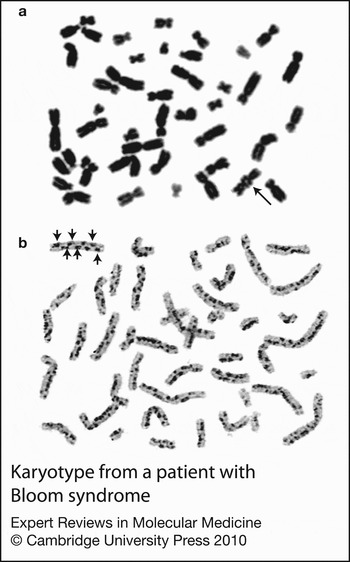
Bloom syndrome is an autosomal recessive disorder characterised by proportionate pre- and postnatal growth deficiency, photosensitive, telangiectatic, hypo- and hyperpigmented skin, predisposition to malignancy and chromosomal instability. There is an increased incidence of diabetes mellitus. Immunodeficiency, although common, is variable and generally not severe (Refs Reference Hütteroth, Litwin and German135, Reference Van Kerckhove136), although life-threatening infection can occur (Ref. Reference German137). Low concentrations of one or more immunoglobulin isotypes are most often found (Refs Reference Hütteroth, Litwin and German135, Reference Van Kerckhove136, Reference Kondo138). Impaired T cell proliferation, diminished CD4+ T-cell numbers and impaired function have been described in Bloom syndrome patients (Refs Reference Hütteroth, Litwin and German135, Reference Taniguchi139). There is a characteristic increase in sister-chromatid exchange seen upon cytogenetic analysis (Fig. 4). Bloom syndrome protein has no role in VDJ recombination (Refs. Reference Babbe13, Reference Hsieh, Arlett and Lieber140), and has only a minor role, if any, in CSR, although microhomology-mediated end joining was observed at Sμ-Sγ3 switch regions, possibly implicating BLM in the resolution phase of CSR (Ref. Reference Babbe14).
Karyotype from a patient with Bloom syndrome. A symmetrical quadridradial (a, arrow), and a large increase in the number of sister chromatid exchanges (b, arrows) are visible. (Reproduced with permission from the Paediatric HSCT Unit, Newcastle General Hospital).

Fanconi anaemia
Fanconi anaemia is a clinically heterogenous autosomal recessive or X-linked disorder characterised by bone marrow failure, skeletal, renal, cardiac and gastrointestinal defects, hypopigmentation and predisposal to malignancy (Ref. Reference Alter141). The majority of immunological problems relate to bone marrow failure, but a few patients present early with significant or prolonged infections, more consistent with immunodeficiency (Ref. Reference Nakanishi75). Although cells from patients show hypersensitivity to agents causing DNA interstrand crosslinks, a few also demonstrate sensitivity to ionising radiation (Ref. Reference Mohseni-Meybodi, Mozdarani and Vosough142). Thirteen genes associated with Fanconi anaemia have been identified to date. Whilst most of the proteins form a core ubiquitin ligase complex, FAND2 is ubquitinated by this complex, and then colocalises to chromatin with other DNA-repair proteins, including the MRN complex (Ref. Reference Nakanishi75). The Fanconi anaemia proteins do not appear to have direct a role in lymphocyte receptor development or modification, and effects on immunity are more likely to be a result of interstrand crosslinks occurring during cellular development, resulting in bone marrow failure.
Diagnosis of DNA-repair defects
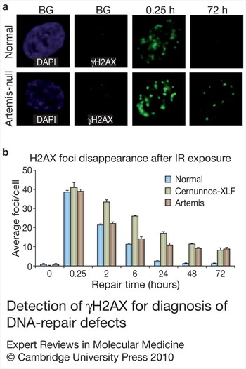
The diagnosis of radiosensitivity is difficult, time consuming and confined to a few laboratories. An index of clinical suspicion is necessary to consider the diagnosis, and diagnostic clues can be gathered from the clinical phenotype (e.g. microcephaly or telangiectasia) and immunological profile (e.g lymphocytopaenia, raised IgM) (Table 2). Cytogenetic analysis might give some clues to the underlying diagnosis by the finding of chromosome 7:14 translocations, seen in ataxia telangiectasia, NBS and other ionising-radiation-sensitivity disorders (Table 2) or an increase in sister chromatid exchanges in Bloom syndrome (Fig. 4). Sensitivity to ionising radiation can also be demonstrated using a clonogenic survival assay in which fibroblasts are irradiated with increasing doses of radiation and the percentage survival of cells is assessed after a fixed period of time (usually 3 weeks) (Ref. Reference Evans98). A similar method is to subject cells to increasing doses of radiation and subsequently stain for γH2AX foci which are present at the site of DSBs but disappear over time, as the damage is repaired (Ref. Reference Riballo1): persistence of such foci is indicative of impaired repair mechanisms (Fig. 5a). Different genetic causes give slightly different results; in artemis-deficient cells, most cells show normal repair, with a small subset showing impairment, whereas C-XLF-deficient cells show a greater degree of impairment to damage repair (Fig. 5b).
Detection of γH2AX for diagnosis of DNA-repair defects. (a) Measurement of γH2AX in fibroblasts. Following ionising radiation, there is an increase over background (BG) in γH2AX foci at 0.25 hours in wild-type and artemis-deficient cell lines. By 72 hours, the wild-type fibroblasts have returned to normal, but the artemis-deficient fibroblasts still manifest γH2AX foci, consistent with defective DNA double-strand break (DSB) repair. (b) Serial measurements of γH2AX foci in wild-type, cernunnos-XLF-deficient and artemis-deficient fibroblasts. γH2AX foci disappear in the normal cells as DSBs are repaired. Cernunnos-XLF-deficient cells show a repair defect that is observable at 2 hours after ionising radiation and remains for up to 72 hours, which indicates a DSB-repair defect that impacts all nonhomologous end joining (NHEJ). The artemis-deficient fibroblasts are completely normal 2 hours after ionising radiation, but show little further repair beyond 24 hours, indicating a subset of breaks that remain unrepaired (approximately 10% of DSBs). (Images courtesy of P.A. Jeggo, Genome Damage and Stability Centre, University of Sussex, UK).

Treatment of primary immunodeficiency associated with DNA-repair disorders
Treatment for DNA-repair-deficient primary immunodeficiencies depends on the clinical presentation, and is generally the same as for similar disorders due to other underlying molecular defects. Patients with B cell deficiency can be treated with prophylactic antibiotics and immunoglobulin replacement therapy. However, patients with severe T cell deficiency require more-aggressive therapy and haematopoietic stem cell transplantation might be indicated, particularly for children with recurrent infection and failure to thrive, and those at risk of developing malignancy. Because of the risk of generalised chromosome damage with the use of radiotherapy, this should be omitted from conditioning regimens. The use of reduced intensity chemotherapy conditioning appears to favour a successful outcome (Refs Reference Gregorek71, Reference Stewart77, Reference Gruhn143, Reference Albert144). Modified chemotherapy regimens for the treatment of lymphoid malignancy are needed, owing to the high level of toxicity using conventional regimens (Ref. Reference Dembowska-Baginska145); radiotherapy should be avoided or restricted. The incidence of secondary malignancies is more frequent in these patients so careful follow-up is required. Gene therapy might be an alternative treatment for some conditions, although clinical trials are not yet in progress (Ref. Reference Benjelloun146). One novel approach to treatment currently in development is the use of antisense oligonucleotides to correct splicing, frameshift and missense mutations and thus convert absent or unstable protein to partially or fully functional protein (Ref. Reference Lai147). Another approach is the use of ribosomal readthrough agents to overcome premature termination codons, and enable some normal protein expression (Ref. Reference Welch148).
Polymorphisms in DNA-repair genes
Increasingly, polymorphisms that are not associated with disease are being found to exert subtle effects. A number of polymorphisms in different DNA-repair genes have been associated with an increased risk of a number of different malignancies (Refs Reference Schuetz149, Reference Margulis150) or increased sensitivity to radiotherapy (Refs Reference Pugh151, Reference Okazaki152). Polymorphisms in LIG4, in combination with a disease-causing mutation lead to a more-severe clinical phenotype than the mutation alone (Ref. Reference Girard153). Counterintuitively, some polymorphisms might also have a protective affect against developing malignancy (Ref. Reference Roddam154).
Outstanding questions
Much has been learnt about the molecular mechanisms of lymphocyte receptor formation, immunoglobulin isotype switching and affinity maturation by the careful study of human primary immunodeficiencies. Conversely, an understanding of the mechanisms has aided discovery of novel genetic immune defects in patients. Much has yet to be discovered, however. The identification of more patients with extremely rare diseases such as LIG1 deficiency, RAD50 deficiency, RIDDLE syndrome and PMS2 deficiency will expand the clinical phenotype, and give greater understanding of the role these proteins have in immune development. Key proteins in the DSB-repair pathway, such as XRCC4, which is known to cause immunodeficiency in animal models, have yet to be linked to human disease. It is likely that careful study of milder antibody deficiencies will reveal further defects in MMR proteins responsible for SHM and antibody affinity maturation. An appreciation of the role that these defects have in immunodeficiency and in the wider biological processes of other cells is likely to lead to improved, more-targeted treatments for these patients. As our understanding of how genetic haplotypes and SNP can influence disease risk, and response to treatment progresses, tailored therapies are likely to become available for specific patient groups.
Acknowledgements
We thank our anonymous peer reviewers for their helpful comments and Professor P.A. Jeggo for providing Figure 5.