INTRODUCTION
Malaria infection is a major health problem to human beings even today. A recent survey from CDC-US has shown that, roughly 2·2 billion people were at risk of P. falciparum infection, with a conservative estimate of 350–500 million individuals who had been infected, causing approximately 1 million deaths per year, mainly in children (CDC-US, 2008). The dwindling effectiveness of frontline affordable drugs and the continuing lack of a vaccine mean that malaria poses a greater problem now than at any time since the failure of the World Health Organization (WHO) eradication drive in the 1950s. It is in this context that malaria research must be focussed.
Malaria infection has variable clinical phenotype, ranging from mild febrile illness to life-threatening severe anaemia, acidosis and end organ failure. A series of studies have proved that both innate and adaptive immune effector mechanisms are required to eliminate malaria parasites (Riley et al. Reference Riley, Wahl, Perkins and Schofield2006). NK cells, essential effector cells of the innate immune system which play an important role in the early phase of host defence against a variety of pathogens, including viruses, bacteria, and parasites (Korbel et al. Reference Korbel, Finney and Riley2004), are involved in early immune mechanisms controlling Plasmodium infection (Roland et al. Reference Roland, Soulard, Sellier, Drapier, Di Santo, Cazenave and Pied2006). Th1 immune effectors are also essential to control the early infection (Wu et al. Reference Wu, Wang, Zheng, Feng, Liu, Ma and Cao2007). The quantitative and qualitative differences in the nature of the anti-malarial immune response have profound effects on disease progression and eventual outcome (Riley et al. Reference Riley, Wahl, Perkins and Schofield2006). Thus, a better understanding of the fine regulation of anti-malaria response is indispensable for the rational development of effective anti-malarial vaccines and novel therapies to alleviate or prevent the symptoms of severe malaria infection.
Rodent malaria parasites provide an available model for investigating the related mechanisms of human Plasmodium infection. In the previous study, we have shown that (i) the outbreak proliferation of blood-stage malaria parasites was the main determinant in the death of P. yoelii 17XL (P.y17XL)-infected BALB/c mice, (ii) acquired resistance in P.y17XL-infected DBA/2 mice needed the induction of an effective pro-inflammatory type 1 response characterized by high levels of IFN-γ and (iii) the frequency of CD4+CD25+Foxp3+regulatory T cells (Tregs) was significantly correlated with the final outcome of malaria infection (Wu et al. Reference Wu, Wang, Zheng, Feng, Liu, Ma and Cao2007). In this study, by depletion of Tregs, we showed that Tregs played an important role in the establishment of protective pro-inflammatory type 1 responses and Tregs regulated pro-inflammatory type 1 responses possibly via induction of the production of IL-10 and CD4+ T cells apoptosis during the early stage of P.y17XL infection.
MATERIALS AND METHODS
Mice, parasites and experimental infection
Female 6 to 8-week-old BALB/c and DBA/2 mice were purchased from Beijing Animal Institute. P.y17XL strain was kindly provided by Dr Motomi Torii (Department of Molecular Parasitology, Ehime University Graduate School of Medicine, Ehime, Japan). Infections were initiated by intraperitoneal (i.p.) injection of 1×10⁶P.y17XL parasitized erythrocytes for BALB/c and DBA/2 mice. Mice used in each experiment were matched for age and sex. Parasitaemia was examined by light microscopy in Giemsa-stained, thin (tail) blood smears. Mortality was monitored daily. All experiments were performed in compliance with local animal ethics committee requirements.
CD25 depletion in vivo
Depletion of Tregs in BALB/c and DBA/2 mice was carried out by i.p. administration of 1mg of anti-mouse CD25 mAb (7D4) one day before parasite challenge and on day 1 p.i. The control group was given the same volume of PBS intraperitoneally. Tregs depletion and CD4+T cell apoptosis were confirmed by flow cytometry. Spleen cell culture supernatants from P.y17XL-infected BALB/c and DBA/2 mice on days 0, 3 and 5 p.i. were used to analyse levels of cytokine IFN-γ and IL-10 after in vivo depletion of Tregs.
Blockade of IL-10R
Blockade of IL-10R in BALB/c mice was carried out by i.p. administration of 20 μg of neutralizing anti-IL-10R Ab (clone 1B1.3a; Biolegend Company) per mouse (Omer et al. Reference Omer, de Souza and Riley2003), from 1 day before parasite inoculation to day 4 p.i. The control group was given the same volume of PBS intraperitoneally. The proportion and absolute numbers of Tregs and CD4+T cell apoptosis were confirmed by flow cytometry. Spleen cell culture supernatants from P.y17XL-infected BALB/c mice on days 0 and 5 p.i. were used to analyse levels of cytokine IFN-γ and IL-10 after in vivo IL-10R blockade.
Spleen cell culture
Spleen cell culture was prepared as previously described (Ma et al. Reference Ma, Zheng, Liu, Guo, Feng, Chen, Li, Wang and Cao2007). Briefly, the spleen was aseptically removed from each mouse and pressed through a sterile fine-wire mesh with 10 ml of RPMI-1640 (Life Technologies) supplemented with 5% heat-inactivated fetal calf serum (FCS) (Hyclone Laboratories, Inc.), 25 mM Hepes (Life Technologies), 0·12% gentamicin (Schering) and 2 mM glutamine (Life Technologies). Cell suspensions were centrifuged at 350 g for 10 min at room temperature (RT). Erythrocytes were lysed with cold 0·17 M NH4Cl and the cells were washed twice with fresh medium. Viability of the spleen cells was determined by trypan blue exclusion and was always >90%. Spleen cells were adjusted to a final concentration of 107 cells/ml in RPMI-1640 supplemented with 10% heat-inactivated FCS. Aliquots (500 μl/well) of the cell suspension were incubated in 24-well flat-bottom culture plates (FALCON) in triplicate for 48 h at 37°C in a humidified 5% CO2 incubator. Then the 24-well plates were centrifuged at 350 g for 10 min at RT, supernatants were collected and stored at −80°C until they were assayed for IFN-γ and IL-10 levels.
Cytokine analysis
Levels of IFN-γ and IL-10 were measured by use of commercial enzyme-linked immunosorbent assay (ELISA) kits according to the manufacturer's protocol (R&D Systems). The OD values were read in a microplate reader at 450 nm. The concentration of cytokines in samples was calculated against the standard curve generated using recombinant IFN-γ and IL-10 respectively.
Cell surface staining and flow cytometry
To assess the apoptosis of CD4+ T cell, spleen cells from BALB/c and DBA/2 mice at different time-points were triple-stained with fluorescein isothiocyanate (FITC)-conjugated anti-CD4 (clone GK1.5) and phycoerythrin (PE)-conjugated anti-Annexin V and 7AAD simultaneously. To assess Tregs depletion, some samples were triple stained with FITC-conjugated anti-CD4, PE-conjugated anti-CD25 (clone 3C7) and allophycocyanin (APC)-conjugated anti-Foxp3 (clone FJK16s) (anti-Foxp3 antibody was from e Bioscience, other antibodies were all from BD Pharmingen). After washing twice with phosphate-buffered saline (PBS) containing 1% FCS and suspending in 300 μl of PBS, the cells were then analysed in a FACS Calibur Flow Cytometer (BD Bioscience) with Cell Quest software. Viable cells were gated by forward and side scattering.
Intracytoplasmic staining
Spleen cells collected from BALB/c and DBA/2 mice at different time-points after infection were previously stimulated with PMA and ionomycin for 2 h at 37°C and then Golgi Stop (BD Bioscience) was added to each reaction (1:500, vol/vol). After continued co-culture for 4 h at 37°C, cells were washed with 3% FCS and resuspended in 100 μl of 3% FCS. FITC-anti-CD4 and PE-anti-CD25 were added for surface staining (Abs were purchased from BD Pharmingen). Then cells were fixed and permeabilized, and intracytoplasmic staining was performed using APC-conjugated anti-Foxp3 (clone FJK16s, eBioscience) and PerCP-Cy5.5-conjugated anti-IL-10 (clone JES5-16E3, e Bioscience). FITC-conjugated rat IgG2b was used as the isotype control.
Statistical analysis
Data were presented as means±standard errors of the means (S.E.). Statistical significance of the differences was analysed by one-way ANOVA (SPSS 12.0). A value of P<0·05 was considered significant.
RESULTS
Comparison of P.y17XL infection course in BALB/c and DBA/2 mice
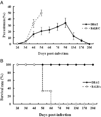
It is well known that BALB/c and DBA/2 mice develop a distinct outcome even when infected with the same strain of malaria parasite. In the present study, both BALB/c and DBA/2 mice were infected with P.y17XL, which was highly virulent to most mice. As expected, P.y17XL was lethal to BALB/c mice with a peak parasitaemia of 48·67±3·4% and all mice died within 6 days. In contrast, P.y17XL infected DBA/2 mice only developed a moderate parasitaemia that increased steadily from 4·57±1·8% on day 3 p.i. to 32±8% on day 9 p.i., followed by a sharp decrease in parasitaemia with survival of all mice (Fig. 1A, B).
Parasitaemia and survival rate of P.y17XL infection in BALB/c and DBA/2 mice. Parasitaemia was calculated by counting the number of parasite-infected erythrocytes per 1000 erythrocytes. Mortality was monitored daily. The data are representative of 2 separate experiments.

Analysis of the effects of Tregs by in vivo CD25 depletion
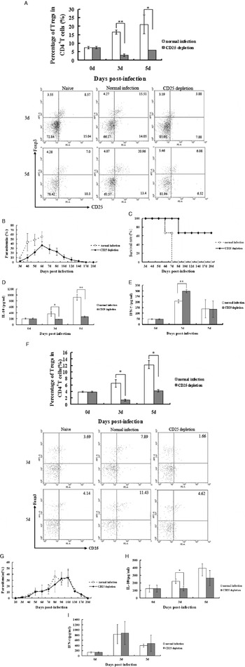
To assess whether Tregs were involved in the inhibition of pro-inflammatory type 1 responses to malaria parasites, Tregs were depleted in vivo by injection of anti-CD25 mAb prior to and post-P.y17XL infection in BALB/c and DBA/2 mice. Tregs depletion was confirmed by flow cytometry (Fig. 2A, F). We compared the difference in frequency of Tregs between the CD25 depletion group and the control group in BALB/c and DBA/2 mice during P.y17XL infection. The proportion of Tregs in the CD4+ T cell population derived from CD25-depleted BALB/c and DBA/2 mice was lower than that of the control group with 81% or 80% reduction on day 3 p.i. and 71% or 66% reduction on day 5 p.i.
Flow cytometric analysis demonstrated Tregs in CD4+ T cell population before and after anti-CD25 mAb treatment. Column diagram (upper) and representative dot plots (lower) of Tregs in the BALB/c and DBA/2 mice without infection (naive), CD25-depleted BALB/c and DBA/2 mice infected with P.y17XL (CD25 depletion) and BALB/c and DBA/2 mice infected with P.y17XL (normal infection) were displayed. The CD4+ cell population was gated for the above dot plots. The proportion and statistics of Tregs in CD4+ T cells, parasitaemia and survival rate in CD25-depleted BALB/c (A, B, C) and DBA/2 (F, G) mice during P.y17XL infection are shown. Parasitaemia was calculated by counting the number of parasite-infected erythrocytes per 1000 erythrocytes. Mortality was monitored daily. Levels of IL-10 (D, H) and IFN-γ (E, I) in spleen cell supernatants in P.y17XL-infected BALB/c and DBA/2 mice during pre-and post-CD25 depletion were measured. Results are presented as arithmetic mean of 3 mice per group±S.E. Single asterisk (*) and double asterisks (**) indicate P<0·05 and P<0·01, respectively, compared with control mice. The data are representative of 2 separate experiments.

In a previous study, we found that dynamic changes in the number of Tregs were significantly correlated with the final outcome of malaria infection. Therefore, to investigate the role of Tregs in malaria infection, parasitaemia, survival rate, and the levels of IL-10, IFN-γ in CD25-depleted BALB/c and DBA/2 mice were evaluated during the process of P.y17XL infection. As shown in Fig. 2B and C, CD25-depleted BALB/c mice demonstrated a weak peak parasitaemia of 29·8% on day 6 p.i. and survival rate of 66·7% in comparison with CD25 non-depleted mice; However, as shown in Fig. 2G, CD25-depleted DBA/2 mice displayed a similar course of parasitaemia compared with CD25 non-depleted mice during the early stage of infection, and all mice survived (data not shown). Consistently, CD25 depletion significantly reversed the production of pro-inflammatory type 1 cytokine, IFN-γ on day 3 p.i. (P<0·01), and obviously decreased the level of IL-10 on days 3 p.i. (P<0·05) and 5 p.i. (P<0·01) in BALB/c mice, respectively (Fig. 2D, E), but not in DBA/2 mice (Fig. 2H, I).
The proportion and absolute numbers of IL-10-secreting Tregs in two mouse strains during P.y17XL infection
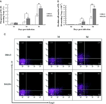
To investigate the possible modulating mechanisms of Tregs, we analysed the proportional changes of IL-10-secreting Tregs by intracytoplasmic staining. The proportion and absolute numbers of IL-10-secreting Tregs were increased on day 3 p.i. in both BALB/c and DBA/2 mice, then went up to 2·7% (absolute cell numbers: 3·2×105) in DBA/2 mice reaching 4·85% (absolute cell numbers: 7·5×105) in BALB/c mice on day 5 p.i.. It should be noted that, although the frequency of IL-10-secreting Tregs increased in DBA/2 mice in response to P.y17XL infection, it was only approximately half of that in BALB/c mice at detected time-points (Fig. 3A, 3B, 3C).
Proportion (A), absolute cell numbers (B) and representative dot plots (C) of IL-10-secreting Tregs in P.y!17XL-infected BALB/c and DBA/2 mice at different time-points. Flow cytometric analysis was performed on spleen cells from BALB/c and DBA/2 mice by quadruple-staining with FITC-anti-CD4, PE-anti-CD25 and intra-cytoplasmic staining with APC-anti-Foxp3 and PerCP-Cy5.5-anti-IL-10. The CD4+CD25+ cell population was gated for the above dot plots. Column diagram of IL-10-secreting Tregs in P.y17XL-infected BALB/c and DBA/2 mice were displayed on days 0, 3 and 5 p.i. Results are presented as arithmetic mean of 3 mice per group±S.E. Single asterisk (*) and double asterisks (**) indicate P<0·05 and P<0·01, respectively, compared with control mice. #P<0·05 and ##P<0·01 indicate the comparison between DBA/2 and BALB/c mice. The data are representative of 2 separate experiments.

The proportion of CD4+ T cell apoptosis in two mouse strains during P.y17XL infection
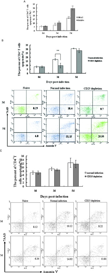
To investigate the immunoregulatory role of Tregs in the development of a Th1 response during early P.y17XL infection, the proportion of apoptotic CD4+ T cells was evaluated. The apoptotic population of CD4+ T cells significantly increased on day 3 p.i. (P<0·01) in both BALB/c and DBA/2 mice, then it gradually increased in resistant DBA/2 mice, while it was rapidly elevated in susceptible BALB/c mice, to approximately twice of that in DBA/2 mice on day 5 p.i. (Fig. 4A). Furthermore, we detected the apoptotic population of CD4+ T cells in P.y17XL-infected BALB/c and DBA/2 mice with Tregs depletion. As expected, Tregs depletion in BALB/c mice also reversed the apoptosis of CD4+ T cells on day 3 p.i. (Fig. 4B). However, there was no significant difference in CD4+T cell apoptosis between Tregs-depleted DBA/2 mice and the control group (Fig. 4C).
CD4+ T cell apoptosis in P.y17XL-infected BALB/c and DBA/2 mice was analysed by FACS at different time-points (A), as well as that in BALB/c (B) and DBA/2 (C) mice after anti-CD25 mAb treatment. Column diagram (upper) and representative dot plots (lower) of CD4+ T cell apoptosis in the BALB/c and DBA/2 mice without infection (naive), CD25-depleted BALB/c and DBA/2 mice infected with P.y17XL (CD25 depletion) and BALB/c and DBA/2 mice infected with P.y17XL (normal infection) were displayed. Flow cytometric analysis was performed on spleen cells from BALB/c and DBA/2 mice by triple-staining with FITC-anti-CD4, PE-anti-Annexin V and 7AAD. The CD4+cell population was gated for the above dot plots. Results are presented as the arithmetic mean of 3 mice per group±S.E. Single asterisk (*) and double asterisks (**) indicate P<0·05 and P<0·01, respectively, compared with control mice. The data are representative of two separate experiments.

Analysis of the effect of IL-10R blockade in vivo
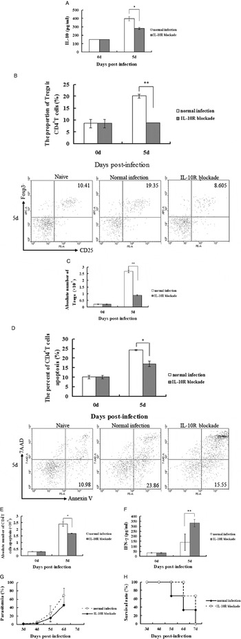
To further investigate the relationship of Tregs and IL-10 in the immunoregulatory mechanisms during malaria infection, IL-10R were blocked in vivo by injection of anti-IL-10R antibody prior to and post-P.y17XL infection in BALB/c mice. In IL-10R-blocked mice, the level of IL-10 obviously decreased compared with IL-10R non-blocked mice on day 5 p.i. (Fig. 5A). In addition, the proportion and absolute numbers of Tregs and CD4+T cell apoptosis, the level of IFN-γ, parasitaemia, and survival rate in IL-10R-blocked mice were evaluated during the process of P.y17XL infection. As shown in Fig. 5B–E, the proportion and absolute numbers of Tregs in IL-10R-blocked mice showed an obvious decrease, accompanied by a lack of CD4+T cell apoptosis compared with IL-10R non-blocked mice. IL-10R blockade significantly restored the production of pro-inflammatory type 1 cytokine, IFN-γ on day 5 p.i. (P<0·01) (Fig. 5F). In addition, a repressed peak parasitaemia was apparent in IL-10R-blocked mice in comparison with IL-10R non-blocked mice (Fig. 5G, H).
Analysis of the effect of IL-10R blockade in P.y17XL-infected BALB/c mice. Levels of IL-10 (A) in spleen cell supernatants in P.y17XL-infected BALB/c mice during pre-and post-IL-10 blockade was measured. Flow cytometric analysis demonstrated the proportion and absolute cell number of Tregs in CD4+ T cells (B, C) and CD4+T cell apoptosis (D, E) in IL-10-blocked mice during P.y17XL infection. Column diagram (upper) and representative dot plots (lower) of Tregs in BALB/c mice without infection (naive), IL-10-blocked BALB/c mice infected with P.y17XL (IL-10 blockade) and BALB/c mice infected with P.y17XL (normal infection) are displayed. The CD4+cell population was gated for the above dot plots. And the level of IFN-γ (F) was also measured. Parasitaemia (G) and survival rate (H) in IL-10-blocked BALB/c are shown. Parasitaemia was calculated by counting the number of parasite-infected erythrocytes per 1000 erythrocytes. Mortality was monitored daily. Results are presented as arithmetic mean of 3 mice per group±S.E. Single asterisk (*) and double asterisks (**) indicate P<0·05 and P<0·01, respectively, compared with control mice. The data are representative of 2 separate experiments.

DISCUSSION
Infection with malaria parasites has long been known to be associated with ‘suppressed’ immune responses to the parasite and to unrelated antigens (Greenwood et al. Reference Greenwood, Bradley-Moore, Bryceson and Palit1972). Tregs is a T-cell subgroup that has immunoregulatory effects different from Th1 and Th2. Tregs were widely accepted as potent suppressors that play important roles in immunotolerance, autoimmunity, and transplantation (Sakaguchi et al. Reference Sakaguchi, Sakaguchi, Asano, Itoh and Toda1995; Shevach et al. Reference Belkaid, Piccirillo, Mendez, Shevach and Sacks2002; Martin et al. Reference Martin, Banz, Bienvenu, Cordier, Dautigny, Bécourt and Lucas2004; Rouse and Suvas, Reference Rouse and Suvas2004; Lee et al. Reference Lee, Sen, Snapper, Lee, Sen and Snapper2005). The suppression of Tregs in malaria infection has been addressed in the rodent model. In this study, we showed that Tregs had an important role in regulating pro-inflammatory type 1 response during early P.y17XL infection through an IL-10-dependent regulatory mechanism and induction of CD4+ T cell apoptosis. More importantly, the intensity of Tregs activation seems to be critical for the outcome of malaria infection.
The characteristic outbreak proliferation of blood-stage malaria parasites leads to the death of the mice infected with P.y17XL. Similar to most other infectious diseases, the immune effector mechanisms are required to eliminate the malaria infection. Indeed, accumulating evidence has indicated that CD4+ T cells are essential to control malaria infection (Taylor-Robinson et al. Reference Taylor-Robinson and Phillips1994; Seixas and Ostler, Reference Seixas and Ostler2005; Couper et al. Reference Couper, Phillips, Brombacher and Alexander2005; Yazdani et al. Reference Yazdani, Mukherjee, Chauhan and Chitnis2006). In particular, the CD4+ Th1 immune effector mechanism is crucial for controlling the outbreak proliferation of malaria parasites during early infection by triggering cellular immune responses (Stevenson et al. Reference Stevenson, Tam, Wolf and Sher1995; Jacobs et al. Reference Jacobs, Radzioch and Stevenson1995; Collins et al. Reference Collins, Jeffery and Roberts2004; Urban et al. Reference Urban, Ing and Stevenson2005). In the previous study, we demonstrated that the resolution of the malaria infection depends on the effective establishment of a Th1 immune response, and changes in the number of Tregs were shown to significantly affect the final outcome of malaria infection (Wu et al. Reference Wu, Wang, Zheng, Feng, Liu, Ma and Cao2007). Here, we further displayed that Tregs depletion with anti-CD25 mAb in P.y17XL-infected BALB/c mice resulted in a higher level of IFN-γ production and a correlated decrease of susceptibility to P.y17XL infection. These data implied that Tregs could significantly suppress the magnitude of P.y17XL-specific pro-inflammatory type 1 response in vivo, which was consistent with the results of Cambos et al. (Reference Cambos, Bélanger, Jacques, Roulet and Scorza2008). These observations also suggested that the regulation of anti-malarial immune responses was as important as their induction in determining the final outcome of infection.
Tregs activation could constitute a mechanism utilized by the parasite to evade host immunity (Hisaeda et al. Reference Hisaeda, Maekawa, Iwakawa, Okada, Himeno, Kishihara, Tsukumo and Yasutomo2004). Tregs depletion led to a modest delay in parasite growth in mice infected with P. berghei NK65 (Long et al. Reference Long, Nakazawa, Onizuka, Huaman and Kanbara2003). Tregs were shown to be required to limit pro-inflammatory immune responses in BALB/c mice to prevent cerebral malaria during secondary infection (Nie et al. Reference Nie, Bernard, Schofield and Hansen2007). In this study, we observed that, in Tregs-depleted BALB/c mice, parasite growth was markedly delayed or decreased. More importantly, a substantial prolongation of survival time was seen in Tregs-depleted BALB/c mice with a survival rate of 66·7% in comparison with Tregs non-depleted mice during the P.y17XL infection. However, in Tregs-depleted DBA/2 mice, only the level of IL-10 production showed a significant decrease on day 3 p.i., while parasitaemia, survival rate and the levels of IFN-γ had no obvious changes compared with normally infected mice. This may correlate with the proliferation and activation pattern of Tregs in DBA/2 mice during the early infection. As shown above, the proportion of Tregs in P.y17XL-infected DBA/2 mice was approximately one half of that in BALB/c mice at detected time-points, which could not exert as much inhibitory effect as those in BALB/c mice. In addition, the capability of IFN-γ production in P.y17XL-infected DBA/2 mice was obviously higher than that in BALB/c mice. Taken together, these observations implied that the development of immune escape was mediated possibly by Tregs via inhibition of the establishment of pro-inflammatory immune responses, thus affecting the course and intensity of immune responses to malaria infection.
Immune-suppressive activity of Tregs can be mediated by a cell-contact-dependent mechanism and through the secretion of suppressive cytokines such as TGF-β and IL-10 (Couper et al. Reference Couper, Blount and Riley2008; Thornton and Shevach, Reference Thornton and Shevach1998; Nakamura et al. Reference Nakamura, Kitani and Strober2001; Asseman et al. Reference Asseman, Mauze, Leach, Coffman and Powrie1999). A human malaria research study reported the close relationship of increased percentages of Tregs and elevated IL-10 responses to antigen of P. falciparum in cord blood mononuclear cells (Brustoski et al. Reference Brustoski, Moller, Kramer, Hartgers, Kremsner, Krzych and Luty2006). During L. major infection the ability of nTreg to limit anti-parasitic effector responses (leading to persistent low level infection) is in part IL-10 dependent (Belkaid et al. Reference Belkaid, Piccirillo, Mendez, Shevach and Sacks2002). Similarly, our data showed that the proportion and absolute numbers of IL-10-secreting Tregs in BALB/c mice were significantly higher than that in DBA/2 mice and IL-10 production was consistent with the Tregs population. These results indicated that the immune-suppressive activity of Tregs can be mediated through the secretion of IL-10. As shown by Goudy et al. (Reference Goudy, Burkhardt, Wasserfall, Song, Campbell-Thompson, Brusko, Powers, Clare-Salzler, Sobel, Ellis, Flotte and Atkinson2003) and Hori and Sakaguchi (Reference Hori and Sakaguchi2004), IL-10 has a wide range of cell sources and affects the differentiation and function of Tregs. We also found experimentally that the majority of IL-10 comes from CD4+CD25+Foxp3− cells. IL-10 from nTreg alone is insufficient to counter the very high levels of inflammation associated with virulent infections (Couper et al. Reference Couper, Blount and Riley2008). Therefore, during the early stages of P.y17XL infection, regulatory effectors of other IL-10-secreting cells, especially CD25+Foxp3− cells, need to be evaluated. We noticed that IL-10 production in CD25-depleted BALB/c mice on day 5 p.i. was higher than that on day 3 p.i.. This phenomenon may be due to recovery of some Tregs on day 5 p.i., and then the percentage of Tregs was higher on day 5 than that on day 3 p.i. in CD25-depleted mice. A related study also found that the numbers and percentages of CD25 (high) cells were transiently reduced after 7D4 treatment within 3 days in Plasmodium yoelii 17X (non-lethal) infection (Couper et al. Reference Couper, Blount, de Souza, Suffia, Belkaid and Riley2007).
In addition, suppressive effects of Tregs may also be mediated, in part, by the induction of apoptosis in the CD4+CD25− effector cells via a contact-dependent mechanism (Gondek et al. Reference Gondek, Lu, Quezada, Sakaguchi and Noelle2005). So we also evaluated immune-suppressive activity of Tregs by measuring the apoptotic CD4+T cells in the spleen. The apoptotic CD4+ T cells in BALB/c mice were higher than that in DBA/2 mice, which was consistent with the frequency of Tregs in both strains of mice. After Tregs were depleted, the apoptotic CD4+ T cells in BALB/c mice markedly decreased in comparison with the control group. Our data also raised the possibility that Tregs were involved in the process of apoptosis of CD4+T cells. Related research proved that malaria parasites also induced apoptosis in CD4+T cells by Fas pathways during infection, but Fas expression alone is not sufficient for apoptosis (Wipasa et al. Reference Wipasa, Xu, Stowers and Good2001). Therefore, other mechanisms of CD4+T cell apoptosis which act through Fas pathways or through the induction of anti-CD25 antibody need to be further studied.
Some studies have specified that systemic over-expression of IL-10 induces a boost in the Tregs population (Goudy et al. Reference Goudy, Burkhardt, Wasserfall, Song, Campbell-Thompson, Brusko, Powers, Clare-Salzler, Sobel, Ellis, Flotte and Atkinson2003) IL-10−/− mice or mice treated with anti-IL-10R or anti-CD25 antibody are able to resolve a strain of Leishmania major (Sd) infection (Anderson et al. Reference Anderson, Mendez and Sacks2005). To support our hypothesis that the immune-suppressive activity and differentiation of Tregs correlated with IL-10 signalling, we evaluated the effect of IL-10R blockade on the inhibitory activity of Tregs. Our results displayed that IL-10R blockade had an effect resembling that of Tregs depletion in P.y17XL-infected BALB/c mice. IL-10R blockade significantly increased the production of IFN-γ, and parasite growth was delayed or decreased. In particular, the proportion and absolute numbers of Tregs was markedly reduced and, at the same time, was accompanied by the loss of CD4+T cell apoptosis compared with IL-10R non-blockaded mice. Although we did not observe a substantial prolongation of survival time in IL-10R-blockaded BALB/c mice, the main reason perhaps was that IL-10-dependent mechanism is only one of the pathways of the immune-suppressive effect of Tregs. Thus, our data further confirmed that Tregs contribute to the suppression of pro-inflammatory type 1 responses, in part, in an IL-10-dependent manner. In addition, we also proved that IL-10 affected the differentiation and function of Tregs.
In summary, pro-inflammatory immune effector mechanisms are essential to control the proliferation of malaria parasites during early P.y17XL infection. Moreover, Tregs have an important role in regulating pro-inflammatory type 1 responses during early rodent malaria infections. The potential regulatory mechanisms likely include the induction of IL-10 and CD4+ T cell apoptosis. Further studies are needed to understand the conditions that induce Tregs differentiation and the required signals for priming and differentiation of Tregs during early P.y17XL infection.
We thank Dr Motomi Torii (Ehime University Graduate School of Medicine, Ehime, Japan) for his guidance in this research and for providing malaria parasite strains of P.y17XL. Critical reading of the manuscript by Drs Hajime Hisaeda and Osamu Kaneko is gratefully acknowledged. This work was supported by Grant 20070159006 from Research Fund for the Doctoral Program of Higher Education of China and Grant 2007CB513100 from National Basic Research Program in China (973 Program).