Introduction
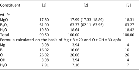
Halurgite is a hydrous magnesium borate mineral discovered in 1959 in cores of several boreholes drilled for boron prospecting in evaporite rocks consisting largely of halite with subordinate boracite, kaliborite, pinnoite and anhydrite. The original description of halurgite (Lobanova, Reference Lobanova1962) included data on occurrence, morphology, optical properties, measured density (2.19 g cm–3), thermal behaviour, chemistry and powder X-ray diffraction (XRD) pattern, but with no geographical information on the locality. Chemical analyses of the mineral carried out on contaminated material gave the simplified formula 2MgO·4B2O3·5H2O (Lobanova, Reference Lobanova1962). Table 1, #1 gives the analysis obtained on material with the fewest impurities. The XRD data reported by Lobanova (Reference Lobanova1962) are of a very poor quality and no attempt was made to characterise the crystallography.
Chemical composition of halurgite*.
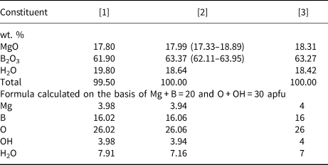
* [1] Wet chemical analysis (type specimen: Lobanova, Reference Lobanova1962); [2] our electron microprobe data for specimen Chelk-5613 (average for four analyses, ranges are in parentheses; H2O is calculated by difference from 100 wt. %); [3] calculated for the idealised formula Mg4[B8O13(OH)2]2·7H2O.
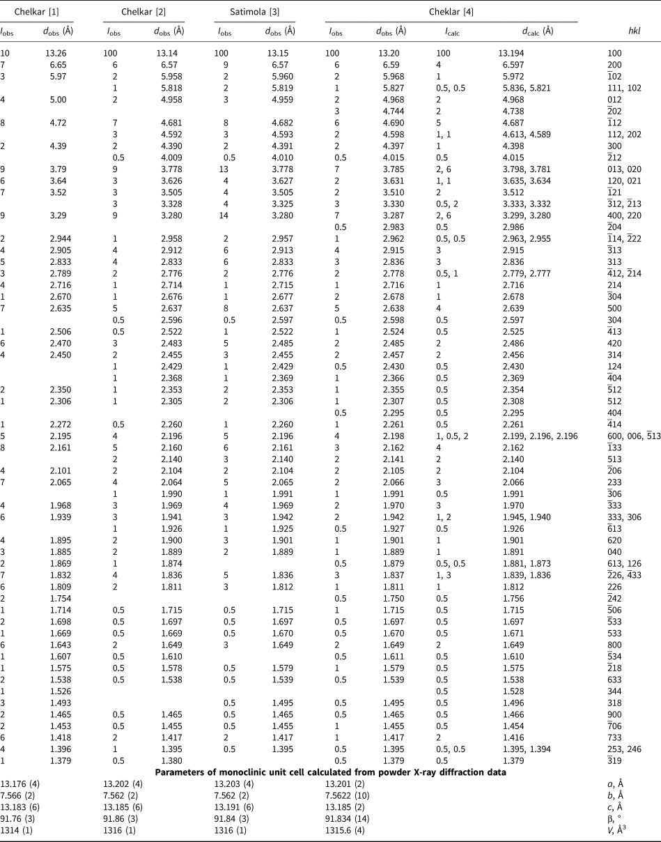
The XRD studies of halurgite were later performed by V.V. Kondrat'eva on the type material provided by V.V. Lobanova. Single-crystal data yielded monoclinic symmetry, possible space group P2/c or Pc and the following unit-cell parameters: a = 13.25(4), b = 7.60(3), c = 13.20(4) Å and β = 92°09(10)°. The density for the simplified formula Mg2B8O14·5H2O (Z = 4) was calculated to be 2.238 g cm–3. The low quality of the single crystals studied precluded determination of the crystal structure, however, it was assumed that the borate polyanion in halurgite could be based on units consisting of two B tetrahedra and two B triangles combined in a finite anionic motif. Kondrat'eva (Reference Kondrat'eva1964) suggested Mg2[B4O5(OH)4]2·H2O as the structural formula and Kondrat'eva (Reference Kondrat'eva1969) obtained and indexed a good-quality powder XRD pattern of halurgite from the same sample (Table 2, #1).
Powder X-ray diffraction data for halurgite*.
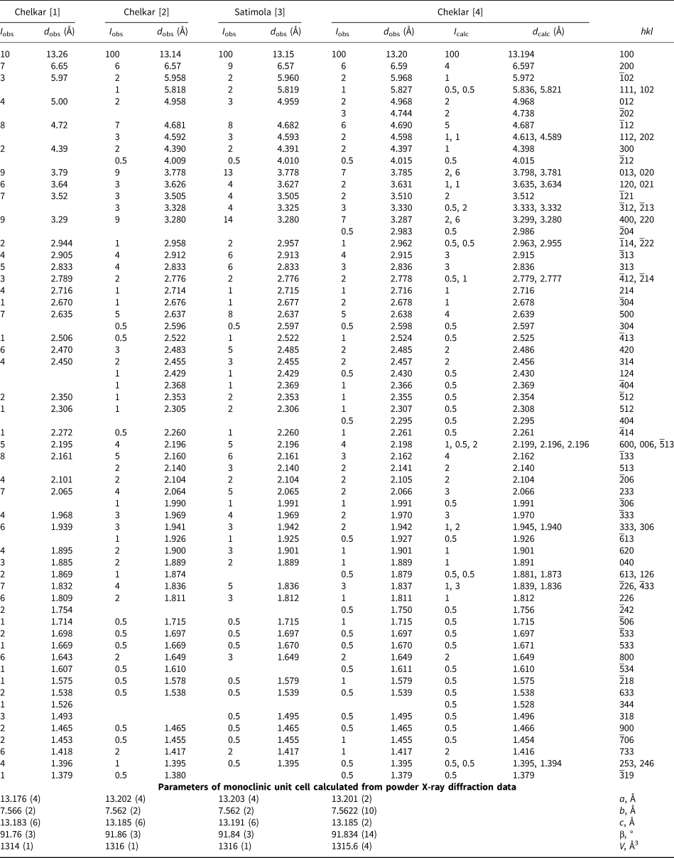
Sources: [1] Kondrat'eva (Reference Kondrat'eva1969); [2–4] this work: [2] type specimen (FMM 69833), [3] specimen FMM ST-7013, [4] specimen with studied crystal structure (Chelk-5613).
Additional data on the occurrence and mineral associations of halurgite were reported by Avrova et al. (Reference Avrova, Bocharov, Khalturina and Yunusova1968). This paper has the first record of the mineral being discovered in Kazakhstan. Its exact type locality, Chelkar salt dome in the North Caspian Region, Western Kazakhstan, and a list of type specimens of halurgite deposited in two museums were published by Pekov (Reference Pekov1998). The infrared (IR) absorption spectrum of the mineral was reported by Chukanov (Reference Chukanov2014). All above-cited works are based on the material extracted from drill cores of boreholes at Chelkar between 1959 and the early 1960s.
A new find of halurgite in the core of a borehole recently drilled in the Chelkar salt dome was briefly described by Korotchenkova and Chaikovskiy (Reference Korotchenkova and Chaikovskiy2016). A single crystal of halurgite suitable for the crystal structure model determination was selected from this sample. Based on this model, the structure was refined on a powder sample using the Rietveld method.
In the present paper, we report a revised formula based on the first crystal-structure determination and chemical analyses of this mineral, an improved powder XRD pattern and IR spectroscopic data. In addition to the new sample, we have re-examined the type material from Chelkar and studied halurgite from the Satimola salt dome, which is also located in the North Caspian Region, Western Kazakhstan. The latter find has not yet been reported in the literature. The XRD and IR spectroscopic characteristics of all studied halurgite samples are nearly identical to one another.
Samples and experimental methods
Description of samples
Three samples of halurgite have been studied in the present work.
Chelk-5613
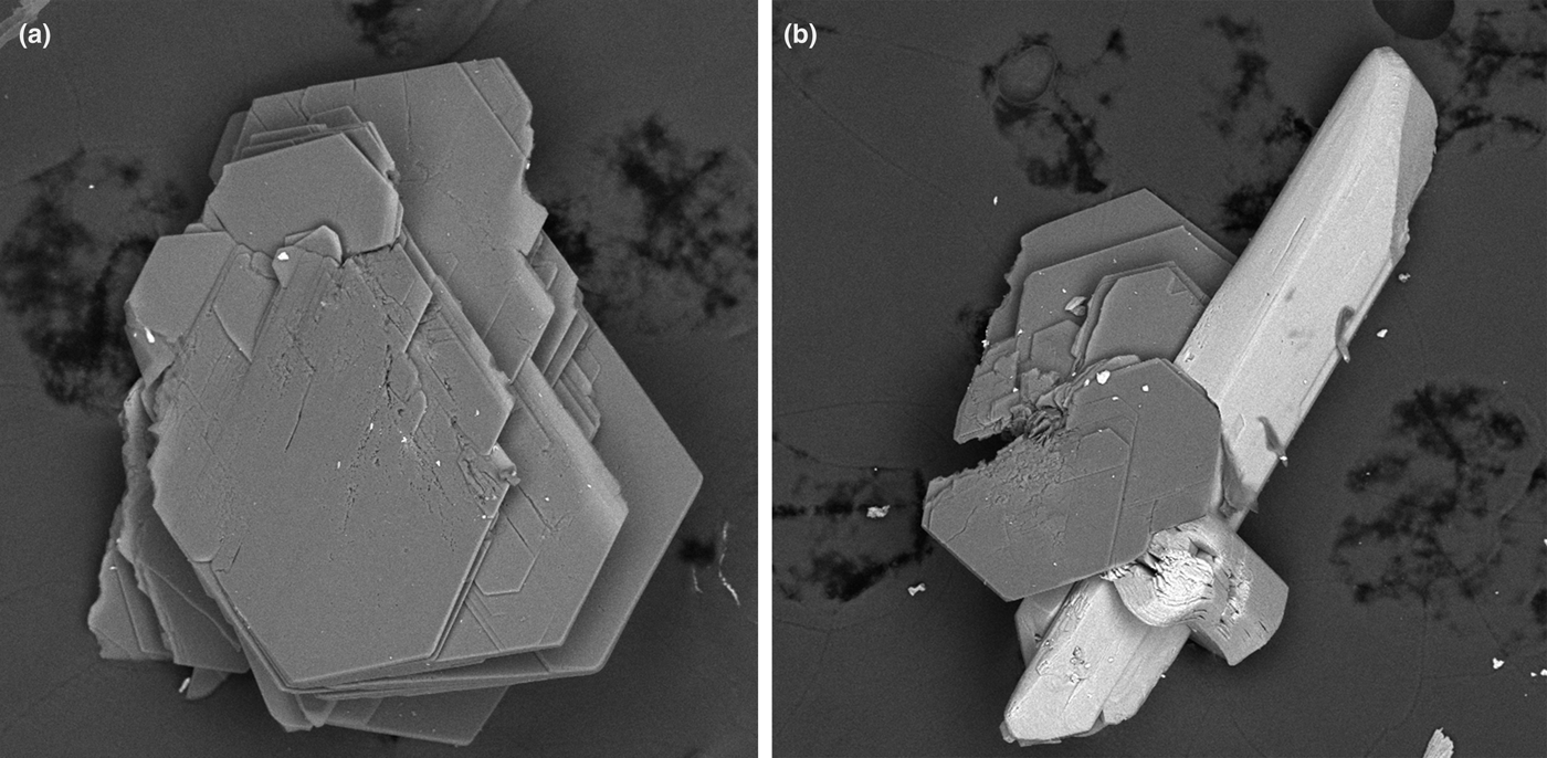
This specimen used for the crystal structure determination was studied previously by Korotchenkova and Chaikovskiy (Reference Korotchenkova and Chaikovskiy2016). Halurgite forms transparent, colourless pseudo-hexagonal lamellar crystals up to 1 mm across, typically in clusters (Fig. 1a) embedded in a halite–carnallite rock and intimately associated with gypsum (Fig. 1b), calcite, boracite, satimolite, kieserite and dashkovaite.
Back-scatter electron images of halurgite from Chelkar (sample Chelk-5613; extracted from halite–carnallite rock after its dissolution in water): (a) cluster of lamellar crystals; and (b) lamellar crystals intergrown with prismatic crystals of gypsum. Field of view width: (a) 0.5 mm; (b) 0.55 mm.

FMM 69833
This is the type specimen of halurgite from Chelkar deposited in the systematic collection of the Fersman Mineralogical Museum of the Russian Academy of Sciences, Moscow, Russia, with catalogue no. 69833 (received from V.V. Lobanova in 1967). Halurgite forms white dense aggregates up to 0.5 cm × 1 cm consisting of split lamellae up to 0.05 mm across. These aggregates are embedded in massive halite and contain fine ingrowths of dashkovaite.
FMM ST-7013
This specimen from Satimola is from the collection of the outstanding Russian mineralogist and mineral collector Victor Ivanovich Stepanov (1924–1988). The collection of V.I. Stepanov was donated by him in 1983 to the Fersman Mineralogical Museum and now it is deposited as a separate part of the museum collections (Pekov et al., Reference Pekov, Lykova and Nikiforov2015). This specimen was collected by I.I. Khalturina, a specialist in the mineralogy of borates, and received by V.I. Stepanov from her in 1968. Halurgite, identified initially in this specimen by I.I. Khalturina, occurs as white sugar-like nodules up to 4 mm across embedded in massive halite and associated with kaliborite.
Chemical composition determination
The chemical composition of halurgite (sample Chelk-5613) was studied using a JEOL JSM-6480LV scanning electron microscope equipped with an INCA-Wave 500 wavelength-dispersive spectrometer (Laboratory of Analytical Techniques of High Spatial Resolution, Department of Petrology, Moscow State University). The wavelength-dispersive spectroscopy mode was used, with an acceleration voltage of 20 kV and a beam current of 10 nA; electron beam was rastered to a 5 µm × 5 µm area to minimise sample damage. The standards used were MgO (Mg) and BN (B). Contents of other elements with atomic numbers higher than oxygen are below detection limits. H2O was not determined because of the scarcity of pure material, and thus its content is given as the difference from 100 wt.%.
The empirical formulae were calculated on the basis of Mg + B = 20 atoms per formula unit (apfu) and O + OH in the borate anion equal to 30 apfu (according to crystal structure data: see below) that allows us to calculate the ratios between O2–, OH– and H2O in halurgite.
Infrared spectroscopy
In order to obtain IR absorption spectra, powdered samples of halurgite were mixed with dried KBr, pelletised, and analysed using an ALPHA FTIR spectrometer (Bruker Optics) with a resolution of 4 cm–1 and 16 scans. The IR spectrum of a pellet of pure KBr was used as a reference.
X-ray diffraction studies and crystal structure determination details
Single-crystal XRD studies of halurgite (sample Chelk-5613) were carried out at room temperature using an Xcalibur S diffractometer equipped with a CCD detector (MoKα radiation, λ = 0.71073 Å). A full sphere of three-dimensional data was collected. Data reduction was performed using CrysAlisPro, version 1.171.37.34 (Agilent Technology, 2014). The data were corrected for Lorentz factor and polarisation effects. A total of 20,819 reflections was obtained in the θ range 2.70 to 28.28°. The crystal structure was solved by direct methods and refined using the SHELX-97 software package (Sheldrick, Reference Sheldrick2015).
Powder X-ray diffraction data of halurgite were collected with a Rigaku R-AXIS Rapid II single-crystal diffractometer equipped with a cylindrical image plate detector (with a radius of 127.4 mm) using Debye-Scherrer geometry, CoKα radiation (rotating anode with VariMAX microfocus optics), 40 kV, 15 mA and exposure 15 min. Angular resolution of the detector is 0.045°2θ (pixel size 0.1 mm). The data were integrated using the software package Osc2Tab (Britvin et al., Reference Britvin, Dolivo-Dobrovolsky and Krzhizhanovskaya2017).
Data treatment and the Rietveld structure analysis were carried out for sample Chelk-5613 using the JANA2006 program package (Petříček et al., Reference Petříček, Dušek and Palatinus2006). The profiles were modelled using a pseudo-Voigt function. The structure was refined in isotropic approximation of atomic thermal displacements, the values of U iso for all atoms of each sort were restricted to be equal. The cation–anion interatomic distances were softly restricted to be close to the values obtained for the single-crystal model.
Results
Chemical composition
Chemical composition of the sample Chelk-5613 is given in Table 1 in comparison with data for the type specimen of halurgite reported by Lobanova (Reference Lobanova1962). Table 1 demonstrates that the B:Mg atomic ratio in both samples is very close to 4:1 and the type specimen contains some more molecular H2O in comparison with the sample Chelk-5613. It is not excluded that this difference is caused by the microporous character of the halurgite structure (see below) that permits a variable amount of weakly bonded water.
Infrared spectroscopic data
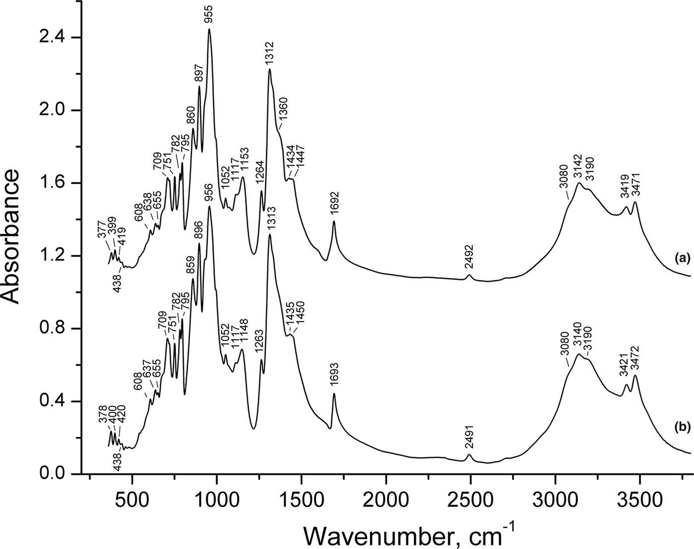
The IR spectra of halurgite from Chelkar (type specimen: FMM 69833) and Satimola (sample FMM ST-7013) are practically identical (Fig. 2). Absorption bands in the range 3000–3500 cm–1 correspond to O–H stretching vibrations (Chukanov and Chervonnyi, Reference Chukanov and Chervonnyi2016). Based on the correlation ν (cm–1) = 3592–304·109·exp[–d(O···O)/0.1321] for hydrogen bonds (Libowitzky, Reference Libowitzky1999), the bands at 3471, 3419, 3190, 3142 and 3080 cm–1 correspond to the d(O···O) distances of 2.86, 2.81, 2.70, 2.69 and 2.67 Å.
Powder infrared spectra of halurgite from (a) Chelkar (the type specimen) and (b) Satimola.
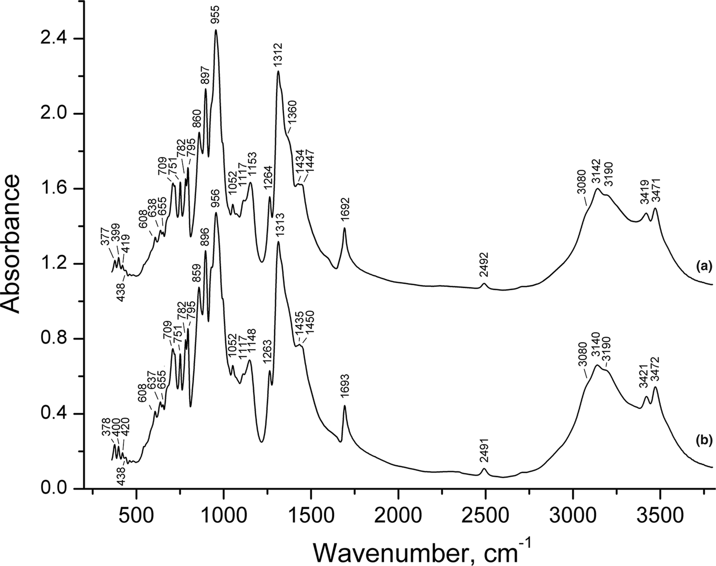
Weak bands in the range from 2200 to 2800 cm–1 correspond to overtones and combination modes. The band at 1692–1693 cm–1 is due to H–O–H bending vibrations of H2O molecules (Jun et al., Reference Jun, Shuping and Shiyang1995). Bands in the ranges 1200–1500 and 850–1160 cm–1 correspond to predominantly [3]B–O stretching and [4]B–O stretching vibrations whereas bands in the range 600–800 cm–1 are due to O–B–O and B–O–B bending vibrations partly combined with symmetric stretching vibrations of BO4 tetrahedra (see Weir, Reference Weir1966; Bermanec et al., Reference Bermanec, Furić, Rajić and Kniewald2003; Zhang et al., Reference Zhang, Li, Wu, Yang and Pan2016). Bands below 600 cm–1 correspond to lattice modes involving Mg–O stretching and H2O librational vibrations (Chukanov and Chervonnyi, Reference Chukanov and Chervonnyi2016).
Taking into account structural data, one can suppose that the bands in the ranges 850–1000 and 1050–1150 cm–1 correspond to stretching vibrations of [4]B–O bonds belonging to the [4]B–O–Mg and [4]B–O–[3]B fragments, respectively, because in the latter case high-force-strength O–[3]B bonds are involved in the vibrations. The bands in the range 600–650 cm–1 are due to out-of-plane vibrations of BO3 triangles (Zhang et al., Reference Zhang, Li, Wu, Yang and Pan2016).
Due to the complexity of the crystal structure of halurgite, its IR spectrum consists of a multitude of overlapping bands that cannot be determined uniquely by analysing the spectral curve. As a result, pairs of individual bands with an intensity ratio of ~1:4 and isotope shift of 4% corresponding to the natural 10B/11B ratio are not observed in the IR spectrum.
The IR spectrum of halurgite is unique and can be used for reliable identification of this mineral.
X-ray crystallography
Single-crystal XRD data showed that halurgite is monoclinic, space group P2/c, with the following unit-cell parameters: a = 13.206(3), b = 7.5551(8), c = 13.1790(12) Å, β = 91.780(15)° and V = 1314.3(4) Å3. Unfortunately, the low quality of the single crystal studied and consequently of the experimental data did not allow good agreement between observed and calculated F values [final R hkl = 0.2697 for 3037 reflections with I > 2σ(I)]. However, reasonable values of interatomic distances, as well as a good agreement between the measured and calculated XRD powder data showed that this model is correct. Further refinement of the structure was performed by the Rietveld method using this model.
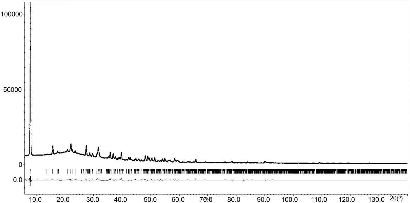
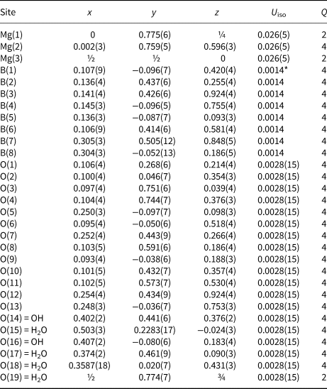
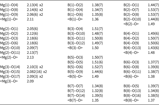
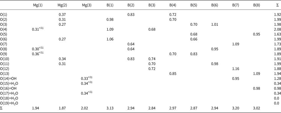
Powder XRD patterns of three above-described samples and the pattern published for the type specimen of halurgite by Kondrat'eva (Reference Kondrat'eva1969), as well as the unit-cell parameters calculated from powder XRD data for all these four samples are very similar to one another (Table 2). The refined unit-cell parameters are: a = 13.201(2), b = 7.5622(10), c = 13.185(2) Å, β = 91.834(14)° and V = 1315.6(4) Å3. The space group is P2/c. Final agreement factors are: R p = 0.0232, R wp = 0.0354 and R obs = 0.0558. The observed and calculated powder XRD diagrams demonstrate very good agreement (Fig. 3). Coordinates and thermal displacement parameters of atoms are given in Table 3 and selected interatomic distances and bond valence calculations [the parameters are taken from Gagné and Hawthorne (Reference Gagné and Hawthorne2015)] in Tables 4 and 5, respectively. The crystallographic information files have been deposited with the Principal Editor of Mineralogical Magazine and are available as Supplementary material (see below).
Observed and calculated powder X-ray diffraction patterns of halurgite. The solid line corresponds to calculated data, the crosses correspond to the observed pattern, vertical bars mark all possible Bragg reflections. The difference between the observed and calculated patterns is shown by curve at the bottom.

Coordinates, isotropic displacement parameters (U iso, in Å2) of atoms and site multiplicities (Q) for halurgite.
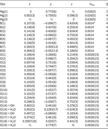
* Fixed in the last stages of the refinement.
Selected interatomic distances (Å) in the structure of halurgite.
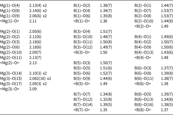
Bond-valence calculations for halurgite*.
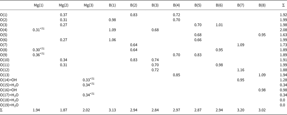
* The bond-valence sums for O(5) and O(7) could be increased due to the possible H bonds with O(16)=OH and O(14)=OH respectively: the corresponding distances are 2.33(5) and 2.42(6) Å. The BVS for O(16) in its turn will be increased due to the possible H bonds with O(15)=H2O and O(19)=H2O and BVS for O(14) will be increased due to the possible H bonds with O(15) = H2O, O(17)=H2O and O(19)=H2O.
Discussion
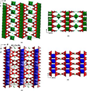
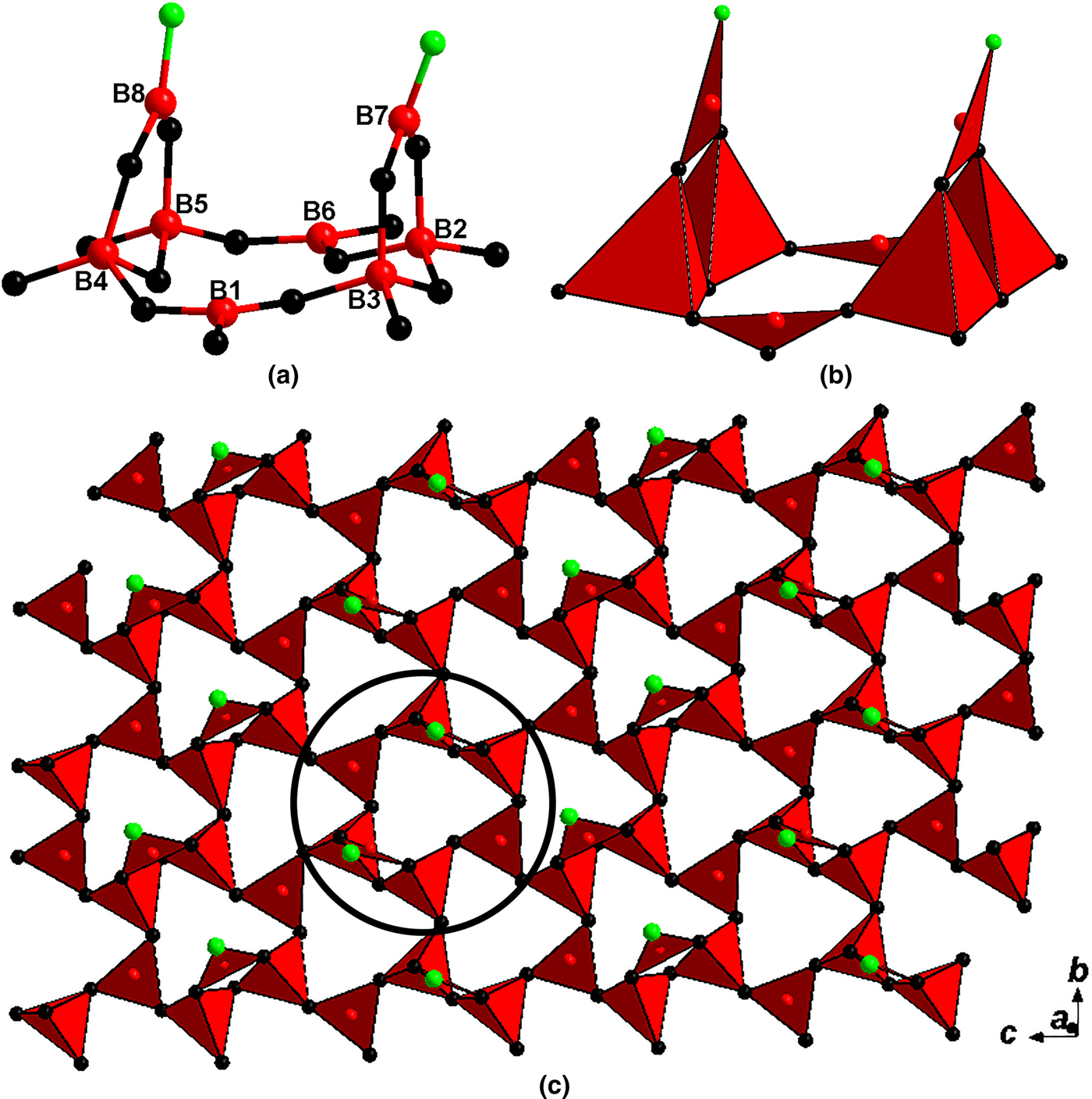
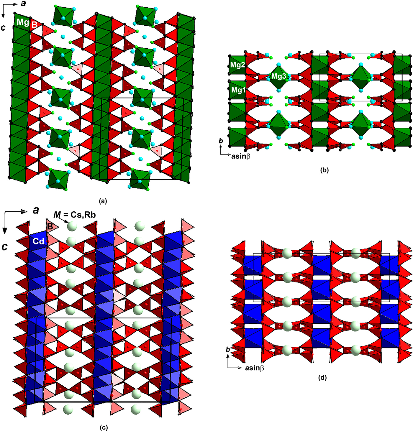
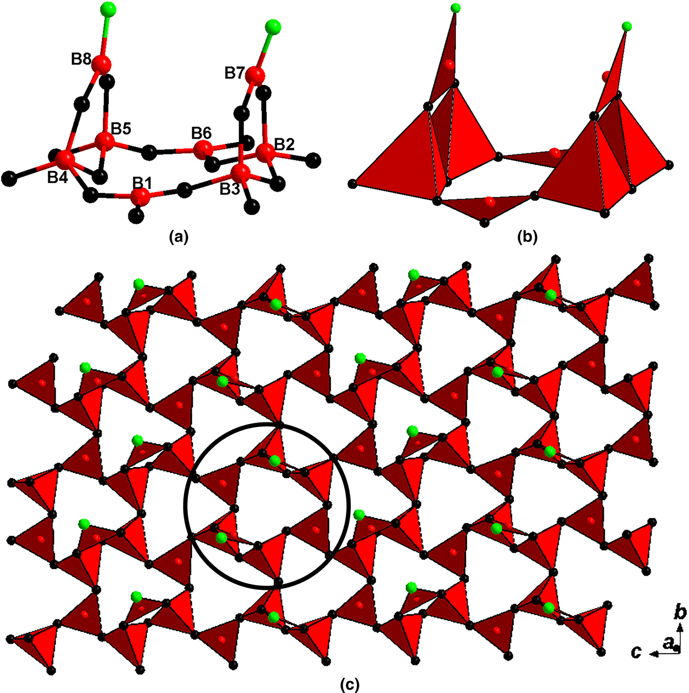
The crystal structure of halurgite (Figs 4a,b) is unique. It is based on the layers of B polyhedra connected via Mg octahedra to form a microporous heteropolyhedral pseudo-framework. There are eight crystallographically non-equivalent B sites adopting both triangular [B(1), B(6), B(7) and B(8) sites] and tetrahedral [B(2), B(3), B(4) and B(5) sites] oxygen coordination. Eight B polyhedra form a fundamental building block (FBB) [B8O16(OH)2] which, according to the classification of FBB in borates (Burns et al., Reference Burns, Grice and Hawthorne1995; Grice et al., Reference Grice, Burns and Hawthorne1999), could be presented as 4Δ4□:<<Δ2□>Δ<Δ2□>Δ>. This means that two three-membered rings (each is formed by two tetrahedra and one triangle) are linked via two triangles. Thus, the six-membered borate ring (formed by two pairs of B tetrahedra and two B-centred triangles) with two additional triangular BO2(OH) groups (each of which is linked to a pair of tetrahedra) is formed (Figs 5a,b). Each [B8O16(OH)2] ring (FBB) links to six adjacent analogous rings to form a [B8O13(OH)2]∞ layer extending in the bc plane (Fig. 5c).
The crystal structures of two heterophylloborates: (a, b) halurgite, along the b and c axes, respectively (positions of O atoms of OH groups are shown as small green circles, O atoms of H2O molecules are given as blue circles); and (c, d) synthetic M 2Cd3B16O28 (M = Rb or Cs) (drawn after Dong et al., Reference Dong, Shi, Zhou, Pan, Yang, Zhang, Yang, Chen and Huang2013), along the b and c axes, respectively. The unit cells are outlined.
Fundamental building block (FBB) [B8O16(OH)2]: (a) ball-and-stick and (b) polyhedral presentations and (c) borate layer [B8O13(OH)2]∞ with outlined FBB in halurgite. For legend see Fig. 4.
A similar FBB was reported in the structures of four synthetic microporous borates: M 2Cd3B16O28 (M = Rb or Cs) (Dong et al., Reference Dong, Shi, Zhou, Pan, Yang, Zhang, Yang, Chen and Huang2013) and M 2Ca3B16O28 (M = Rb or Cs) (Zhang et al., Reference Zhang, Li, Wu, Yang and Pan2016) in which the [B8O18] FBBs are connected to form [B8O15]∞ layers very similar to those found in halurgite. However, unlike the latter, in these synthetic H-free compounds two face-to-face [B8O15]∞ layers are further bridged by corner-sharing O atoms to build a 3D open framework [B8O14]∞ (Figs 4c,d), whereas in halurgite, these sites are occupied by O atoms of OH groups and do not form B–O–B bridges. Thus, the borate layer in halurgite can be considered as a half of the double-layer in the above mentioned anhydrous compounds. The similarity of the (100) borate layers in halurgite, M 2Cd3B16O28 and M 2Ca3B16O28 (all four synthetic borates are monoclinic, space group C2/c) results in similar values for the b, c and β unit-cell parameters: in halurgite b = 7.5622(10), c = 13.185(2) Å and β = 91.780(15)° whereas in these synthetic borates b = 7.6820(11)–7.731(9), c = 13.3694(8)–13.456(5) Å and β = 91.068(8)–91.705(3)°.
In halurgite the linkage between adjacent face-to-face borate layers is via Mg(3) octahedra: each Mg(3) octahedron shares two O(14)=OH vertices with two B(7) triangles belonging to neighbouring borate layers. There are three crystallographically non-equivalent Mg sites in the halurgite structure. All Mg cations are octahedrally coordinated in the mineral, however, different Mg octahedra play different roles in the structure: Mg(1–2)O6 octahedra are isolated from each other and bridge the borate layers from the non ‘face-to-face’ sides sharing all vertices with B polyhedra whereas Mg(3)(OH)2(H2O)4 octahedra link the layers from the ‘face-to-face’ side (Figs 4a,b). The arrangement of Mg(1–2)O6 octahedra is very close to those found for CdO6 octahedra in M 2Cd3B16O28 (M = Rb or Cs) (Dong et al., Reference Dong, Shi, Zhou, Pan, Yang, Zhang, Yang, Chen and Huang2013) and Ca polyhedra (which could be reduced to octahedra if elongated Ca–O distances are omitted) in M 2Ca3B16O28 (M = Rb or Cs) (Zhang et al., Reference Zhang, Li, Wu, Yang and Pan2016), as shown in Figs 4c,d.
There are four crystallographically non-equivalent sites of O atoms of H2O molecules in halurgite. Two of them [O(15) and O(17)] participate in the coordination of Mg(3) cations and two more [O(18–19)] are located in the channels of the microporous heteropolyhedral pseudo-framework and are weakly bonded (Table 5).
The simplified crystal chemical formula of halurgite, thus, is Mg4[B8O13(OH)2]2·7H2O (Z = 2). The calculated density for the sample Chelk-5613 based on the structure refinement data is 2.176 g cm–3 that is close to the value measured for the type specimen: 2.19 g cm–3 (Lobanova, Reference Lobanova1962).
In the structures of synthetic M 2Cd3B16O28 and M 2Ca3B16O28 (M = Rb or Cs) (Dong et al., Reference Dong, Shi, Zhou, Pan, Yang, Zhang, Yang, Chen and Huang2013; Zhang et al., Reference Zhang, Li, Wu, Yang and Pan2016) large M cations occupy tunnels in the borate double layers (Figs 4c,d). In halurgite Mg cations and H2O molecules are located between the single borate layers (Figs 4a,b), which could be considered as a half of the double layer in M 2Cd3B16O28 and M 2Ca3B16O28 (M = Rb or Cs). The presence of such voids and tunnels which can host large cations and H2O molecules determines the microporous character of the compounds.
The crystal structures of halurgite and related synthetic borates can also be described using another approach, which was developed for heterophyllosilicates containing three-layer HOH modules in their structures. In the HOH abbreviation O means octahedral layer sandwiched between two heteropolyhedral (H) layers. In heterophyllosilicates the H layers are built up by Si tetrahedra together with five- or six-coordinated polyhedra commonly occupied by Ti, Nb or Fe, whereas in the O layer octahedral sites are usually occupied by Mn, Fe, Mg, Ti or Na. Representatives of the structural types of astrophyllite, lamprophyllite, bafertisite, lomonosovite and related layered silicate minerals belong to the family of heterophyllosilicates (Ferraris et al., Reference Ferraris, Ivaldi, Pushcharovsky, Zubkova and Pekov2001; Ferraris and Gula, Reference Ferraris, Gula, Ferraris and Merlino2005; Pakhomovsky et al., Reference Pakhomovsky, Panikorovskii, Yakovenchuk, Ivanyuk, Mikhailova, Krivovichev, Bocharov and Kalashnikov2018 and references therein).
For borates, this approach can be modified slightly in part of the H layers of the HOH module. Unlike silicates in which Si is only tetrahedrally coordinated by O ligands, borates can contain B oxygen triangles and tetrahedra. Thus, in a borate a heteropolyhedral module can be built by BO3 triangles and BO4 tetrahedra only, whereas H layers in a heterophyllosilicate necessarily contain five- or six-coordinated polyhedra occupied by metal cations together with Si tetrahedra.
In halurgite the HOH module consists of two heteropolyhedral borate H layers [B8O13(OH)2]∞ formed by B triangles and tetrahedra (Fig. 5c) and sandwiched between them interrupted O layer composed by Mg(1–2)O6 octahedra isolated from each other. The distance between borate tetrahedra belonging to the H layers within the HOH module is distinctly less than that between tetrahedra belonging to the H layers of adjacent HOH modules in halurgite (Figs 4a,b) and synthetic borates structurally related to this mineral (Figs 4c,d). This difference results in the voluminous Mg(3)(OH)2(H2O)4 complex and additional, weakly bonded H2O molecules [O(18–19): Tables 3 and 5] being located between the HOH modules in halurgite; similarly the large Rb and Cs cations are located between the HOH modules in the above-mentioned synthetic borates.
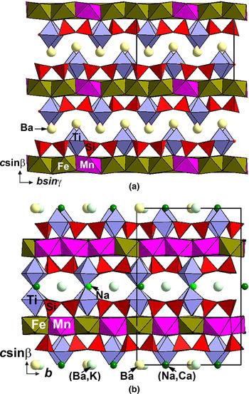
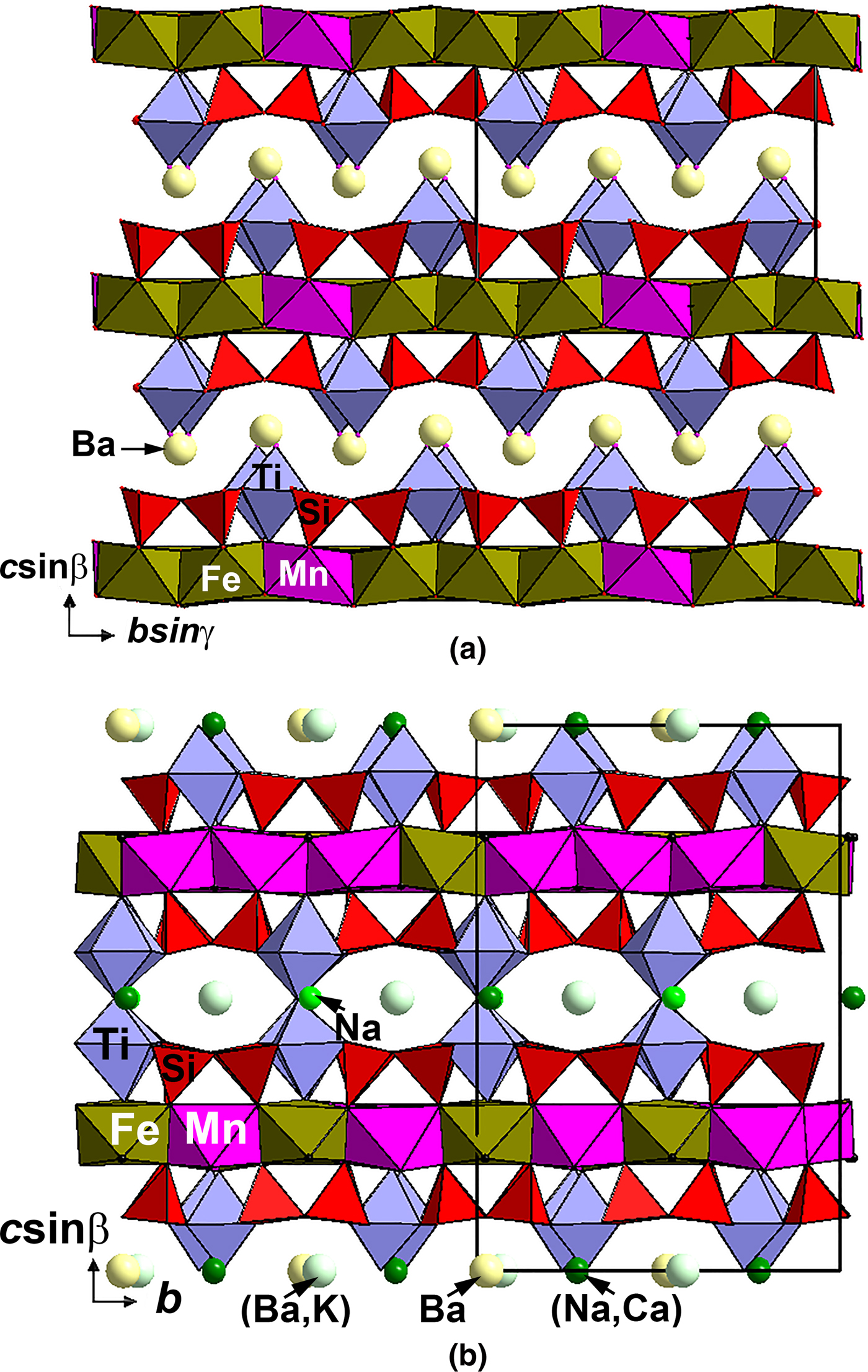
In terms of this approach, the relationship between halurgite and synthetic M 2Cd3B16O28 and M 2Ca3B16O28 (M = Rb or Cs) (Dong et al., Reference Dong, Shi, Zhou, Pan, Yang, Zhang, Yang, Chen and Huang2013; Zhang et al., Reference Zhang, Li, Wu, Yang and Pan2016) in which topologically the same HOH modules are connected via oxygen vertices of BO3 triangles of H layers belonging to adjacent HOH modules, is comparable with the relationship between the heterophyllosilicates bafertisite Ba2(Fe,Mn)4Ti2(Si2O7)O2(OH,F)2F2 (Cámara et al., Reference Cámara, Sokolova, Abdu and Pautov2016) and perraultite (Na,Ca)(Ba,K)(Mn,Fe)4(Ti,Nb)4Si4O16(OH,F,O)3 (Yamnova et al., Reference Yamnova, Egorov-Tismenko and Pekov1998). Their structures are based on HOH modules composed by an O layer of Fe- or Mn octahedra which is sandwiched between two H layers formed by Si2O7 groups and TiO6 octahedra. In bafertisite, adjacent HOH modules are linked only via Ba polyhedra (Fig. 6a) whereas in perraultite the linkage between neighbouring HOH modules is via common oxygen vertices of TiO6 octahedra (Fig. 6b). Thus, the former topologically resembles halurgite while the latter – its synthetic relatives.
The crystal structures of the related heterophyllosilicate minerals (a) bafertisite (drawn after Cámara et al., Reference Cámara, Sokolova, Abdu and Pautov2016) and (b) perraultite (drawn after Yamnova et al., Reference Yamnova, Egorov-Tismenko and Pekov1998). The unit cells are outlined.
Taking into account the above-discussed crystal chemical features, halurgite and synthetic M 2Cd3B16O28 and M 2Ca3B16O28 (M = Rb or Cs) can be named ‘heterophylloborates’, by analogy with heterophyllosilicates.
Conclusion
Three samples of halurgite were re-examined: two from the Chelkar salt dome in North Caspian Region, Western Kazakhstan, the type locality, including the type specimen deposited in the Fersman Mineralogical Museum of the Russian Academy of Sciences, Moscow, Russia and one from the Satimola salt dome located in the same region. The symmetry and unit-cell dimensions determined for this mineral by Kondrat'eva (Reference Kondrat'eva1964) have been confirmed. The crystal structure of halurgite has been solved for the first time. The mineral is monoclinic, space group P2/c, a = 13.201(2), b = 7.5622(10), c = 13.185(2) Å, β = 91.834(14)° and V = 1315.6(4) Å3. The idealised crystal chemical formula is Mg4[B8O13(OH)2]2·7H2O (Z = 2). The structure comprises [B8O13(OH)2]∞ layers connected via MgO6 and Mg(OH)2(H2O)4 octahedra into a microporous heteropolyhedral pseudo-framework.
The crystal structure of halurgite can be also described in terms of an approach developed earlier for heterophyllosilicates containing three-layer HOH modules (HOH means octahedral layer O sandwiched between two heteropolyhedral layers H: Ferraris and Gula, Reference Ferraris, Gula, Ferraris and Merlino2005). In halurgite the HOH module consists of two heteropolyhedral (BO3 triangles + BO4 tetrahedra) borate H layers [B8O13(OH)2]∞ and a central interrupted O layer composed by compact MgO6 octahedra between them while a more voluminous Mg(OH)2(H2O)4 octahedral complex and additional H2O molecules are located between HOH modules. In the light of this approach, halurgite and four related synthetic H-free borates M 2Cd3B16O28 and M 2Ca3B16O28 (M = Rb or Cs) (Dong et al., Reference Dong, Shi, Zhou, Pan, Yang, Zhang, Yang, Chen and Huang2013; Zhang et al., Reference Zhang, Li, Wu, Yang and Pan2016) can be considered as microporous heterophylloborates.
Supplementary material
To view supplementary material for this article, please visit https://doi.org/10.1180/mgm.2019.36
Acknowledgements
We thank Peter Leverett, Charles A. Geiger, Edward S. Grew and anonymous referee for valuable comments and editorial work. This study was supported by the Russian Foundation for Basic Research, grants 18-05-00332 (structure determination) and 18-29-12007 (crystal chemical analysis). The technical support by the SPbSU X-Ray Diffraction Resource Center in the powder XRD studies is acknowledged.