Introduction
Globalization is bringing social, economic, cultural and technological benefits to human beings, but also facilitates biological invasions of alien species worldwide (Reaser et al., Reference Reaser2007). Exotic, or alien, species can produce a variety of outcomes in the new environments in which they are introduced. Invasive species are species that have spread outside their natural distribution and threaten biodiversity (Convention on Biological Diversity, 2018). The lack of natural predators in new environments is recognized as a key factor that promotes the success of invasive species (Fridley and Sax, Reference Fridley and Sax2014). In addition to harming new environments, invasive organisms may potentially cause economic problems and impact public health (Beck et al., Reference Beck2008; Lowry et al., Reference Lowry2013; Marbuah et al., Reference Marbuah, Gren and McKie2014). Biological invasion may cause two main effects on the epidemiology of parasites and infectious diseases: (1) spill-over of co-introduced parasites, and/or (2) spill-back of native parasites that infect introduced species (Dunn et al., Reference Dunn2012; Morand, Reference Morand, Vila, Hulme and Ruiz2015).
New Guinea flatworms, Platydemus manokwari De Beauchamp 1963 (Platyhelminthes: Geoplanidae), are a known predator of land snails, slugs and other soil invertebrates (Kaneda et al., Reference Kaneda, Kitagawa and Ichinohe1990). They were successfully introduced into several places for use as biological control against the giant African snail, Achatina fulica, and have spread across many regions worldwide (Sugiura, Reference Sugiura2009; Justine et al., Reference Justine2015). Two haplotypes based on the mitochondrial cytochrome c oxidase subunit I gene (COI) have been identified: the Australian haplotype, which is found in Australia and the Solomon Islands (the two countries located closest to Western New Guinea, the island of origin of P. manokwari), and the world haplotype, which is found in New Caledonia, French Polynesia, the Solomon Islands, Singapore, Florida, Puerto Rico and France (Justine et al., Reference Justine2015). The latter haplotype has been widely dispersed throughout the world, and was probably transported by humans. This flatworm species is one of several examples of harmful invasive species that cause biodiversity reduction and extinction of native snail species on many islands in the tropical Pacific Ocean (Ohbayashi et al., Reference Ohbayashi2007; Sugiura and Yamaura, Reference Sugiura and Yamaura2009). Consequently, the International Union for Conservation of Nature added P. manokwari to the list of 100 of the world's worst invasive alien species (Lowe et al., Reference Lowe2000).
In addition to its impact on ecosystems, P. manokwari has been also recognized as a potential carrier of zoonotic parasites. This land planarian species potentially acts as paratenic (transport) host that harbours infective stage larvae (L3) of the rat lungworm, Angiostrongylus cantonensis (Asato et al., Reference Asato2004), which is the causative agent of angiostrongyliasis in the Pacific Islands and several Asian countries (Alicata and Jindrak, Reference Alicata and Jindrak1970; Graeff-Teixeira et al., Reference Graeff-Teixeira, Aramburu da Silva and Yoshimura2009; Eamsobhana, Reference Eamsobhana2013). More than 2,800 registered cases of angiostrongyliasis have been documented worldwide, and many cases have certainly gone unreported or unrecognized (Wang et al., Reference Wang2008). Thailand is a hotspot of this disease, with over 1,300 cases recorded during 2000–2009 (Eamsobhana, Reference Eamsobhana2013). Patients become infected by the nematode as a result of ingesting L3 larvae, by consuming raw molluscs or crustaceans in traditional food, or fresh vegetables, fruits or water contaminated with infected snails and slugs. Because humans are not a definitive host for this parasite, ingested larvae are unable to develop into adults, as happens in rats. The larvae usually migrate to extremities of the nervous system (e.g. the brain or eyes) and then die off. The main cause of illness is the effect of inflammatory granulomatous reaction around the dead parasites in the brain, which manifests as eosinophilic meningoencephalitis (Alicata and Jindrak, Reference Alicata and Jindrak1970; Wang et al., Reference Wang2008). Additionally, worms moving around or migrating to the brain could result in physical neurological damage (Wang et al., Reference Wang2008).
In October 2017, P. manokwari was reported for the first time in Thailand. The flatworms were first seen preying on a giant African snail in a house garden in Lam Luk Ka District, Pathum Thani Province, and were first officially reported in Thailand in a press release of the Thailand Biodiversity Conservation Group (Panitvong, Reference Panitvong2017). Subsequently, the presence of New Guinea flatworms became a hot topic in newspapers, television programmes and online media. The presence of this invasive flatworm species together with the news broadcast by local mass media caused severe societal concern; people started to worry about effect of the alien flatworm species on public health, particularly because it is a potential cause of eosinophilic meningoencephalitis.
The invasion status and public health impact of New Guinea flatworms in Thailand are still not known, and further investigation is required. Accordingly, the objectives of this study were to (1) confirm New Guinea flatworm identification based on genetic information; (2) assess the current distribution and invasion status of the New Guinea flatworm in Thailand; and (3) investigate whether the invasive New Guinea flatworm is able to harbour Angiostrongylus nematodes and plays a role in angiostrongyliasis epidemiology.
Materials and methods
Platydemus manokwari occurrence records in Thailand
Information regarding P. manokwari occurrence in Thailand was extracted from the nationwide social network portals associated with the Thailand Biodiversity Conservation Group (i.e. the Siamensis.org Facebook page and New Guinea Flatworm Line ID @sde5284v). Platydemus manokwari observations across Thailand were reported by members of the two social network groups. From October 2017 onwards, citizens were asked to submit photographic or video clip evidence and information on the location (province or district) where they encountered the alien flatworm species. Platydemus manokwari was identified morphologically from the submitted evidence by the social network group administrators and flatworm taxonomists to verify the records from non-taxonomist citizens. Only the records that clearly showed morphological characteristics of P. manokwari (see Justine et al., Reference Justine2014) were included in the database (supplementary table S1). The submitted images or video clips that were not of sufficient quality for accurate identification were excluded from further analysis.
To illustrate potential invasion of P. manokwari across the country, a flatworm distribution map was generated using the maptools package (Bivand and Lewin-Koh, Reference Bivand and Lewin-Koh2017) implemented in R v3.3.0 (R Core Team, 2016).
Platydemus manokwari specimen collection
To obtain fresh flatworm samples, our field research team first contacted citizens who had reported P. manokwari occurrence on the social network portals. Then, an appointment was made by the field research team to visit where the samples were collected, or arrangement was made for those citizens to send the flatworm specimens by post. In the latter case, the specimen providers were instructed to prepare live flatworms in a plastic container with the lid closed tightly, and include cotton wool/tissue paper soaked in water to maintain a high moisture level during transportation (see the laboratory rearing method described in Kaneda et al., Reference Kaneda, Kitagawa and Ichinohe1990). GPS coordinates were recorded for each sampling location where P. manokwari was obtained.
Investigation of angiostrongylid larvae in Platydemus manokwari
Parasitic infection was investigated from P. manokwari tissue using the artificial tissue digestion technique (pepsin digestion) and microscopic examination. Whole flatworm bodies were digested individually in 35 ml of 1% pepsin–HCl solution for 1 h at 37°C. Angiostrongylus larvae (L3) were counted and isolated by classic sedimentation technique, as follows. The samples were washed in 0.85% normal saline solution to remove and stop pepsin activity, and then let stand for 20 minutes to allow sedimentation of L3 larvae. For each sample, supernatant was discarded, to retain only sediment for subsequent microscopic inspection. The prevalence, mean abundance and range of angiostrongylid larvae infection in P. manokwari were estimated using Quantitative Parasitology v3.0 (Rozsa et al., Reference Rozsa, Reiczigel and Majoros2000).
Molecular procedures
Genetic materials of P. manokwari were obtained from living samples from each location. They were extracted individually using a DNeasy Blood & Tissue kit (QIAGEN, Hilden, Germany). A small piece of each flatworm was cut (c. 0.5 cm3) and crushed with pellet pestles (polypropylene stick) in a 1.5 ml microcentrifuge tube that contained ATL buffer and proteinase K solution. All of the subsequent steps followed the manufacturer's protocol.
The partial mitochondrial COI sequence was selected as a gene target for polymerase chain reaction (PCR) with the primers COI-ASmit1 and COI-ASmit2 (Littlewood et al., Reference Littlewood, Rohde and Clough1997; Justine et al., Reference Justine2014). PCR amplification was carried out in 20 μl reactions that contained 1 μl of DNA template, 2× KAPA Taq ReadyMix (KAPA Biosystem, Cape Town, South Africa), 3 mm MgCl2, and 0.2 μm of each primer. PCR was run as follows: initial denaturation at 95°C for 5 minutes; 40 cycles of 95°C for 30 s, 48°C for 40 s and 72°C for 50 s; and finishing with final extension at 72°C for 7 minutes. PCR products were visualized in 1.5% agarose gel electrophoresis at 150 V for 30 minutes.
To confirm the nematode parasite species, L3 angiostrongylid larvae from flatworm hosts were arbitrarily selected for molecular identification using the mitochondrial COI marker. The larvae were pooled (five individuals) in absolute ethanol. Under stereomicroscope, larvae were submerged in 10 μl nuclease-free water (Life Technologies, Carlsbad, California, USA), and cut into small pieces using the tip of a 21G needle. Subsequently, genomic DNA was prepared using the HotSHOT DNA extraction method following the protocol described by Montero-Pau et al. (Reference Montero-Pau, Gómez and Muñoz2008).
To confirm genetic information of the isolated larvae, PCR amplification of angiostrongylid mitochondrial COI DNA was performed in a 50 μl reaction mixture that included c. 1 ng of genomic DNA template of the L3 larvae, 5 units/μl of TaKaRa Ex Taq (Clontech, Palo Alto, California, USA), 1× Ex Taq Buffer, 2 mm MgCl2, 0.2 mm dNTPs mixture, and 1 μm of each primer: LCO1490: 5′-GGTCAACAAATCATAAAGATATTGG-3′ and HC02198: 5′-TAAACTTCAGGGTGACCAAAAAATCA-3′ (Folmer et al., Reference Folmer1994). PCR was run as follows: initial denaturation at 94°C for 1 minute; 5 cycles of 94°C for 1 minute, 45°C for 90 s, 72°C for 90 s; 35 cycles of 94°C for 1 minute, 50°C for 90 s, and 72°C for 1 minute; and a final extension at 72°C for 5 minutes. The PCR products were visualized by 1.5% agarose gel electrophoresis at 150 V for 30 minutes.
PCR products of P. manokwari and angiostrongylid larvae were cleaned and subjected to DNA sequencing on an ABI 3730xl DNA Analyzer (Bioneer, Daejeon, South Korea). Sequencing data were edited using Bioedit v7.1 (Hall, Reference Hall2011) and compared using the sequence database on Nucleotide Basic Local Alignment Search Tool with BLASTn online platform (https://blast.ncbi.nlm.nih.gov). DNA sequences were aligned with CLUSTALX v2.0 (Larkin et al., Reference Larkin2007). Maximum likelihood phylogenetic trees were then constructed based on the Hasegawa–Kishino–Yano substitution model (for P. manokwari) and Tamura–Nei substitution model (for angiostrongylid larvae) with 1000 pseudo-replications in MEGA v7.0 (Kumar et al., Reference Kumar, Stecher and Tamura2016).
Results
Platydemus manokwari distribution records in Thailand
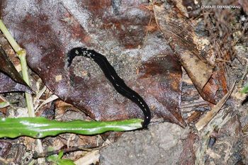
The number of P. manokwari observations was reported with photographic evidence taken by volunteer members of the online social networks: the Siamensis Facebook group and New Guinea Flatworm Line ID @sde5284v, throughout Thailand (fig. 1). In total, there were 66 verified cases in 40 districts of 31 provinces (Bangkok, Buriram, Chaiyaphumi, Chanthaburi, Chaing Mai, Chonburi, Chumphon, Kamphaeng Phet, Kanchanaburi, Khon Kaen, Lampang, Loei, Nakhon Ratchasima, Nakhon Si Thammarat, Narathiwat, Nonthaburi, Pathum Thani, Phetchaburi, Phitsanulok, Phuket, Prachin Buri, Rayong, Samut Prakarn, Samut Sakorn, Sara Buri, Songkhla, Surat Thani, Trat, Ubon Ratchathani, Udon Thani and Yala) (supplementary table S1). This flatworm was frequently reported in several districts in Bangkok and its neighbouring provinces, and provinces on the east coast (i.e. Chonburi, Rayong, Chanthaburi and Trat). In addition to the mainland, this flatworm was also reported on islands (i.e. Muang and Kathu Districts, Phuket Province and Koh Kut District, Trat Province). The oldest evidence of P. manokwari occurrence in Thailand was a photograph of the flatworm taken at Kasetsart University, Chatuchak District, Bangkok on 7 January 2010 (credit: Mr Chayajit Deekrachang) (fig. 2).
Distribution of the New Guinea flatworm (Platydemus manokwari) in Thailand. Mapping data are based on photographic evidence of P. manokwari reported by public volunteers on the online social network portals the Siamensis.org Facebook page and New Guinea Flatworm Line ID@sde5284v (orange dots); red dots indicate L3 Angiostrongylus-positive P. manokwari specimens in the present study. The grey scale bar represents the altitudinal gradient.

The oldest photographic evidence of Platydemus manokwari occurrence in Thailand (credit: Chayajit Deekrachang, 2010). This New Guinea flatworm specimen was found in a small garden around a monument at Kasetsart University, Bangkok.

Platydemus manokwari collection
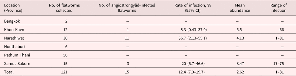
A total of 121 living P. manokwari samples were collected from six localities in Thailand, including Bangkok (Saimai District, latitude: 13.901853 and longitude: 100.681765), Khon Kaen (Kranuan District, latitude: 16.707427 and longitude: 103.079975), Narathiwat (Cho-airong District, latitude: 6.2422833 and longitude: 101.426388), Nontaburi (Bang Bua Thong District, latitude: 13.899101 and longitude: 100.428332), Pathum Thani (Lam Luk Ka District, latitude: 13.949563 and longitude: 100.738588) and Samut Sakorn (Ban Phaeo District, latitude: 13.591572 and longitude: 100.111636) (table 1).
Angiostrongylid (L3) infection rate, mean abundance, and range in Platydemus manokwari from various locations in Thailand.

The flatworms were uniform in external appearance, long and flattened, and dark brown or black in body colour, with a light brown line running dorsally from the anterior tip to posterior end. The ventral part was a pale cream, grey or light brown, with a white cylindrical pharynx located at the middle part of the body. The collected flatworms differed in size, and ranged from 12–98 mm long and 1–8.5 mm wide. All flatworms were collected alive and survived until arrival at the laboratory for further investigation.
Genetic confirmation of Platydemus manokwari in Thailand
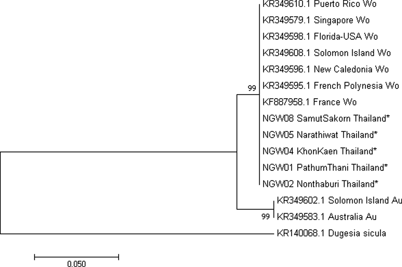
COI sequences (424 bp) of the flatworms from five localities were analysed, except the specimens from Bangkok, as there were only two individuals and those were used in the experiment for angiostrongylid larva investigation. The COI sequences from all analysed specimens were 100% identical (E-value = 0) to P. manokwari sequences in the NCBI database (e.g. accession nos KR349595.1, KF887958.1 and KR349610.1). Phylogenetic relationships showed that the five representative Thai P. manokwari samples grouped in the world haplotype clade alongside other P. manokwari sequences from France (KF887958.1), Florida (KR349598.1), Puerto Rico (KR349610.1), Singapore (KR349579.1), French Polynesia (KR349595.1), New Caledonia (KR349596.1) and the Solomon Islands (KR349608.1) (fig. 3).
Phylogenetic analysis based on mitochondrial COI sequences of Platydemus manokwari collected from Thailand compared with the world (Wo) and Australian (Au) haplotypes from previous studies (Justine et al., Reference Justine2014, Reference Justine2015). A sequence of an aquatic planarian, Dugesia sicula, was used as the outgroup. The phylogenetic tree was constructed using the maximum likelihood method based on the Hasegawa–Kishino–Yano model. The tree is drawn to scale, with branch lengths indicating the number of substitutions per site.

Evidence of Angiostrongylus larvae in Platydemus manokwari
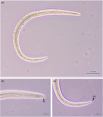
In total, 317 L3 larvae of Angiostrongylus nematodes were recovered from the New Guinea flatworms. All of the larvae microscopically exhibited the key characteristics of Angiostrongylus larvae: a pair of chitinous rods at the cephalic part, and caudal constriction (tail-pointed tip) (fig. 4). The larvae were 468–489 mm long and 25.9–32.6 mm wide (n = 10), and resembled the L3 stage of Angiostrongylus spp. (Alicata and Jindrak, Reference Alicata and Jindrak1970). The overall rate of infection was 12.4% (15/121 flatworms). Up to 81 larvae were found in an infected flatworm (total mean abundance 2.62). The infected P. manokwari were from Narathiwat, Samut Sakorn and Khon Kaen Provinces, with infection rates of 36.7%, 20% and 8.3%, respectively (see table 1 and fig. 1). The COI sequence (579 bp) of pooled Angiostrongylus larvae was translated based on translation table 5 (the invertebrate mitochondrial DNA code) in MEGA v7.0 (Kumar et al., Reference Kumar, Stecher and Tamura2016), and there were no premature stop codons, which ensured that the sequence was not a pseudogene. In addition, we compared the sequence to COI sequences from the complete mitochondrial genome samples in GenBank (i.e. NC_013065, A. cantonensis; NC_030332, A. malaysiensis). The maximum likelihood phylogenetic tree showed that the L3 larvae of Angiostrongylus nematodes grouped with the AM6 isolate of A. malaysiensis (accession no. KT947979) (fig. 5).
Microscopic illustration of an infective stage larva (L3) of Angiostrongylus sp. isolated from P. manokwari after a tissue digestion experiment. The whole body (a), and cephalic (b) and caudal ends (c) of the larva are presented with key morphological characteristics (CR = chitinous rods, TPT = tail-pointed tip) of angiostrongylid larvae.

Phylogenetic analysis of mitochondrial COI sequences of pooled L3 Angiostrongylus isolated from P. manokwari in Samut Sakorn Province, Thailand (AL3NGW43-PL). Molecular analysis confirmed larval identification as A. malaysiensis. The phylogenetic tree was constructed using the maximum likelihood method based on the Tamura–Nei model. The tree is drawn to scale, with branch lengths indicating the number of substitutions per site.

Discussion
The invasive New Guinea flatworm is reported here for the first time in Thailand. Specimens collected from several places were confirmed to be P. manokwari by mitochondrial COI sequence. This species occurs widely throughout the country. There was also evidence of this flatworm harbouring infective-stage larvae of Angiostrongylus nematodes. Here, distribution of this flatworm species and its potential impacts on the environment and public health are discussed.
This is also the first report of P. manokwari in mainland South-east Asia, although it was previously found in two island countries of this region: the Philippines and Singapore (Justine et al., Reference Justine2014, Reference Justine2015). It appears that P. manokwari is already distributed throughout Thailand, and was found on both the mainland and islands (i.e. Koh Kut District, Trat Province; Koh Samui District, Surat Thani Province; and Phuket Province) (fig. 1). The oldest photographic evidence dates back to 2010 (fig. 2), which indicates that P. manokwari was introduced to Thailand at least 8 years ago, but was not publicly reported until October 2017 (Panitvong, Reference Panitvong2017). There is some speculation regarding the possible ways that this flatworm was introduced into Thailand. International commercial trade appears to be the main route that has facilitated invasion of this flatworm (the worm itself, or its fragment or cocoon) across several countries and continents. Materials, particularly pottery and commercially imported plants (either the soil or the plants themselves), could be contaminated (Sugiura et al., Reference Sugiura, Okochi and Tamada2006; Sugiura and Yamaura, Reference Sugiura and Yamaura2009; Justine et al., Reference Justine2014). Subsequent within-country movement of contaminated plants and other types of materials associated with soil can easily transport the flatworm to new places (Sugiura, Reference Sugiura2009).
Interestingly, P. manokwari occurrences tended to be well aggregated in/around the capital city, Bangkok, and nearby provinces, and some other industrialized cities and ports on the east coast (i.e. Chonburi and Rayong) (fig. 1). These cities could be hypothesized to be the first portals of P. manokwari introduction via seaports or international airports. Alternatively, this could be interpreted as possible bias from using a citizen science approach. The majority of the records may come from highly developed areas where people have easy access to technology (e.g. they more commonly use smart phones or the internet) compared with rural areas.
In addition, local observations revealed that P. manokwari occurrences seemed to be limited to human-made habitats (e.g. orchards, military camps, private home gardens, kitchen walls and bathrooms, including toilets). There is still no information about occurrence of this flatworm in rural agriculture lands, woodlands, community forests, national parks and other protected areas. Elucidating the occurrence of these species in more rural areas could help reveal if this species was accidentally imported into the country by humans. However, this invasive flatworm species may not be able to survive in these environments for unknown reasons, such as unsuitable environmental conditions (biotic and abiotic resistance), propagule pressure, unknown native resident predation, genetic constraint, or lack of mutualism (Blackburn and Duncan, Reference Blackburn and Duncan2001; Keane and Crawley, Reference Keane and Crawley2002; Zenni and Nuñez, Reference Zenni and Nuñez2013). Further investigation is required to more thoroughly understand P. manokwari invasion and associated consequences.
Mitochondrial COI sequences of the flatworm specimens confirmed P. manokwari taxonomic identification. The five representative P. manokwari in Thailand all grouped in the world haplotype, with low genetic variation. This finding could result from limitations of using a partial single gene marker (424 bp of COI in this case) that may be too low in resolution to draw conclusions regarding the population structure of this species, and/or this species is currently in the early stage of invasion. This flatworm may not be able to establish populations easily, particularly in the tropical mainland, where high biodiversity or environmental heterogeneity potentially act as ecological buffers that determine response to external pressures (Melbourne et al., Reference Melbourne2007; Freestone et al., Reference Freestone, Ruiz and Torchin2013). Further fine-scale investigation of population genetics (e.g. DNA microsatellite markers) and ecological parameters in these areas would help elucidate the population structure of the invasive New Guinea flatworm in Thailand.
Among the P. manokwari specimens collected in Thailand, infective stage larvae (L3) of Angiostrongylus had a total infection rate of 12.4%. The infection rate in Thailand was quite high, but slightly lower than those in other areas, such as A. cantonensis larvae in P. manokwari (14.1%) from Okinawa, Japan (Asato et al., Reference Asato2004). In Hawaii, Qvarnstrom et al. (Reference Qvarnstrom, Bishop and da Silva2013) reported very high prevalence of A. cantonensis infection (67%, 8/12 specimens examined) in land planarian species (some of which were P. manokwari). Epidemiological research on angiostrongyliasis in its endemic areas, including Thailand, has largely focused on the risk of infection from ingesting raw or undercooked intermediate host snails (i.e. Pila spp., Pomacea canaliculata, Cryptozona siamensis, and Achatina fulica) (Pipitgool et al., Reference Pipitgool1997; Tesana et al., Reference Tesana2009; Vitta et al., Reference Vitta2011, Reference Vitta2016). However, information regarding the potential risk of terrestrial flatworms, such as P. manokwari as a paratenic host (carrier) of the parasite, is limited, particularly in Thailand. Transmission of L3 Angiostrongylus to humans includes both eating intermediate or paratenic hosts, and ingesting vegetables or water contaminated with infected slugs or snails (Heyneman and Lim Reference Heyneman and Lim1967; Alicata and Jindrak Reference Alicata and Jindrak1970; Ash, Reference Ash1976; Alto, Reference Alto2001; Cowie, Reference Cowie2013). Shedding of L3 Angiostrongylus in the mucus of intermediate/paratenic hosts (e.g. snails, slugs and land planarians) potentially results in parasite contamination of water and food, particularly in leafy salad, vegetable juice, and fruit. Damaged or decayed intermediate hosts could also release numerous L3 Angiostrongylus that can survive up to 72 hours in a moist environment (Cheng and Alicata, Reference Cheng and Alicata1964; Cowie, Reference Cowie2013) and subsequently contaminate water and vegetables. Qvarnstrom et al. (Reference Qvarnstrom, Bishop and da Silva2013) also speculated that the flatworms could hide in and contaminate fresh vegetables with the parasitic nematode through their secretions and/or when they are chopped during food preparation. Moreover, with their exceptional capacity to survive (e.g. their regeneration ability) and expand their populations quickly and globally as an invasive species, P. manokwari could potentially act as an important reservoir that harbours and spreads Angiostrongylus to new places via rat infection. Based on previous literature and the results of our study, we emphasize the important role of P. manokwari in angiostrongyliasis epidemiology in Thailand and nearby countries.
Based on genetic information, our results showed that L3 Angiostrongylus recovered from P. manokwari in Thailand were A. malaysiensis. Although A. cantonensis is believed to be the main agent of angiostrongyliasis, which causes eosinophilic meningitis worldwide, the closely related A. malaysiensis may also be a human pathogen in endemic areas, including Thailand and other countries in South-east Asia (Prociv et al., Reference Prociv, Spratt and Carlisle2000; Rodpai et al., Reference Rodpai2016). Angiostrongylus malaysiensis can cause neurological disease in monkeys under experimental conditions (Cross, Reference Cross and Cross1979). Reports from Malaysia, Indonesia and Thailand indicate that A. malaysiensis also potentially causes human disease (Carney and Stafford, Reference Carney, Stafford and Cross1979; Lim and Ramachandran, Reference Lim, Ramachandran and Cross1979; Rodpai et al., Reference Rodpai2016). However, A. malaysiensis has still not been isolated from humans (Prociv et al., Reference Prociv, Spratt and Carlisle2000).
Recent evidence revealed that A. cantonensis and A. malaysiensis widely co-occur throughout the countries of the Indochinese Peninsula (Rodpai et al., Reference Rodpai2016; Dusitsittipon et al., Reference Dusitsittipon2017; Eamsobhana et al., Reference Eamsobhana2018). Apart from the two Angiostrongylus spp. mentioned above, there are also some other metastrongylid nematodes in wild murine rodents from this region (i.e. A. siamensis, Thaistrongylus harinasutai and Malayometastrongylus diardinematus) (Ohbayashi et al., Reference Ohbayashi, Kamiya and Bhaibulaya1979; Gibbons and Krishnasamy Reference Gibbons and Krishnasamy1986; Veciana et al., Reference Veciana2014). L3 infective stage larvae of these metastrongylids ideally circulate in the environment and are picked up by their mollusc intermediate hosts, or even paratenic hosts like P. manokwari, to continue their life cycle. In angiostrongyliasis epidemiology, it is still largely unknown whether L3 of these metastrongylids, similar to the well-known agent A. cantonensis, can cause eosinophilic meningoencephalitis in humans; additional collaborative studies on disease surveillance in the environment and clinical studies are needed to elucidate this issue. Improvement of clinical (particularly molecular) diagnostic methods would also help improve our understanding of angiostrongyliasis epidemiology in South-east Asia.
In ecology, host–parasite relationships have been recognized as important interactions in ecosystem functioning, with various effects of individuals on populations and communities (Morand, Reference Morand, Vila, Hulme and Ruiz2015). There are two main epidemiological hypotheses regarding the role of invasive host species in parasite transmission: (1) parasite spill-over, in which invasive host species co-introduce novel parasites to local native hosts; and (2) parasite spill-back, in which a native parasite infects competent invasive host species, which produces a wider host range that promotes parasite survival (Kelly et al., Reference Kelly2009; Strauss et al., Reference Strauss, White and Boots2012; Morand, Reference Morand, Vila, Hulme and Ruiz2015). In the present case of a new invasive species in Thailand, P. manokwari, the invasive flatworms appeared to be infected by A. malaysiensis as a result of their predation on native intermediate host molluscs. This finding indicates potential evidence of parasite spill-back. The invasive flatworms may act as a new host for Angiostrongylus spp., which could potentially amplify the transmission of the parasite in the invaded areas.
Globally, humans have entered into the “Information Age”, with increasing use of modern digital technologies for communication. This technological revolution has resulted in changes in communication methods in a wide range of societal disciplines, including ecological and environmental sciences (Arts et al., Reference Arts, van der Wal and Adams2015). For example, volunteers that participate in a biodiversity exploration campaign report their local information via digital devices, such as smart phones and related technology (a citizen science approach), and this is rapidly becoming an alternative method in ecological research and nature conservation (Dickinson et al., Reference Dickinson, Zuckerberg and Bonter2010; Conrad and Hilchey, Reference Conrad and Hilchey2011). Gathering data in this manner would promptly and cost-effectively produce information on the distribution of the invasive flatworm species at the national level. However, the information obtained would require careful verification, particularly the identification of flatworm species by non-taxonomist volunteers, as some endemic organisms (e.g. land planarians: Bipalium javanum, Diversibipalium haasei and Anisorhynchodemus spp.; and slugs: Pamarion spp. and Semperula spp.) could easily be misidentified, and could be eliminated unintentionally.
In summary, the globally invasive flatworm P. manokwari was reported for the first time in Thailand. The flatworms were also found harbouring infective stage larvae of Angiostrongylus malaysiensis. These findings provide updated information on the role of this invasive species in the life cycle of a parasitic nematode. Continued monitoring of the distribution status of this invasive flatworm and additional research on its biology would help provide an enhanced understanding of infection by this nematode in its endemic areas and contribute to our understanding of the importance of invasive species in biodiversity conservation worldwide.
Supplementary material
To view supplementary material for this article, please visit https://doi.org/10.1017/S0022149X18000834
Acknowledgements
We thank Dr Jean-Lou Justine and Dr Leigh Winsor for their invaluable help identifying the flatworms from photographs, and providing an illustrated list of land planarians found in Thailand and the key to distinguish Platydemus manokwari from other flatworm species. In addition, we are grateful for their comments and suggestions that greatly improved the manuscript. We also thank all of the volunteers who reported evidence of the invasive flatworm occurrence on the Siamensis.org Facebook page (https://www.facebook.com/groups/122260101128398/) and New Guinea Flatworm Line ID @sde5284v). We acknowledge Mr Apinun Prasert, Mr Mongkol Untachai, Mr Saran Sittisamran, Mr Pathai Sricharoon and Mr Preeda Poomchan for their help with local collection of the flatworm specimens. We also thank Mr Chayajit Deekrachang for allowing us to use his P. manokwari photograph in this publication. We thank Mallory Eckstut, PhD, from Edanz Group (www.edanzediting.com/ac) for editing a draft of this manuscript in English.
Financial support
This research was supported financially by the internal research budget of the Department of Helminthology, Faculty of Tropical Medicine, Mahidol University.
Conflict of interest
None.