Introduction
Thaumaleidae is a family of Diptera with roughly 190 described species and, as their common name seepage or madicolous midges suggests, thaumaleids are restricted to thin films of vertically flowing water (Vaillant Reference von Röder1956; Pivar et al. Reference Pivar, Moulton and Sinclair2018). This specialised environment and poor dispersal ability of adults have led them to be poorly collected and consequently understudied. Found along cascading streams, rock face seeps, and margins of waterfalls, thaumaleid adults are best collected by sweeping above the madicolous habitat and adjacent riparian vegetation, while immatures can be plucked from the substrate using forceps or flushed into a white pan (Sinclair and Saigusa Reference Sinclair and Stuckenberg2002).
The Southern Hemisphere fauna consists of four genera: Afrothaumalea Stuckenberg, Austrothaumalea Tonnoir, Niphta Theischinger, and Oterere McLellan. Before this study, six described species were recorded from South America, all from Chile and Argentina. Edwards (Reference Edwards1930) described five species now assigned to Austrothaumalea (two species), Niphta (two species), and Oterere (one species), while Schmid (Reference Shorthouse1970) described an additional species of Austrothaumalea. An undescribed thaumaleid species from Ecuador was reported by Röder (Reference Sæther1886), but this record remains unverified as the specimen has not been located despite multiple attempts by the authors. A revision of Chilean and Argentinean fauna by R.J.P. was well underway when material of Neothaumalea atlantica new genus, new species, described herein, was discovered and made available. This species represents the first record of Thaumaleidae in Brazil and first east of the Andes Mountains. Both sexes and preimaginal stages are described.
Materials and methods
Terms used for adult structures follow Cumming and Wood (Reference Cumming and Wood2017), except wing venation where the interpretation of Sinclair (Reference Sinclair and Saigusa2015) is accepted. Homology of the male terminalia follows Sinclair (Reference Sinclair1992). Terms used for larval and pupal structures follow those of Courtney et al. (Reference Courtney, Sinclair and Meier2000) and Borkent (Reference Borkent2012), respectively. The numbering system for larval head capsule setae and sensory pits follows Sinclair and Stuckenberg (Reference Theischinger1995). The distribution map was created using SimpleMappr (Shorthouse Reference Sinclair2010).
Thaumaleidae were collected directly into 95% ethanol and later dried using hexamethyldisilazane, as outlined by Brown (Reference Brown1993). Adult genitalia were cleared using hot, 85% lactic acid. Representative males and larvae were also cleared with the GeneJET Genomic DNA Purification Kit #K0722 (ThermoScientific, Waltham, Massachusetts, United States of America) in order to study structures cleared less aggressively while simultaneously extracting DNA for future molecular study. The lysate preparation protocol was followed and cleared voucher specimens were stored in 70% ethanol. Specimens were viewed under a Meiji Techno RZ stereomicroscope mounted with a Progres Gryphax® Naos camera (Jenoptik, Jena, Germany) to take light micrographs of pinned adults, and immatures in alcohol. Images were taken using iSolution Lite x64 (Focus Precision Industries, Victoria, Minnesota, United States of America) and stacked using Helicon Focus 6.7.1 (HeliconSoft, Roseau Valley, Dominica). Cleared terminalia and larval head capsules in glycerine were viewed with an Olympus BH-2 compound microscope and images were taken following the protocol described above. Line drawings were first traced from stacked micrograph images taken on the Olympus microscope (micrographs on their own failed to clearly illustrate critical characters), then inked and scanned for publication. Samples for scanning electron microscopy were dried using hexamethyldisilazane, mounted on gold-plated metal supports, sputter-coated, and scanned using a JEOL JSM-6390LV scanning electron microscope (Akishima, Tokyo, Japan) at the Central Laboratory of Electronic Microscopy. Cleared terminalia were preserved either in slide mounts made following the procedures outlined by Sæther (Reference Schmid1969) and mounted in Euparal, or placed in glycerine filled microvials pinned beneath the specimen.
Label data for primary types are presented exactly as they appear. Data are listed from the top downward on the staging pin, with data from each label enclosed in quotation marks; lines are delimited by a forward slash mark. The repository is given in parentheses. Specimens are deposited in the following repositories: Canadian National Collection of Insects, Ottawa, Ontario, Canada (CNC), Coleção Entomológica da Universidade Federal de Santa Catarina, Brazil (UFSC), Museu Nacional do Rio de Janeiro, Brazil (MNRJ), Museu de Zoologia da Universidade de São Paulo, Brazil (MZSP), the National Museum of Natural History, Smithsonian Institution, Washington, District of Columbia, United States of America (USNM), and the University of Tennessee, Knoxville, Tennessee, United States of America (UTK).
Neothaumalea Pivar, Moulton, and Sinclair, new genus
Type species. Neothaumalea atlantica new species.
Etymology. From the Greek “neo-” (new) in reference to the discovery of a new genus from the New World.
Diagnosis. Eye bridge narrow, tapered to two to three facets; head clothed in setae of uniform length. Mesoscutum with prominent antealar ridge; scutum clothed in short, undifferentiated setulae; proepisternal setae absent. Wing with R2+3 crossvein absent. Male abdomen with sternites 4–6 modified with medial heavily setose, sclerotised flap-like projections; female abdomen unmodified. Male terminalia with hypandrium narrow, paired parameres and membranous gonocoxal plate.
Neothaumalea atlantica Pivar and Pinho, new species
Type material. Holotype: slide-mounted male with larval and pupal exuviae labelled: “BRAZIL, Santa Catarina State, Grão Pará / Parque Nacional de São Joaquim, Serra do Corvo Branco / 28°03'21"S 49°22'01"W, 1245.m a.s.l. / 10.ix.2016, seepage, L.C. Pinho leg.” (MZSP). Allotype: female, same data as holotype except: “L.C. Pinho & A.P. Amaral leg.” (MZSP). Paratypes: Brazil: Santa Catarina State: Alfredo Wagner, BR282 km 111, 27°44'00"S 49°22'02"W, 915 m, 29.ix.2016, L.C. Pinho (two pupal exuviae (slide mount), UFSC); Brazil: Santa Catarina State: Grão Pará, Parque Nacional de São Joaquim, Serra do Corvo Branco, 28°03'21"S 49°22'01"W, 1245 m, 10.ix.2016, sweeping, L.C. Pinho and A.P. Amaral (two males, CNC; two males, MNRJ; two males, UFSC; two males, USNM; one male, UTK); Brazil: Santa Catarina State: Grão Pará, Parque Nacional de São Joaquim, Serra do Corvo Branco, 28°03'21"S 49°22'01"W, 1245 m, 7.x.2014, seepage, L.C. Pinho and A. Pitaluga (three larvae, MNRJ; two females (with larval and pupal exuviae), four larvae (slide mount), UFSC); Brazil: Santa Catarina State: Grão Pará, Parque Nacional de São Joaquim, Serra do Corvo Branco, 28°03'21"S 49°22'01"W, 1245 m, 28.ix.2016, seepage, L.C. Pinho and A. Pitaluga (10 larvae, one pupa, CNC; seven larvae (slide mount), MZSP); Brazil: Santa Catarina State: Lauro Müller, Serra do Rio do Rastro, 28°23'57"S 49°32'54"W, 1326 m, 29.ix.2016, seepage, L.C. Pinho (25 larvae, six pupae, CNC; one pupal exuviae, UFSC); Brazil: Santa Catarina State: Urubici, Parque Nacional de São Joaquim, Rio Italianinho, Cachoeira, 28°08'49"S 49°37'06"W, 1530 m, 23.viii–5.ix.2014, malaise, L.C. Pinho et al. (one female, UFSC); Brazil: Santa Catarina State: Urubici, SC370, São Pedro, 28°03'14"S 49°25'22"W, 980 m, 10.ix.2016, seepage, L.C. Pinho and A.P. Amaral (two pupae (slide mount), MNRJ).
Recognition. Neothaumalea atlantica males are distinguished from other genera by sternites 4–6 bearing densely setose flap-like extensions, hypandrium narrow, and gonostyli bearing five long, apical, spine-like setae. Both sexes also have an antealar ridge, a narrow eye bridge comprising only two to three facets, as well as uniform lengthened setae on both the head and scutum.
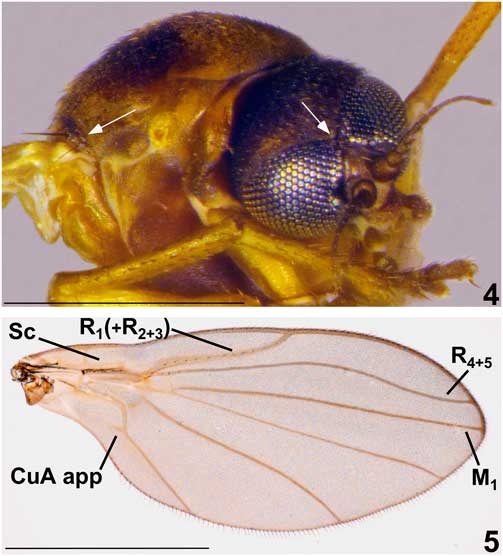
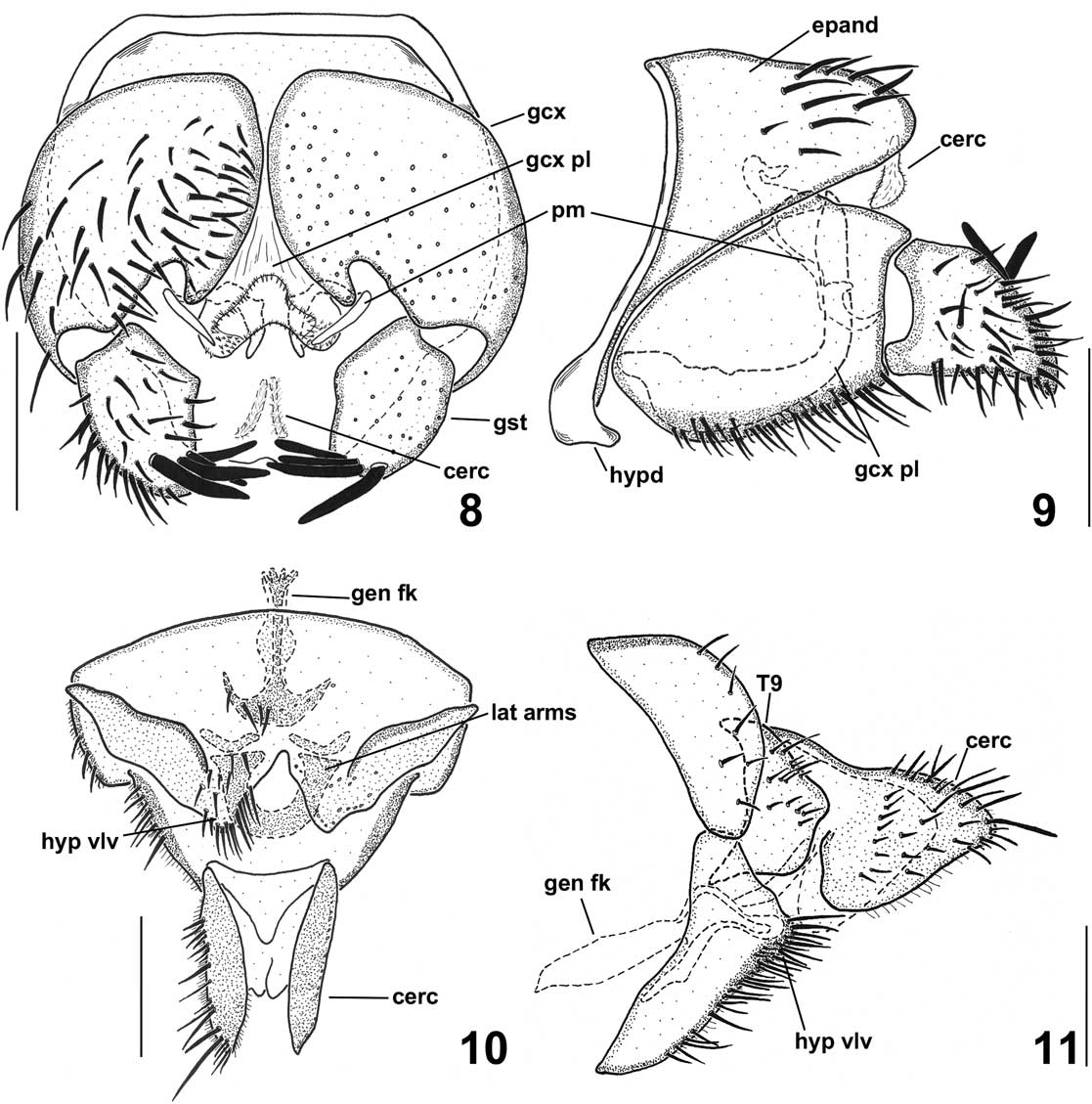
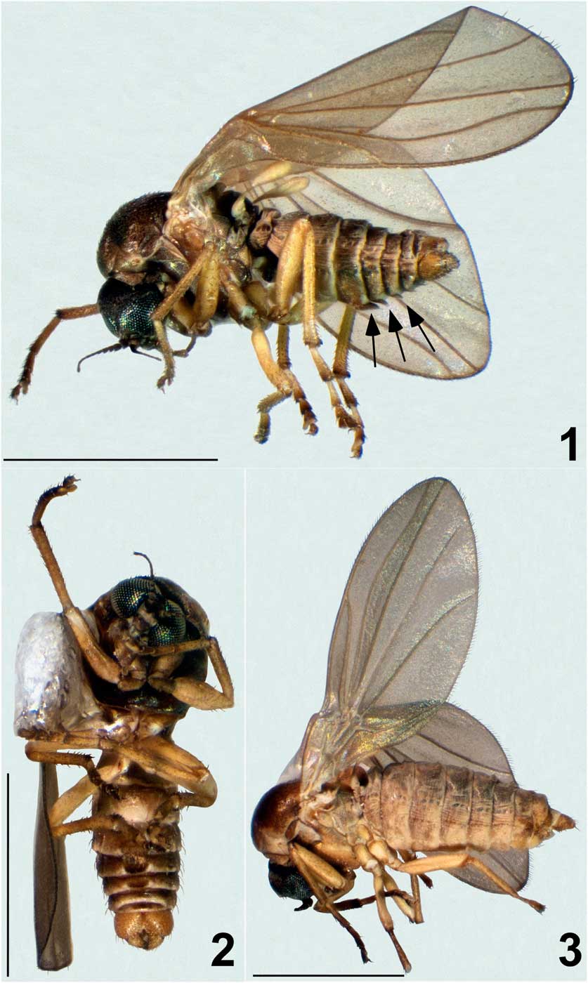
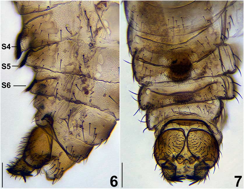
Description of male. n=10 (Figs. 1–2). Length. 2.00–2.05 mm. Colouration. Head dull, brown; pronotum light brown; postpronotum brown; postpronotal lobe light brown; mesonotum and pleura brown and somewhat shiny; scutellum and mediotergite brown, shiny; katepisternum light brown; halter pale and legs pale brown, legs becoming slightly darker on apical tarsomeres; abdomen brown; terminalia pale brown. Head. Eyes above antennae closely approximated, tapered to two to three facet width, exposing triangular frons above antennae (Figs. 4, 12). Clothed in setae of uniform length, lacking long orbital setae. Thorax. Mesoscutum with prominent antealar ridge, bearing strong medial seta flanked by one weak seta above and two below (Fig. 4). Scutum clothed in short, undifferentiated setulae; scutellum with row of marginal setae. Proepisternal setae absent. All legs with fourth tarsomere bilobed. Wing. (Fig. 5): Wing length: 2.03–2.32 mm. Slightly infuscate throughout, apex somewhat narrowed; Sc incomplete; R1 and R1(+R2+3) with uniseriate row of microtrichia along entire length, remaining veins bare; R flexed into cell br; R2+3 crossvein absent; bend in R4+5 well defined; R4+5 and M1 running parallel toward margin; M1 straight, terminating at wing apex; CuA with short basal appendage. Abdomen. (Figs 6, 7, 16–17): Abdominal sternite 1 narrow, spectacle shaped; sternite 2 reduced to slender median sclerite, a few setae restricted to laterad on posterior third; sternite 3 rectangular, weakly sclerotised, bearing setae on laterad and middle areas of median third; sternites 4–6 rectangular, weakly sclerotised with lateral setae, posterior margins modified with medial heavily setose, sclerotised flap-like projection, projection on sternite 4 overlapping sternite 5, projection on sternite 6 smallest; sternite 7 rectangular, weakly sclerotised, with row of anterior marginal setae and few setulae medially; sternite 8 trapezoidal, with setae and more sclerotised than preceding sternites. Male terminalia. (Figs. 8–9, 13–15): Epandrium with posterior margin rounded, short, not extending past gonostyli; without lobes or projections. Hypandrium narrow, same width as gonostylar spine-like setae. Gonocoxites bulbous, not much longer than wide; closely approximated medially. Gonostylus 1.5 times longer than wide, conical; bearing five long apical spine-like setae. Parameres paired, blade like; short, not extending past gonocoxal plate, diverging towards apex in ventral view. Gonocoxal plate membranous with cleft, setose posterior margin; basally not fused to hypandrium. Aedeagus membranous, sac-like. Cercus narrow, transverse band.
Light micrographs of adult Neothaumalea atlantica habitus: 1, male lateral view with arrows indicating abdominal modifications; 2, male ventral view; 3, female lateral view. Scale bar=1.0 mm.
Light micrographs of adult Neothaumalea atlantica: 4, arrows pointing to antealar ridge and narrow eye bridge (scale bar=0.5 mm); 5, right wing (scale bar=1.0 mm). CuA app, cubital vein with appendage; M, medial vein; R, radial vein; Sc, subcosta.

Light micrographs of cleared abdomen of Neothaumalea atlantica to illustrate modified sternites 4–6: 6, lateral view; 7, ventral view. Scale bar=0.1 mm. S, sternite.
Illustrations of Neothaumalea atlantica genitalia: 8, male ventral view; 9, male lateral view; 10, female ventral view; 11, female lateral view. Scale bar=0.1 mm. Cerc, cercus; epand, epandrium; gen fk, genital fork; gcx, gonocoxite; gcx pl, gonocoxal plate; gst, gonostylus; hypd, hypandrium; hyp vlv, hypogynial valve; lat arms, lateral arms; pm, paramere; T, tergite.

Scanning electron micrographs of adult male Neothaumalea atlantica: 12, anterior view of head; 13, ventral view of genitalia; 14, caudal view of genitalia; 15, caudal view of left gonostylus; 16, ventral view of abdomen illustrating modified sternites 4–6; 17, close-up of modified sternite 4. Scale bars: Figs. 12–14, 16=0.1 mm; Figs. 15, 17=0.05 mm. Epand, epandrium; gcx, gonocoxite; gcx pl, gonocoxal plate; gst, gonostylus; S, sternite.

Description of female. n=4 (Fig. 3). Similar to male except as follows: Length: 2.2 mm. Abdominal sternites 4–6 unmodified. Female terminalia (Figs. 10–11): Hypogynial valve short, not projecting beyond tergite 9; divergent in ventral view; densely setose. Tergite 9 subquadrate in lateral view, longer than tergite 8, lacking lateral projections. Sternite 9 (genital fork) slender, anchor shaped, swollen medially; lateral arms forming sclerotised semicircle. Cercus triangular, slightly scalloped on ventral margin toward apex, longer than wide, projecting posteroventrally; bearing numerous setae, single strong seta at apex. Spermathecal ducts not observed.
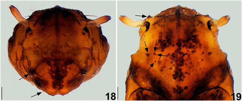
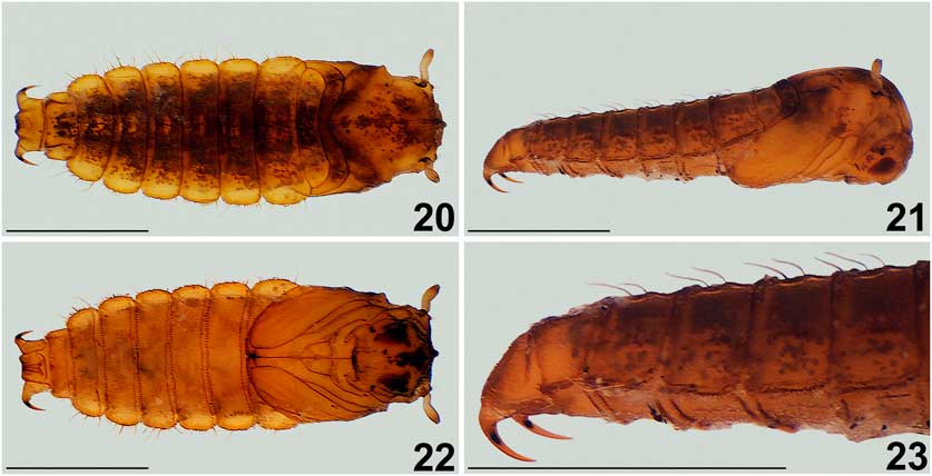
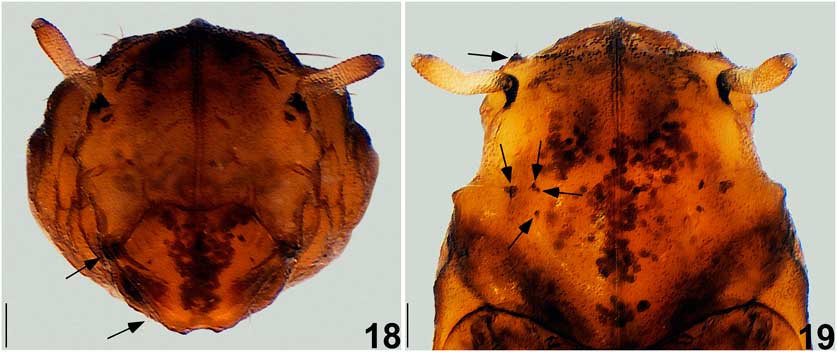
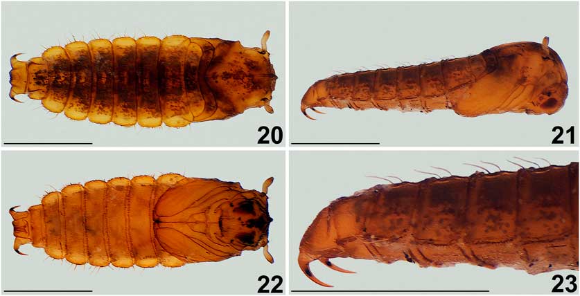
Description of pupa. n=14 (Figs. 18–23). Length. 2.85–3.80 mm. Colouration. Light brown; black spot above eyes in developing adult; caudal hook with black medial spot. Head. Maxillary sheath short, posteromedially directed, apex truncate; three short, slender setae on small tubercle above black spot over eye; single slender seta below inner margin of eye in ventral aspect. Thorax. Prothoracic and mesothoracic leg sheaths projecting straight and shorter than wing sheaths; metathoracic leg projecting beyond wing sheath and touching medially. Wing sheaths extending nearly to posterior margin of abdominal sternite 2. Respiratory organ three times as long as maxillary sheath; cylindrical with medial arch; spiracular openings encircling apex. Three slender setae on small tubercle anterior to respiratory organ. Mesothorax with three pairs of short, slender dorsocentral setae and one pair of long setae; dorsolateral tubercle bare. Abdomen. Dorsoventrally flattened; broadest at segment 2. Spiracles very weakly developed, most distinct on segments 5–7, forming slight swelling, not projecting. Segments 1–7 with two pairs of dorsal setae on raised keel projecting posteriorly; lateral margins crenulate, bearing numerous small blade-like setae and four interspersed long, hook-tipped setae. Segment 8 with dorsolateral keel, posterior margin ending in small tubercle bearing pair of slender setae; posterior lateral margins bearing three slender setae; lateral margins crenulate, not as expanded as preceding segments, bearing numerous blade-like setae and two interspersed long setae. Caudal segment truncate, terminating in pair of large, stout anteroventrally curved hooks; caudal hooks with pair of sub-basal fine setae on outer margin. Sternites 2–7 with row of setulae complete around margin; sternite 8 with row of setulae incomplete along posterior margin, faint along anterior margin; caudal sternite without setulae.
Light micrographs of Neothaumalea atlantica pupa. Arrows depicting location of setae: 18, anterior view of head; 19, dorsal view of thorax and respiratory organ. Scale bar=0.1 mm.
Light micrographs of Neothaumalea atlantica pupa: 20, dorsal view; 21, lateral view; 22, ventral view; 23, close-up of caudal segments and hooks. Scale bar=1.0 mm.
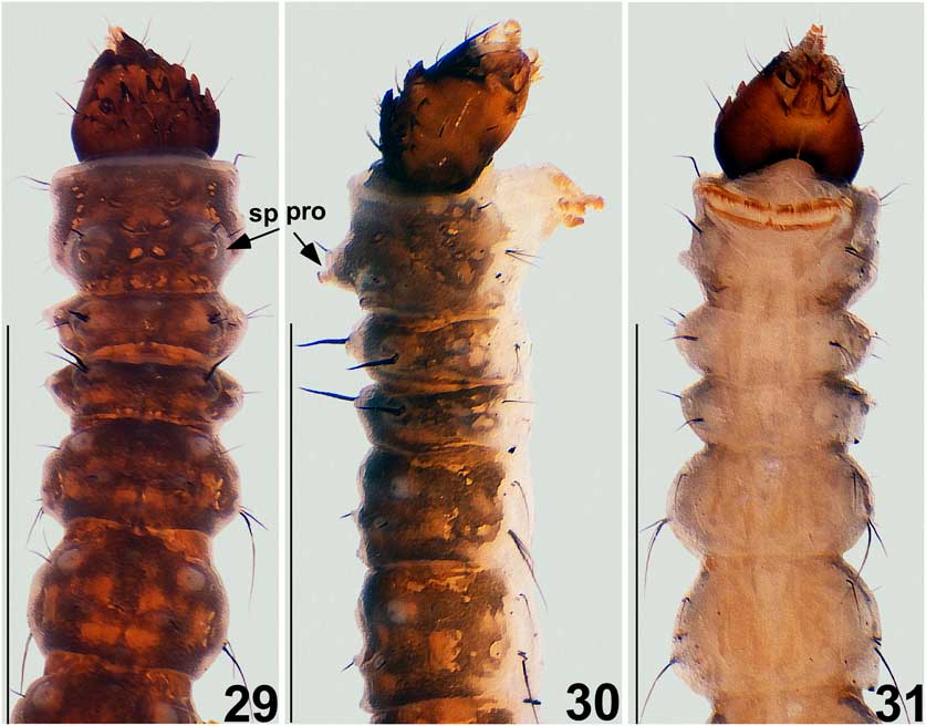
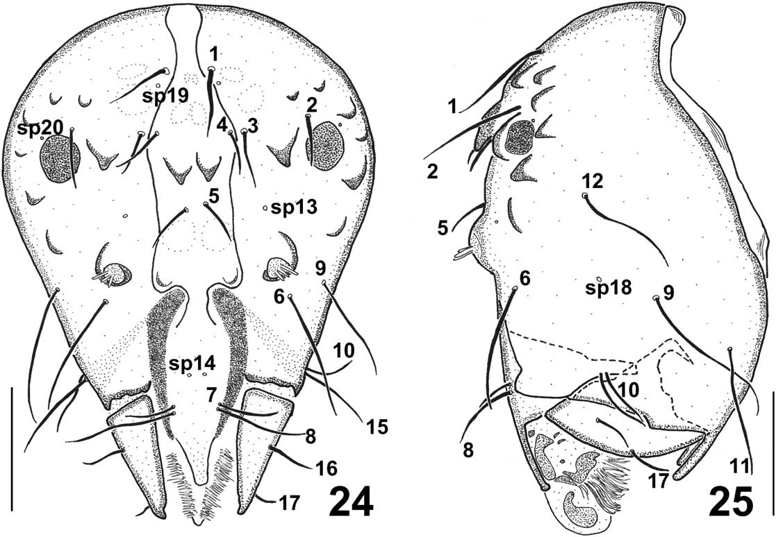
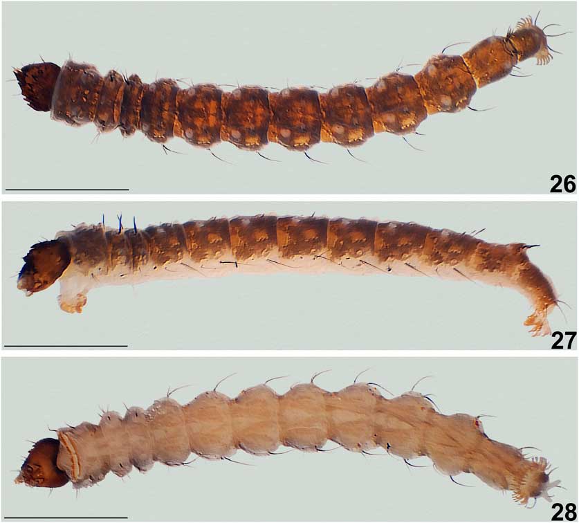
Description of larva. n=44 (Figs. 24–34). Length of final instar. 4.66–5.33 mm. Colouration. Head capsule varies, may be pale brown to dark brown. Body brownish red dorsally, with creamy protuberances; cream coloured ventrally. Head capsule. Two large, circular eye spots; antenna on tubercle, with three finger-like processes; with 10 tubercles, length of which may vary between individuals from short (Fig. 24) to long (Fig. 29); 15 pairs of unbranched setae; five sensory pits (13, 14, 18, 19, 20), sensory pit 13 above antennal tubercle. Thorax. Prothorax with single pair of protuberances bearing single spiracle; spiracular protuberance bearing one pair of dorsal setae anterior to spiracle and single dorsolateral seta; single long mid-lateral seta; group of four anterolateral setae situated near head capsule, three long and one short; three closely approximated setae near base of prothoracic leg (Keilin’s organ). Mesothorax and metathorax with one pair of lateral and dorsolateral protuberances and an unpaired dorsomedial protuberance; dorsomedial protuberance naked; dorsolateral protuberance bearing two setae, anterior seta thickened; lateral protuberance bearing five setae, three short, two long; single antero-ventrolateral seta and one pair of mid-ventrolateral setae; mesothorax with additional seta between dorsolateral and lateral protuberance. Abdomen. Ventral abdominal sternites flat, smooth. Segments 1–7 with many protuberances and setae; unpaired small dorsomedial protuberance on anterior and posterior margins, not bearing setae; anterodorsolateral pair large, with single short seta beneath; dorsolateral pair small, bearing single short seta; five ventrolateral setae. Segment 8 with unpaired small dorsomedial protuberance, not bearing setae; pair of large dorsolateral protuberances flanking posterior spiracular plate, bearing single small medial seta and single long seta at base; three short posterolateral setae and two long ventrolateral setae; ventral sternite bearing two pairs of setae. Posterior spiracular plate with sclerite encircling procerci; procercus shorter than length of spiracular plate, less than twice as long as wide, bearing pair of single small plumose seta and four long setae; without cone-like protuberance on either side of cerci. Terminal segment with single small dorsomedial protuberance, not bearing seta; four short lateral setae, one long; single long ventrolateral seta; two pairs of long setae on posterior margin, above pair of anal papillae; ventral sternite bearing single pair of setae.
Illustrations of Neothaumalea atlantica larval head capsule: 24, anterior view; 25, lateral view. Scale bar=0.1 mm. Sp, sensory pit.
Light micrographs of Neothaumalea atlantica larva: 26, dorsal view; 27, lateral view; 28, ventral view. Scale bar=1.0 mm.
Light micrographs of Neothaumalea atlantica larva, anterior half: 29, dorsal view; 30, lateral view; 31, ventral view. Scale bar=1.0 mm. Sp pro, spiracular protuberance.

Light micrographs of Neothaumalea atlantica larva, posterior half: 32, dorsal view; 33, lateral view; 34, ventral view. Scale bar=1.0 mm.

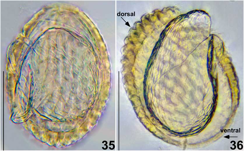
Description of egg. (Figs. 35–36). Broadly ovate; ventral surface thin, half width of dorsal surface, smooth and flattened; dorsal surface thickened, reticulate. Embryo seemingly exposed.
Light micrographs of Neothaumalea atlantica egg: 35, dorsal view; 36, cross section. Scale bar=0.1 mm.

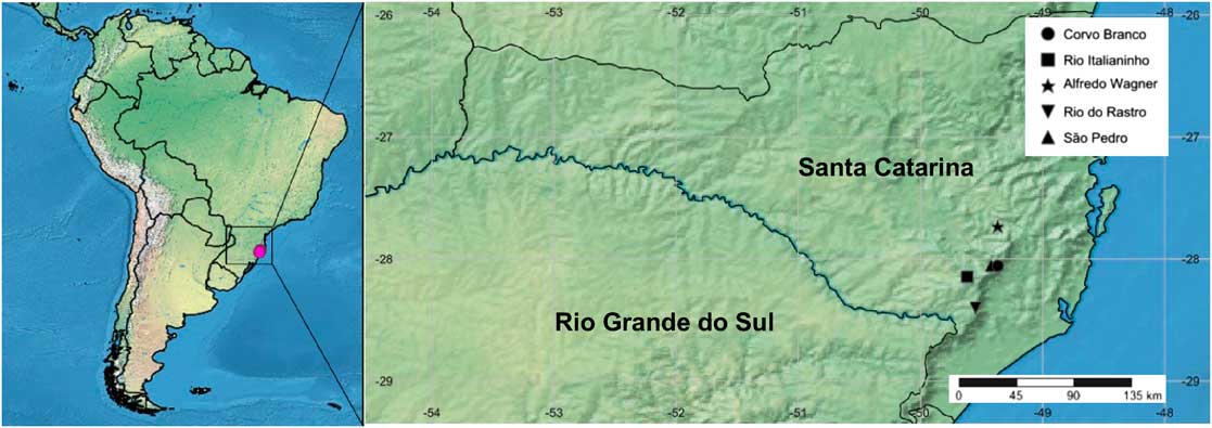
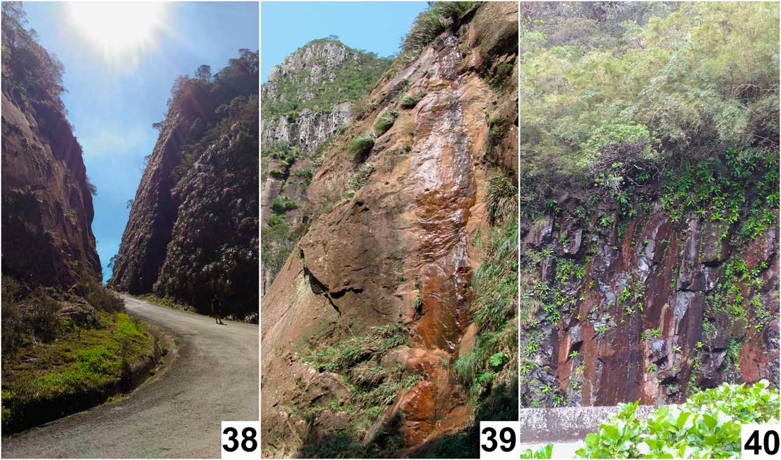
Distribution. (Figs. 37–40) This species is currently only known from five localities in Brazil’s southern state of Santa Catarina, at elevations ranging from 915 to 1530 m in Aparados da Serra Geral, a mountain range with steep slopes running parallel to the Atlantic coastal plain. Aparados da Serra Geral, the coldest region in Brazil, is carved from volcanic rocks that date from the Mesozoic era (Wildner et al. Reference Wildner, Orlandi and Giffoni2004). A possible record of the genus was recently discovered in Serra do Caraça (Minas Gerais), however this is based only on a single pupal exuviae and additional material is required to confirm this determination.
Distribution map of Neothaumalea atlantica.

Habitat of Neothaumalea atlantica: 38, Serra do Corvo Branco, Santa Catarina, Brazil (28°03'21"S, 49°22'01"W); 39, rock face seepage, Serra do Corvo Branco site; 40, Serra do Rio do Rastro site (28°23'57"S, 49°32'54"W).

Etymology. The specific epithet atlantica is in reference to the Atlantic Forest where this species is found. The name is to be regarded as a noun in apposition.
Bionomics. The larvae of this species are somewhat flattened ventrally compared to typical thaumaleid larvae. This is perhaps an adaptation to the madicolous zone of streams subject to rapid changes in water level and velocity; i.e., regions exposed to more variable seasonal rainfall. The recurved caudal pupal hooks could also aid in anchoring the pupa during changes in flow conditions.
Discussion
Neothaumalea is most similar to the Gondwanan genus Niphta based on morphological characters. Both genera share a developed antealar ridge, which is absent in all other Southern Hemisphere genera (Theischinger Reference Vaillant1986). This ridge is present, however, in the Holarctic genus Trichothaumalea Edwards. Both Neothaumalea and Niphta also lack proepisternal setae adjacent to the anterior spiracle present in all other genera. Neothaumalea atlantica differs from Niphta by the presence of paired parameres (fused in Niphta) and hypandrium (absent in Niphta).
Among the few species with associated immature stages, the long hook-tipped pupal abdominal setae found in Neothaumalea atlantica are also present in Austrothaumalea and Niphta (Sinclair Reference Sinclair2000). Recurved pupal caudal hooks are present in Neothaumalea atlantica and Niphta collessi Theischinger (Sinclair Reference Sinclair2000), but are absent among known Chilean species of Niphta (R.J.P., unpublished data). The pupa is dorsoventrally flattened, as in Trichothaumalea and Afrothaumalea stuckenbergi Sinclair (Sinclair Reference Sinclair and Saigusa2015). Unfortunately the immature stages of most thaumaleid species remain unknown and confident phylogenetic trends remain elusive.
Neothaumalea atlantica has several unique characteristics. Perhaps the most obvious of these are the heavily setose, sclerotised, flap-like projections on male abdominal sternites 4–6. These projections represent the first abdominal modifications described in Thaumaleidae and the second sexually dimorphic character in Thaumaleidae to be described; the other being the enlarged first palpal segment of male Androprosopa larvata Mik (Schmid Reference Schmid1958, Figs. 34–36). While their function remains unknown, the presence of these projections only on males indicates that they may be used during mating. Neothaumalea atlantica also has a very narrow eye bridge that reveals a small triangular frons, unlike any other species in the family. In all remaining genera of Thaumaleidae, the eyes meet broadly, though a small frons may still be visible in some species. The holoptic condition of the eyes in both sexes has been viewed as a family level synapomorphy, but it remains uncertain whether the reduced holoptic condition (i.e., narrowed eye bridge) is a secondary reduction or plesiomorphic compared to the fully holoptic condition in the remaining genera. Neothaumalea is also the only genus in the Southern Hemisphere to have setae of uniform length both on the head and the scutum; although Trichothaumalea also exhibits this trait, as well as on the scutellum. The large, stout setae on the gonostyli are unique among South American thaumaleids, but this feature appears to be somewhat homoplastic given the presence or absence of stout setae among species of other genera (e.g., Afrothaumalea; see Sinclair Reference Sinclair and Saigusa2015).
The above characters support the erection of a genus for this new thaumaleid from Brazil. Future studies should focus on the collection of material from the southern region of Brazil and adjacent localities. Though this represents the first record of thaumaleids east of the Andes Mountains, this is unlikely to be the only species present given the extensive area that likely holds suitable habitat. Future collection attempts throughout South America are strongly encouraged and will undoubtedly yield many new discoveries.
Acknowledgements
The authors would like to thank Carlos A.H. Flechtmann, São Paulo State University, for his generous help in facilitating the export of material from Brazil. This allowed authors to study specimens firsthand to better understand their morphology, as well as enable its inclusion in molecular studies aimed at elucidating phylogenetic relationships among world Thaumaleidae. Thanks are also due to Erika M. Shimabukuro, São Carlos Federal University, for notifying the authors of the potentially congeneric pupa from Serra do Caraça. Also, the Smithsonian’s S.W. Williston Diptera Research Fund for funding a recent trip to Chile, giving the authors the opportunity to collect sufficient material to conduct comparative studies between genera and reach our conclusions. This research was supported in part by National Science Foundation award DEB-1146290 (J.K.M.), the University of Tennessee Hatch Project TEN00479, the University of Tennessee Department of Entomology and Plant Pathology, and by the Research Program in Biodiversity – Atlantic Forest (Programa de Pesquisa em Biodiversidade, Mata Atlântica, 457841/2012-9). The authors would also like to thank technicians of the Central Laboratory of Electronic Microscopy and Multiuser Laboratory of Biology Studies, both from the Federal University of Santa Catarina, Brazil. Rüdiger Wagner (Kassel, Germany) and an anonymous reviewer kindly reviewed and commented on the manuscript.