Introduction
Since the commercialisation of the poultry industry producers have faced the major issue of mycotoxin contamination in poultry feed, which causes abnormalities in the birds. Among all the animals used for human consumption, ruminants have the ability to inactivate and detoxify mycotoxins in ruminal fluid which contains a diverse ecosystem of microflora and microfauna. A recent review by Abidin and Khatoon (Reference ABIDIN and KHATOON2012a) explains this phenomenon, however; birds lack such control mechanisms internally, and are susceptible to adverse effects of mycotoxins present within feed.
The history of mycotoxicosis is ancient, and includes ergotism as stated in old Testament, the decline of Etruscan civilization by zearalenone and/or T-2 toxin and ochratoxin contamination found in Egyptian tombs causing deaths of investigating archaeologists (Abidin and Khatoon, Reference ABIDIN and KHATOON2012a). Mycotoxicosis remains a major problem in the current era.
Different prevention strategies have been adapted to avoid fungal contamination in feed but these cannot ensure the prevention of mycotoxins in the food chain. Once such toxins get entry into feed their complete removal is not possible (Abidin and Khatoon, Reference ABIDIN and KHATOON2012b). There are 300 chemically different mycotoxins currently identified, but there may be many others yet to be discovered. The most important among them are aflatoxins, ochratoxins, zearalenone, trichothecenes, fumonisins and patulin. Mycotoxins cause economic losses either directly due to animal's death or indirectly due to suppressed/decreased productive efficiency of the animal. From the list of identified mycotoxins, ochratoxins and aflatoxins are considered to be the most important as far as poultry industry is concerned (Perrone et al., Reference PERRONE, SUSCA, COZZI, EHRLICH, VARGA, FRISVAD, MEIJER, NOONIM, MAHAKARNCHANAKUL and SAMSON2007). One species of fungi/mould can produce various mycotoxins within feed (Agawane and Lonkar, Reference AGAWANE and LONKAR2004). Despite research done on the effects of ochratoxin in poultry there is a lack of an extensive review in the available literature where all published findings have been comprehensively described which is the purpose of the present review.
Ochratoxins
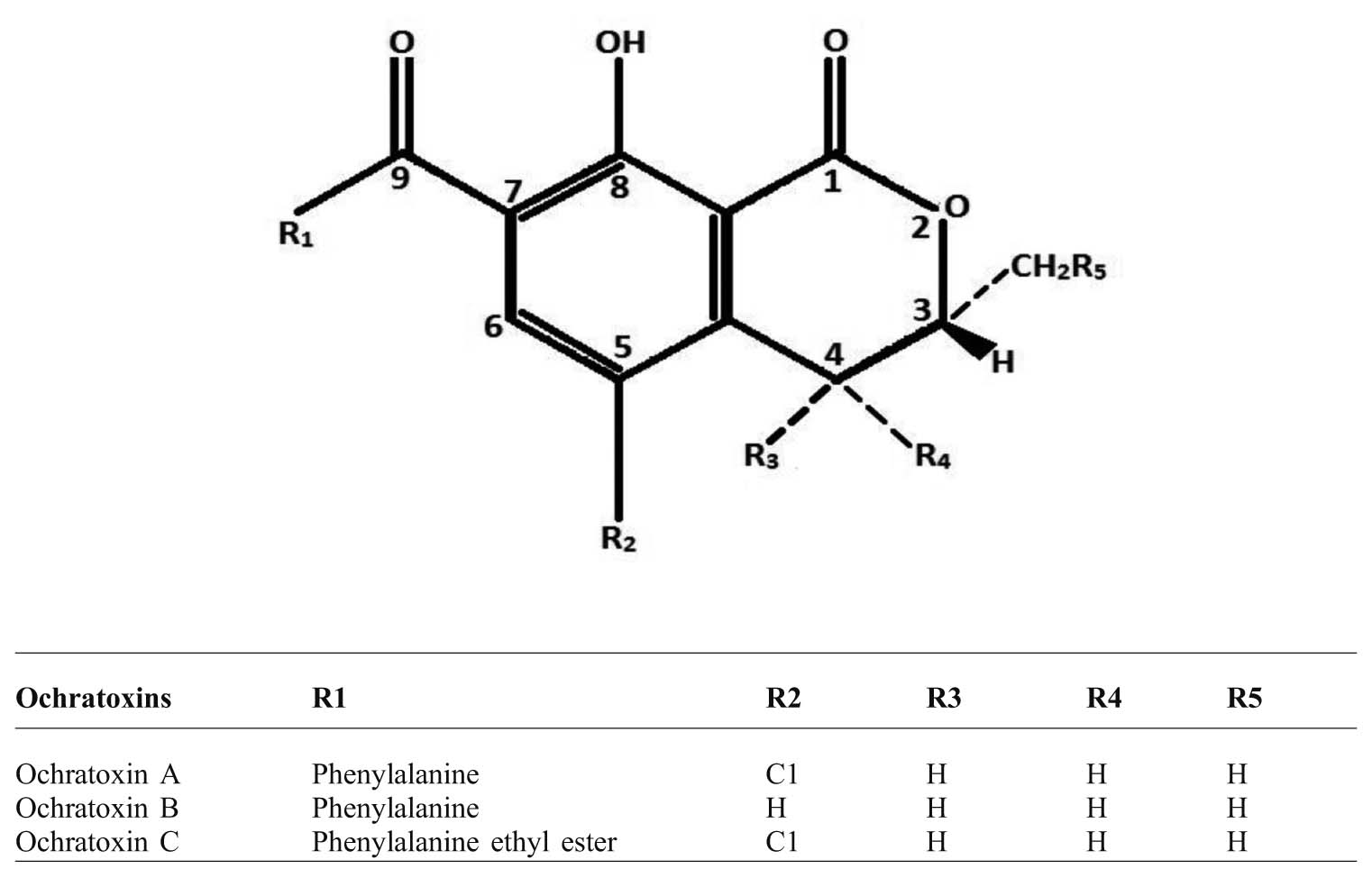
Ochratoxins are primarily produced by several species of Aspergillus and Penicillium fungi with Aspergillus ochraceus being the major producer, and are chemically weak organic acids. The structure of ochratoxin shows that they are pentaketides having di-hydro-isocoumarin linked by an amide bond with L-β-phenylalanine through a 7-carboxy group. On the basis of minor structural differences, ochratoxins have been divided into three subtypes namely ochratoxin A (OTA), B (OTB) and C (OTC), with OTA being the most toxic due to its chlorinated analogue followed by OTB having moderate toxicity and OTC being non-toxic due to phenylalanine ethyl ester structure (O'Brien and Dietrich, Reference O'BRIEN and DIETRICH2005; Abidin et al., Reference ABIDIN, KHATOON and NUMAN2011). When hydrolysed OTA and OTB become ochratoxin α and ochratoxin β which are the non-toxic products due to absence of the phenylalanine moiety (Moss, Reference MOSS1996; Xiao et al., Reference XIAO, MADHYASTHA, MARQUARDT, LI, VODELA, FROHLICH and KEMPPAINEN1996; Li et al., Reference LI, MARQUARDT, FROHLICH, VITTI and CROW1997).
Regarding OTA and OTB, the R1 region is same while there are differences in the R2 region. Similarly, OTC contains phenylalanine ethyl ester in the R1 region instead of a simple phenylalanine moiety in the R1 region as observed in OTA (Figure 1).
Chemical structure of different ochratoxins.

OTA is a white, crystalline odourless solid molecule with a molecular weight of 403.82 with the empirical formula of C20H18O6NCl. It has a melting point is 168-172ºC while the melting point of its benzene derivative is 90ºC. OTA emits maximum fluorescence at 428 nm in absolute alcohol and at 467 nm in 96% ethanol. It is slightly soluble in water but shows extreme solubility in organic solvents. OTA shows high stability and can be stored at -20ºC for several years when preserved in methanol or ethanol (Valenta, Reference VALENTA1998).
OTA has been classified as a Group 2B pathogen due to its carcinogenicity by the International Agency for Research on Cancer (IARC, 1993). The species Penicillium verrucosum is the major OTA producer in cold and temperate climatic regions while, in tropical climatic zones, the major OTA producers are Aspergillus ochraceus, A. carbonarius, A. melleus and A. niger (Abarca et al., Reference ABARCA, ACCENSI, BRAGULAT and CABAÑES2001; Rosa et al., Reference ROSA, PALACIOS, COMBINA, FRAGA, REKSON, MAGNOLI and DALCERO2002; Reference ROSA, RIBEIRO, FRAGA, GATTI, CAVAGLIERI, MAGNOLI, DALCERO and LOPES2006).
Presence of ochratoxin A in poultry feed and ingredients
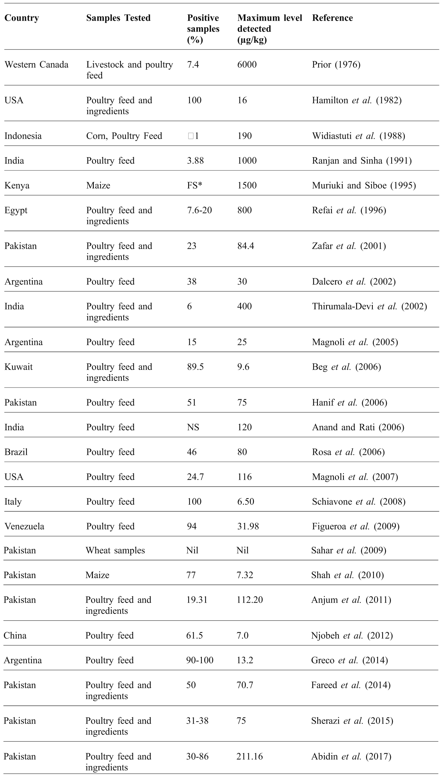
Ochratoxins are important contaminants which can be detected in poultry feed samples, raw materials and other ingredients. It was first detected in 1969 as a natural contaminant of corn samples (Shotwell et al., Reference SHOTWELL, HESSELTINE and GOULDEN1969). Up until now, it has been detected as a contaminant of different food, feeds and ingredients all over the World. It was reported by Bacon et al. (Reference BACON, SWEENEY, ROBBINS and BURDICK1973) that temperatures up to 30ºC along with high moisture conditions increased OTA production in feed from Aspergillus ochraceus, while low temperature (15 to 22ºC) along with low moisture conditions favoured the production of penicillic acid. The presence of OTA in poultry feed and its ingredients are presented in Table 1. However, regarding the prevalence of OTB and OTC, no reports are available in published literature regarding their detection in poultry feed or ingredients.
Different regulatory levels for OTA are available for various feed raw materials and animal by-products. As per the European Commission Recommendation (2006, 576) the maximum tolerable level (MTL) for complete feedstuffs is 100 µg/kg (Denli and Perez, Reference DENLI and PEREZ2010). The oldest study from Western Canada (Prior, Reference PRIOR1976) gave maximum detected OTA levels (6000 µg/kg) which was made before the approval of legislation for the safe levels of OTA contamination. A recent study by Abidin et al. (Reference ABIDIN, KHATOON, AROOJ, HUSSAIN, ALI, MANZOOR and SALEEMI2017) reported levels beyond MTL however feed samples collected in hot and humid environments showed higher levels of OTA beyond the MTL and vice versa. Predisposing factors like hot and humid environments, poor storage conditions, pre-harvest and harvest conditions are important in the production of OTA within poultry feed and its ingredients.
Occurrence of ochratoxin A (OTA) in poultry feed and feed ingredients.

*FS=Field study
Mode of action of OTA
There is a hypothesis that OTA, due to its great resemblance to the amino acid phenylalanine, might be used as a substrate and cause strong inhibitory effects on enzymes, ultimately leading to inhibition of protein synthesis along with lipid peroxidation in different biological systems (Dirheimer, Reference DIRHEIMER1996). However, in vitro studies have revealed that OTA toxicity was not reduced when L-phenylalanine was given (Bruinink et al., Reference BRUININK, RASONYI and SIDLER1997) demonstrating that OTA did not affect phenylalanine dependent processes (Bruinink and Sidler, Reference BRUININK and SIDLER1997). Rather, it might be indirectly related to aminoacyl-tRNA formation by inhibiting the production of ATP in mitochondria instead of binding with phenylalanyl-tRNA synthetase (Fink-Gremmels, Reference FINK-GREMMELS1999). OTA causes suppression of respiration in the mitochondria along with inhibition of t-RNA synthetase enzyme by acting as a competitive inhibitor of the carrier protein situated in the inner lining of mitochondria, altering energy metabolism and leading to lipid peroxidation in the mitochondria of kidney cells (Marquardt and Frohlich, Reference MARQUARDT and FROHLICH1992; Gentles et al., Reference GENTLES, SMITH, KUBENA, DUFFUS, JOHNSON, THOMPSON, HARVEY and EDRINGTON1999). Moreover, OTA is considered responsible for causing lipid peroxidation and increased oxidative stress in exposed individuals. Reactive oxygen species (ROS) are produced as a result of normal aerobic biotransformation of certain xenobiotics. Under normal conditions, there is a homeostatic balance between the production and scavenging of ROS in tissues. Oxidative stress occurs where production exceeds the cellular antioxidant capacity to scavenge ROS. Tissue injury associated with higher ROS concentration may occur due in critical cellular targets like lipids, DNA or proteins (Jacobson, Reference JACOBSON1996). Oxidative stress caused by OTA has been well documented by Hoehler and Marquardt (Reference HOEHLER and MARQUARDT1996) and Hoehler et al. (Reference HOEHLER, MARQUARDT and ROHLICH1997). However, no information about the details of biotransformation of OTA in poultry is currently available (Wu et al., Reference WU, DUAN, WANG and WANG2011).
Ochratoxicosis in poultry
The levels and durations of OTA exposure are the main pre-disposing factor regarding severity of alterations and adverse effects in birds. The discussions below relate to the alterations induced by OTA in different poultry species regarding growth parameters (body weight, feed intake and relative organ weights), production performance and histopathological alterations in different visceral organs.
growth performance
OTA-associated decreased body weight gain is not caused by the direct effect of OTA, rather is related to reduced feed intake (Elaroussi et al., Reference ELAROUSSI, MOHAMED, EL BARKOUKY, ATTA, ABDOU and HATAB2006). OTA-associated growth retardation in birds might occur due to its ability to impair protein metabolism. In vitro studies show that OTA acts as a competitive inhibitor of carboxypeptidase A and this mechanism might cause inhibition of protein metabolism in vivo (Pitout and Nel, Reference PITOUT and NEL1969). Competitive attachment of OTA on phenylalanine binding sites may be a reason for the inhibition of protein synthesis (Creppy et al., Reference CREPPY, STORMER, ROSCHENTHALER and DIRHEIMER1983). OTA-associated growth suppression and weakness disturbs mineral homeostasis leading to skeletal changes and/or abnormalities (Duff et al., Reference DUFF, BURNS and DWIVEDI1987). Growth alterations induced by OTA in poultry as observed by different researchers are presented in Table 2.
relative organ weights
OTA causes an increase in the relative organ weights of liver and kidney and a reduction in thymus, size and bursa of Fabricius. Increase in the size of kidney and liver occurs due to enlargement of epithelium and hyperaemia occurring probably in an effort to eliminate OTA from body. These degenerative changes and enlargement in the epithelial cells of kidney and liver are probably due to exposure to OTA during elimination through kidney and liver, as the hepatic portal system along with enterohepatic recirculation are being involved in elimination of toxic materials from the body (Fuchs, Reference FUCHS1988). However, direct effects of OTA on these organs might have a role in producing these alterations. In a feeding trial with turkeys in a four-week study, no increases in relative liver weight were observed, which might be due to lower susceptibility of turkeys to OTA as compared to broilers (Dwivedi and Burns, Reference DWIVEDI and BURNS1984a). Similarly, no enlargement in the kidneys of turkeys was observed in birds exposed to higher doses of OTA for three weeks (Chang et al., Reference CHANG, DOERR and HAMILTON1981). This again reinforces the reduced susceptibility of OTA in turkeys compared to other poultry species.
OTA caused decreased relative weights of thymus, bursa of Fabricius and spleen probably due to a reduction of lymphoid tissue in these organs along with other degenerative alterations (Creppy et al., Reference CREPPY, STORMER, ROSCHENTHALER and DIRHEIMER1983). Another theory behind this is that OTA might delay the division of rapidly dividing cells in the lymphoid tissue (Stoev et al., Reference STOEV, STEFANOV, RADIC, DOMIJAN and PERAICA2004). On the whole, the decrease in certain lymphoid organs could be a possible reason for observed immunosuppression in birds. A summary of the alterations in the relative organ weights of poultry by different scientists is presented in Table 2
Ochratoxin A (OTA) associated alterations of growth parameters and relative organ weights poultry.

*NMF=Naturally mouldy feed; **=Slaughter house-based study; WL=White Leghorn;
OTA effects on performance of laying hens
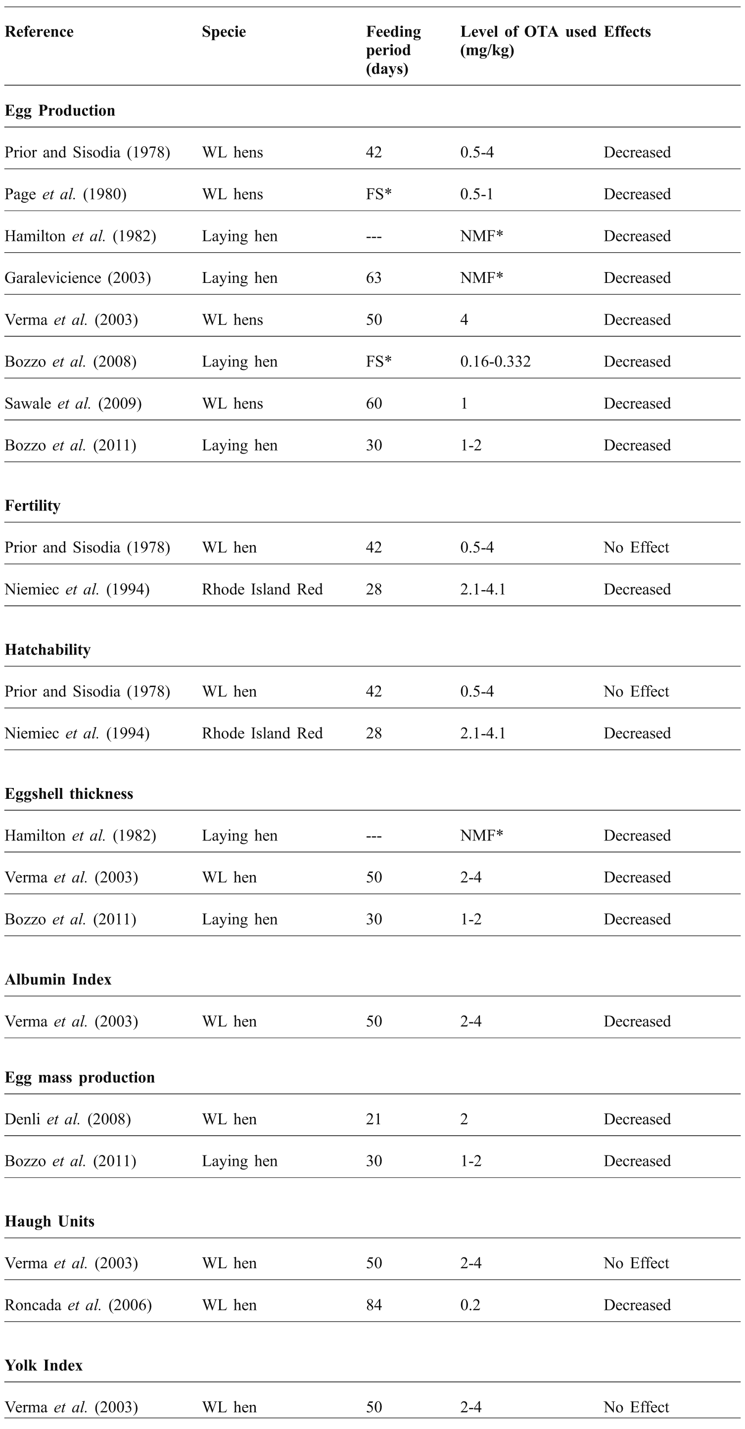
OTA adversely affects the productivity of hens in terms of deteriorated egg quality, poor hatchability and reduced egg size. It causes a decline in egg laying (clutch size), shell damage, reduced shell thickness and delayed sexual maturation of hens along with alterations in the ultrastructure of shell and changes in external/internal shell membranes (Choudhury et al., Reference CHOUDHURY, CARLSON and SEMENIUK1971; Scholtyssek et al., Reference SCHOLTYSSEK, NIEMIEC and BAUER1987; Niemiec et al., Reference NIEMIEC, CYWA-BENKO, STEPINSKA, WEZYK and SWIERCZEWSKA1993a; Reference NIEMIEC, GOLINSKI, BORZEMSKA, SWIERCZEWSKA and STEPINSKA1993b). It may be assumed that disturbances in mineral metabolism can be attributed to the thinner egg shells observed (Niemiec et al., Reference NIEMIEC, BORZEMSKA, GOLINSKI, KARPINSKA, SZELESZCZUK and CELEDA1994). Table 3 summarises some of the studies showing altered productivity parameters due to OTA in birds.
Ochratoxin A (OTA) associated alterations in production parameters of poultry.

*NMF=Naturally mouldy feed; FS=Field Study; WL=White Leghorn
Histopathological alterations
OTA produces deleterious alterations in almost all the organs of birds. Discussed below are some of the notable effects of OTA in birds.
liver
This is the organ most severely affected by OTA, and histological alterations include congested parenchyma, presence of hydropic/vacuolar degeneration within the cytoplasm of hepatocytes, pyknotic nuclei of hepatocytes, necrosis of individual hepatocytes, biliary hyperplasia, cell swelling and congestion, hazy vacuoles in cytoplasm of hepatocytes and congested sinusoidal spaces. The severity of these lesions depends upon the dose and timing of OTA exposure. Such lesions have been reported in several experimental trials where OTA contamination in feed ranged from 0.5-20 mg/kg for variable time intervals (21-42 days) in white Leghorn hens (Page et al., Reference PAGE, STEWART, WYATT, BUCH, FLETCHER and BROWN1980), broilers (Kumar et al., Reference KUMAR, JINDAL, SHUKLA, ASRANI, LEDOUX and ROTTINGHAUS2004), broiler chicks (Sandhu et al., Reference SANDHU, SINGH and SINGH1995; Solcan et al., Reference SOLCAN, COMAN and SOLCAN2008) and white Leghorn cockerels (Abidin et al., Reference ABIDIN, KHAN, KHATOON, SALEEMI and KHAN2016).
kidney
The adverse effects of OTA on kidneys include atrophy and degeneration of proximal as well as distal convoluted tubules, interstitial fibrosis, pyknotic nuclei in tubular epithelial cells, congested glomeruli, vacuolar degeneration in tubular epithelia, complete necrosis of tubules, congested glomeruli along with cellular infiltration, condensed nuclei of epithelial cells, proteinaceous material in urinary spaces and congested urinary spaces of Bowman's capsules. These alterations are dose dependent with maximum severity seen in birds exposed to higher levels. Different trials using 0.5-20 mg/kg OTA for three to six weeks have reported such findings in white Leghorn hens (Page et al., Reference PAGE, STEWART, WYATT, BUCH, FLETCHER and BROWN1980), broilers (Sandhu et al., Reference SANDHU, SINGH and SINGH1995; Kumar et al., Reference KUMAR, JINDAL, SHUKLA, ASRANI, LEDOUX and ROTTINGHAUS2004; Solcan et al., Reference SOLCAN, COMAN and SOLCAN2008) and white Leghorn cockerels (Abidin et al., Reference ABIDIN, KHAN, KHATOON, SALEEMI and KHAN2016).
bursa of Fabricius
This is the main lymphoid organ present in birds and is severely affected by exposure to OTA. The bursa associated alterations include increased interfollicular connective tissue, reduced diameter of the cortex as compared to the medulla, increased number of pyknotic nuclei in both the cortex and medulla, formation of cysts in surface epithelium, lymphoid cells depletion and degenerative changes in the germinal centre. These changes may be linked to the immunosuppression in the birds associated with OTA exposure. Levels of OTA ranging from 0.13 to 8 mg/kg feed for a period two to 10 weeks are thought to be responsible for such alterations in broiler chickens (Chang et al., Reference CHANG, DOERR and HAMILTON1981; Dwivedi and Burns, Reference DWIVEDI and BURNS1984a; Kozaczynski, Reference KOZACZYNSKI1994; Stoev et al., Reference STOEV, ANGUELOV, IVANOV and PAVLOV2000; Reference STOEV, DASKALOV, RADIC, DOMIJAN and PERAICA2002a; Reference STOEV, DJUVINOV, MIRTCHEVA, PAVLOV and MANTLE2002b; Kumar et al., Reference KUMAR, JINDAL, SHUKLA, ASRANI, LEDOUX and ROTTINGHAUS2004; Koynarsky et al., Reference KOYNARSKY, STOEV, GROZEVA, MIRTCHEVA, DASKALOV, MITEV and MANTLE2007), turkeys (Dwivedi and Burns, Reference DWIVEDI and BURNS1985) and Babcock-300 chicks (Doupnik and Peckham, Reference DOUPNIK and PECKHAM1970).
thymus
This is another important lymphoid organ in birds primarily involved in the production of T-cells. Histologically the lesions associated with OTA in the thymus result in depletion of lymphocytes, congestion, vacuolation within cortical region with multiple pyknotic nuclei and increased number of Hassel's corpuscles within the medullary region when OTA is administered at the rates of 0.13-5 mg/kg feed for two to six weeks (Kozaczynski, Reference KOZACZYNSKI1994; Stoev et al., Reference STOEV, ANGUELOV, IVANOV and PAVLOV2000; Reference STOEV, DASKALOV, RADIC, DOMIJAN and PERAICA2002a; Reference STOEV, DJUVINOV, MIRTCHEVA, PAVLOV and MANTLE2002b; Kumar et al., Reference KUMAR, JINDAL, SHUKLA, ASRANI, LEDOUX and ROTTINGHAUS2004).
spleen
The spleen is affected by the administration of OTA to birds, whereby lesions cause the depletion of lymphocytes, congestion and haemorrhages in white pulp, loss of demarcation between red and white pulp along with significant congestion and hyperplasia of splenic parenchyma (Kozaczynski, Reference KOZACZYNSKI1994; Stoev et al., Reference STOEV, ANGUELOV, IVANOV and PAVLOV2000; Reference STOEV, DASKALOV, RADIC, DOMIJAN and PERAICA2002a; Reference STOEV, DJUVINOV, MIRTCHEVA, PAVLOV and MANTLE2002b; Kumar et al., Reference KUMAR, JINDAL, SHUKLA, ASRANI, LEDOUX and ROTTINGHAUS2004) when 0.13-5 mg/kg OTA in feed are given for a period of up to six weeks.
lungs
OTA associated microscopic lesions in the lungs of birds cause congestion of the parenchyma, infiltration of mononuclear cells (MNC), serous exudate in alveoli and prominent interlobular septa due to oedema. Kumar et al. (Reference KUMAR, JINDAL, SHUKLA, ASRANI, LEDOUX and ROTTINGHAUS2004) has reported such alterations when 2 mg/kg OTA in feed was given for a period of 21 days
heart
In terms of microscopic lesions, the heart is considered to be the least affected organ by OTA ingestion in birds. No specific lesions except mild oedema of inter-muscular bundle spaces and infiltration of mononuclear cells (MNC) have been observed with OTA levels of 2 mg/kg feed given for 21 days (Mohiuddin et al., Reference MOHIUDDIN, REDDY and AHMED1992; Kumar et al., Reference KUMAR, JINDAL, SHUKLA, ASRANI, LEDOUX and ROTTINGHAUS2004).
Conclusions
From this review, the presence of ochratoxin A (OTA) in poultry feed adversely affects growth and performance of birds, and once OTA enters the body, it affects the functioning of each and every organ of the body. This can be seen in histopathological alterations associated with OTA administration. Similarly, the growth and laying performance of birds is seriously affected by OTA contamination in feed, which subsequently compromises the overall economy of the poultry farm.