Background
Cardiomyopathies represent a heterogeneous group of diseases characterised by structural and functional abnormalities of the myocardium that impair the heart’s ability to pump blood effectively, ultimately leading to symptoms of heart failure. Although these conditions can affect individuals at any age, childhood-onset cardiomyopathies are rare with an estimated annual incidence of 0.7 to 1.3 per 100,000 person-yearsReference Lipshultz, Sleeper and Towbin1–Reference Arola, Jokinen and Ruuskanen3 are poorly characterised and often associated with poor outcomes despite advances in medical therapy. In cases of end-stage heart failure, ventricular assist devices and heart transplantation remain the primary therapeutic options.Reference Burch, Siddiqi, Celermajer, Scott, Bull and Deanfield4–Reference Willerson7
Primary cardiomyopathies are frequently of genetic origin. With the decreasing cost and turnaround time of whole-exome sequencing, an increasing number of genetic loci associated with these disorders have been identified. Variants are typically found in genes encoding proteins related to the sarcomere, cytoskeleton, desmosome, nucleus, or mitochondria, leading to structural and/or functional disturbances in key myocardial processes. In paediatrics, five morpho-functional phenotypes have been described: dilated cardiomyopathy, hypertrophic cardiomyopathy, restrictive cardiomyopathy, arrhythmogenic cardiomyopathy, and left ventricular non-compaction cardiomyopathy.Reference Lipshultz, Sleeper and Towbin1,Reference Lipshultz, Law and Asante-Korang8–Reference Maron, Towbin and Thiene11
Although genetic testing continues to reveal new variants associated with cardiomyopathies, data from South America remain limited. Most of the existing knowledge originates from European and North American cohorts. Despite ongoing advances, more than half of all primary cardiomyopathy cases still lack an identifiable genetic cause.Reference Spracklen, Keavney, Laing, Ntusi and Shaboodien12
The clinical course of each patient is influenced by the type of cardiomyopathy, the interval between symptom onset and diagnosis, genotype–phenotype correlations, the number of identified pathogenic variants, and potential environmental modifiers. Previous studies have demonstrated that the number of pathogenic variants present in an individual can significantly impact the clinical phenotype: the greater the number of variants, the more severe the structural involvement and the worse the clinical prognosis.Reference Spracklen, Keavney, Laing, Ntusi and Shaboodien12,Reference Wang, Wang and Zou13
The objective of this study was to describe the clinical characteristics of a paediatric population with primary cardiomyopathies, with a particular emphasis on aetiology and short- and long-term outcomes.
Materials and methods
Study design
This was a retrospective descriptive study including patients under 18 years of age diagnosed with primary cardiomyopathy between 2009 and 2024 at two tertiary care centres in Medellín, Colombia. The study data were obtained from the patient’s medical history during the care process. The study was conducted in accordance with the principles outlined in the Declaration of Helsinki and was approved by the Institutional Review Board (approval number 25/2023).
Patient population
Patients were eligible for inclusion if they were diagnosed with primary cardiomyopathy during the study period. Primary cardiomyopathies were defined as a group of disorders primarily affecting the myocardium, resulting in structural and functional abnormalities in the absence of congenital heart disease, valvular disease, hypertension, coronary artery disease, infectious causes (e.g., myocarditis), or systemic conditions (e.g., autoimmune diseases or involvement of other organs). The five recognised cardiomyopathy subtypes: dilated cardiomyopathy, hypertrophic cardiomyopathy, restrictive cardiomyopathy, arrhythmogenic cardiomyopathy, and left ventricular non-compaction cardiomyopathy were included. Left ventricular non-compaction cardiomyopathy was diagnosed in patients who fulfilled echocardiographic and/or cardiac magnetic resonance criteria for ventricular non-compaction, in the presence of associated ventricular dysfunction, a family history of cardiomyopathy, clinical symptoms, major cardiovascular events, and/or a pathogenic or likely pathogenic genetic variant related to the condition.
For each case of primary cardiomyopathy, aetiology was classified as genetic, familial, or idiopathic. A genetic aetiology was assigned when a pathogenic or likely pathogenic variant was identified through genetic testing in the cardiomyopathy or exome panel and correlated with the observed phenotype. Genetic variants classified as of uncertain significance, but which correlated with the phenotype and were supported by databases as being causative of disease, were also included as genetic variants. A familial aetiology was defined by the presence of affected first- or second-degree relatives across multiple generations, in cases where genetic testing was negative or inconclusive. Idiopathic cardiomyopathy was diagnosed when, despite thorough imaging (echocardiography, cardiac MRI), laboratory evaluation, genetic testing, and histological analysis (when available), no identifiable cause was determined. Phenocopies associated with mitochondrial disorders, syndromic conditions, or neuromuscular diseases were excluded due to their distinct clinical course and prognosis.Reference Marino, Digilio, Toscano, Giannotti and Dallapiccola14–Reference Lipshultz, Orav and Wilkinson17
Variables
The study collected sociodemographic variables (age, sex, weight, and height), diagnostic variables (cardiomyopathy subtype, presenting symptoms, and ventricular function), aetiological data including genetic variants, treatment-related variables (medical therapy, interventional procedures, or surgery), and outcome measures (death, heart transplantation, and device implantation). Patients were followed from their initial presentation to the most recent clinical record available in the participating institutions.
Statistical analysis
Continuous variables were expressed as mean ± standard deviation and/or as median with interquartile range, depending on the distribution assessed using the Kolmogorov–Smirnov test for normality. Categorical variables were presented as absolute numbers and percentages. Statistical analyses were performed using SPSS version 27.0 (IBM Corp., Armonk, NY).
Results
A total of 76 patients from the two participating centres met the inclusion criteria. The mean age at diagnosis was 6.3 ± 5.6 years, with a slight female predominance (56.6%). A first- or second-degree family history of cardiomyopathy was documented in 31.6% of patients.
Subtypes of cardiomyopathy and baseline characteristics
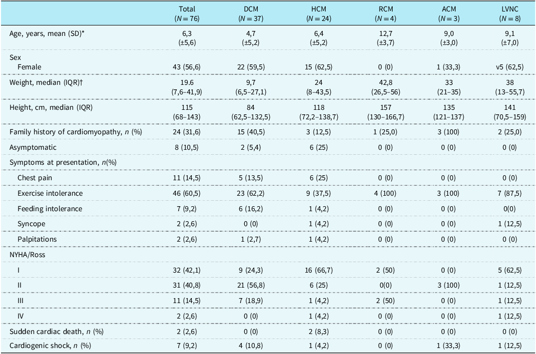
Dilated cardiomyopathy was the most common subtype (n = 37, 48.6%), followed by hypertrophic cardiomyopathy (n = 24, 31.5%), left ventricular non-compaction cardiomyopathy (n = 8, 10.5%), restrictive cardiomyopathy (n = 4, 5.2%), and arrhythmogenic cardiomyopathy (n = 3, 3.9%). Baseline characteristics by cardiomyopathy subtype are summarised in Table 1. The most frequent indication for initial cardiologic evaluation was the presence of clinical symptoms (68.5%), followed by family screening (28.9%), and sudden cardiac arrest (2.6%).
Baseline and clinical characteristics by cardiomyopathy subtype

*SD = standard deviation. †IQR = interquartile range.
Clinical characteristics
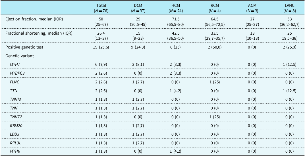
A total of 89.5% of patients presented symptoms during follow-up, with symptoms being more common in patients with restrictive cardiomyopathy and arrhythmogenic cardiomyopathy. The most frequent symptom was exercise intolerance (60.5%), followed by chest pain (14.5%) and feeding intolerance (9.2%). At the time of diagnosis, most patients were in NYHA/Ross functional class I or II (82.9%). Cardiogenic shock occurred in 9.2% of patients at the onset of cardiomyopathy. Left ventricular ejection fraction was reduced at diagnosis in patients with dilated cardiomyopathy (29% [interquartile range 20.5–45]) and arrhythmogenic cardiomyopathy (27% [interquartile range 25–27]), as shown in Table 2.
Echocardiographic and genetic characteristics by cardiomyopathy subtype

Genetic testing and aetiology
A family history of cardiomyopathy was present in 31.6% of the cohort. The three patients who exhibited an arrhythmogenic cardiomyopathy phenotype were siblings, all demonstrating phenotypic characteristics consistent with Carvajal syndrome. However, genetic confirmation could not be obtained due to early mortality and limited follow-up. Among the remaining subtypes, the most frequent aetiology was idiopathic, as illustrated in Figure 1.
Aetiology of primary cardiomyopathy by cardiomyopathy subtype.
Note: ACM cases were excluded, as their shared sibling aetiology precluded classification and could bias the results.

A variant as a cause of disease
A disease-causing variant was identified in 19 patients (25.6%, n = 19/76), with the highest frequency observed in patients with restrictive cardiomyopathy (50%), followed by those with hypertrophic cardiomyopathy (25%), left ventricular non-compaction cardiomyopathy (25%), and dilated cardiomyopathy (24.3%). The most frequently affected gene was MYH7 (n = 6, 7.9%), which was identified as a disease-associated variant in dilated cardiomyopathy, hypertrophic cardiomyopathy, and left ventricular non-compaction cardiomyopathy, followed by TTN (n = 2, 2.6%), MYBPC3 (n = 2, 2.6%), and FLNC (n = 2, 2.6%). Of the patients with a genetic mutation, six had variants of uncertain significance in other structural genes located in cardiomyocytes, which had no relevant clinical impact on outcomes. Table 2 outlines the genetic variants associated with each type of cardiomyopathy. Figure 2 illustrates the geolocation of the mutations, shown on a map of Colombia.
Geolocation of genetic variants by birthplace and cardiomyopathy subtype.

Treatment at the time of the study
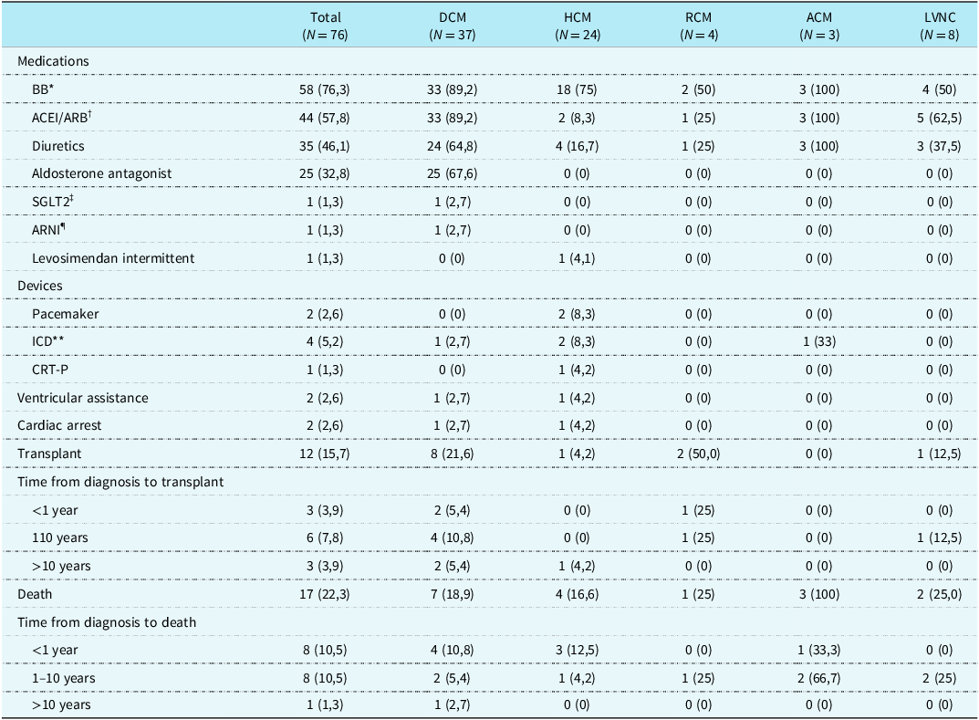
A total of 72 patients (94.7%) received one or more cardiovascular medications at the time of the study. The description of the medications received by cardiomyopathy subtype is shown in Table 3. The most common medication type was beta-blockers (n = 58, 76.3%), followed by angiotensin-converting enzyme inhibitors (n = 44, 57.8%) and diuretics (n = 35, 46.1%). In two patients with dilated cardiomyopathy, neprilysin inhibitors and sodium-glucose cotransporter 2 inhibitors were used.
Treatment and outcomes for each subtype of cardiomyopathy

*BB = beta-blocker. †ACEI/ARB II = angiotensin-converting enzyme inhibitor/angiotensin receptor blocker. ‡SGLT2 = sodium-glucose cotransporter 2 inhibitor. ¶ARNI = angiotensin receptor/neprilysin inhibitors. **ICD = implantable cardioverter defibrillator. ††CRT-P = cardiac resynchronisation therapy pacemaker.
In the group with hypertrophic cardiomyopathy, four patients (16.6%) required septal myectomy due to severe left ventricular outflow tract obstruction. Of these patients, two (50%) required a pacemaker due to complete atrioventricular block as a complication of septal myectomy. Of the entire cohort, four patients required defibrillator therapy (2 hypertrophic cardiomyopathy, 1 dilated cardiomyopathy, and 1 arrhythmogenic cardiomyopathy), and one patient required cardiac resynchronisation therapy (1 dilated cardiomyopathy). Two patients received ventricular assist device therapy as a bridge to heart transplantation (1 dilated cardiomyopathy, 1 hypertrophic cardiomyopathy, see Table 3.
Follow-up and outcomes
During the follow-up period, 12 patients (15.7%) received a heart transplant, with the highest incidence in restrictive cardiomyopathy (50%) and dilated cardiomyopathy (21.6%). The time interval from diagnosis to heart transplant in the study population was less than 10 years in all of them. During the study period, 17 patients (22.3%) died, with the following distribution by cardiomyopathy type: arrhythmogenic cardiomyopathy (n = 3, 100%), left ventricular non-compaction cardiomyopathy (n = 2, 25%), restrictive cardiomyopathy (n = 1, 25%), and dilated cardiomyopathy (n = 7, 18.9%). See Table 3.
Discussion
This study represents the largest paediatric cardiomyopathy registry in Colombia, demonstrating a high prevalence of genetic, familial, and idiopathic cardiomyopathies, along with a significant symptom burden that requires medications, implantable devices, surgical interventions, and heart transplantation.
Clinical characteristics of the population
In line with data published in paediatric registries from North AmericaReference Lipshultz, Sleeper and Towbin1 and Australia,Reference Nugent, Daubeney and Chondros2 the leading cause of cardiomyopathy in this study was dilated cardiomyopathy (48.6%). In contrast, the European registry reported hypertrophic cardiomyopathy as the most common cause. This difference may be explained by the exclusion of patients younger than 1 year of age and the high referral rate of patients with hypertrophic cardiomyopathy to specialised centres, given the perceived risk of genetic disease.Reference Kaski, Norrish and Gimeno Blanes10
The rates of presentation for other cardiomyopathy subtypes, such as restrictive cardiomyopathy, left ventricular non-compaction cardiomyopathy, and arrhythmogenic cardiomyopathy, were consistent with those reported by Ware et al. Reference Ware, Wilkinson and Tariq18 In the present study, a female predominance was observed across all cardiomyopathy subtypes, except for arrhythmogenic cardiomyopathy, which aligns with the findings of Ware et al. Reference Ware, Wilkinson and Tariq18 Other North American and Australian registries, however, reported a higher incidence in males.Reference Lipshultz, Sleeper and Towbin1,Reference Nugent, Daubeney and Chondros2 Recent studies on adults have determined that sex influences the prevalence, morpho-functional manifestations, and clinical course of cardiomyopathies. Although the underlying mechanisms remain poorly understood, genes involved in the expression of various ion channels, ventricular remodelling, and disease modifiers may influence outcomes, alongside epigenetic factors (social and environmental complexities between the sexes), potentially leading to greater disadvantages for women with this conditionReference Argiró, Ho and Day19,Reference Xu, Chen and Tillman20 These findings have not been extensively studied in the paediatric population, presenting an opportunity for future research.
Familial disease and genetics
The proportion of patients with a familial history was estimated at 31.6%, with variations observed according to the subtype of cardiomyopathy. This was most common in patients with arrhythmogenic cardiomyopathy, where the reported cases represented a familial cluster, followed by those with dilated cardiomyopathy. In contrast, the rate of family history was lowest in patients with hypertrophic cardiomyopathy, which contrasts with previous studies where familial disease was more prevalent in this cardiomyopathy subtype.Reference Kaski, Norrish and Gimeno Blanes10 This discrepancy may be explained by the fact that family screening in our healthcare system is complex. There are a few cardiogenetic programmes available, and screening is sometimes incomplete or performed only once without follow-up. In addition, administrative limitations prevent the direct issuance of referrals or screening medical orders for the family members of the index case.
Genetic diagnosis and variants
The overall genetic diagnosis rate was 25.6%, with the highest rate observed in restrictive cardiomyopathy (50%), hypertrophic cardiomyopathy (25%), left ventricular non-compaction cardiomyopathy (25%), and dilated cardiomyopathy (24.3%). The diagnostic yield of exome sequencing was 71.4%. The rate of genetic diagnosis with pathogenic and likely pathogenic variants was similar to that reported by Ware et al., Reference Ware, Wilkinson and Tariq18 which found a rate of 32%. However, the yield was higher in other registries, such as the European Cardiomyopathy and Myocarditis RegistryReference Kaski, Norrish and Gimeno Blanes10 (60%) and the North American Paediatric Cardiomyopathy RegistryReference Ware, Wilkinson and Tariq18 (48%). This discrepancy may reflect selection bias from expert centres with easier access to genetic testing. This contrasts with the population in the present study, where the retrospective nature of the research limited the ability to perform additional genetic studies or reanalyse previously conducted genetic tests in a controlled manner.
As determined in both adult and paediatric studies, sarcomeric genes are predominantly responsible for cardiomyopathy across different subtypes.Reference Kaski, Syrris and Esteban21–Reference Ware, Bhatnagar and Dexheimer24 In this study, the most common pathogenic or likely pathogenic variants across the cardiomyopathy subtypes were in MYH7, TTNI3, TNNT2, MYBPC3, and FLNC. The MYH7 gene was the most frequent cause in dilated cardiomyopathy, while MYBPC3 was most commonly seen in hypertrophic cardiomyopathy, consistent with internationalReference Kaski, Syrris and Esteban21–Reference Bagnall, Singer and Wacker23 and nationalReference Castro-Oróstegui and Beltrán-Durán25 reports on genotyping in paediatric cardiomyopathies. However, these findings suggest that genotype-phenotype correlations specific to each cardiomyopathy subtype may provide a more refined understanding of the molecular underpinnings of the disease and its association with presentation, clinical progression, and outcomes. Therefore, when an index case is identified, family cascade screening should be initiated due to the importance of early detection, especially in patients associated with the autosomal dominant inheritance pattern of these cardiomyopathies.
Although in recent years—particularly in adult populations—excessive trabeculations have been increasingly recognised as a morphological phenotype within dilated and hypertrophic cardiomyopathies, this concept has not yet gained wide acceptance in paediatric cardiology. The main reasons include genetic heterogeneity, overlap between genotype and phenotype, the lack of diagnostic imaging criteria validated for children, and heterogeneous findings observed in longitudinal follow-up of paediatric patients with left ventricular non-compaction cardiomyopathy. Moreover, the prognostic significance of excessive trabeculation in this group remains uncertain. Therefore, multicentre studies and an international consensus specifically focused on the paediatric population are needed to refine diagnostic accuracy and improve risk stratification and outcomes in these patients.Reference Petersen, Friedrich and Ho26,Reference van Waning, Caliskan and Hoedemaekers27
The potential impact of the number of variants on clinical symptoms and the outcomes of different cardiomyopathies has been explored.Reference Xie, Yang and Gong28,Reference Walsh, Thomson and Ware29 In this study, six patients presented pathogenic variants associated with variant of uncertain significance in other proteins. One patient, with a pathogenic mutation in MYH6 and variants of uncertain significance in SCN5A, developed complete atrioventricular block after the cardiomyopathy diagnosis. However, no differences in mortality, transplantation, or use of ventricular assist devices were observed when compared to those with a single variant. Another aspect to consider is the role of digenic inheritance, which suggests that mutations in more than one gene may contribute to disease development. This complicates the diagnosis and understanding of cardiomyopathy pathogenesis and remains an area of active research.Reference Parikh, Day and Lakdawala30,Reference Liu, Yang, Gong, Wang, Yao and Xie31
Treatment
A high proportion of patients in this study received cardiovascular medications, which may indicate bias due to the severity of their presentation upon admission, as also noted in the European Registry of Cardiomyopathy and Myocarditis. One in nineteen patients received implantable cardioverter defibrillator therapy, which is consistent with other paediatric studies.Reference Maron, Spirito and Ackerman32 The proportion was higher in patients with arrhythmogenic cardiomyopathy (33% vs. 60%) compared to other registries of both adults and children.Reference Kaski, Norrish and Gimeno Blanes10–Reference Marino, Digilio, Toscano, Giannotti and Dallapiccola14,Reference Wilkinson, Lowe and Salbert16–Reference Charron, Elliott and Gimeno33 In patients with hypertrophic cardiomyopathy, the proportion requiring septal myomectomy (16.6% vs.7.5%) was higher than in other studies despite the fact that in our cohort, we did not include patients with phenocopies, in whom the prevalence of left ventricular outflow tract obstruction is higher.Reference Wilkinson, Lowe and Salbert34–Reference Jia, Meng, Tang, Sun, Peng and Zhang36
Outcomes
The overall mortality rate in the present registry was 22.3%, occurring within the first 10 years after diagnosis. This rate varied substantially when analysing the subtypes separately, being higher in our patients with arrhythmogenic cardiomyopathy (100%), which was explained by a particular severe form of the disease in a familial presentation.Reference Kaski, Norrish and Gimeno Blanes10,Reference Erolu, Akalın, Saylan Çevik and Yücelten37
Heart transplantation was a common treatment option in some of the cardiomyopathy subtypes, and it was more frequent within the first 10 years after diagnosis. One in two patients with restrictive cardiomyopathy required a heart transplant, followed by one in five patients with dilated cardiomyopathy, consistent with previous studies reported in this patient subpopulation.Reference Towbin, Lowe and Colan38 A ventricular assist device was required in two patients in the entire cohort (1 dilated cardiomyopathy, 1 hypertrophic cardiomyopathy) as a bridge to heart transplantation.
Limitations
Although this study represents one of the largest series of primary cardiomyopathy in our country, it has several limitations. As in other publications, the data obtained may not reflect the overall paediatric cardiomyopathy population, as they were collected from two referral centres. Therefore, the complexity of cases and the severity of outcomes may be overestimated. The descriptive and retrospective nature of the study limited the recording of some variables during follow-up, such as biomarkers and myocardial strain measurements. Some cardiomyopathy subtypes, such as restrictive cardiomyopathy and arrhythmogenic cardiomyopathy, included a low number of patients, and their results should thus be interpreted with caution. Due to the small number of patients and events, the study lacked sufficient statistical power to perform direct comparisons between cardiomyopathy subtypes; hence, all analyses were purely descriptive.
Conclusions
Primary cardiomyopathies are rare in infants and children but are associated with significant morbidity and mortality. The genetic basis of cardiomyopathies and genotype–phenotype correlations play a key role in timely diagnosis, clinical follow-up, cascade screening of first-degree relatives, primary and secondary prevention of sudden cardiac death, and long-term outcomes. Expanding our knowledge of paediatric cardiomyopathies both nationally and globally is a growing need and a driving force for future research.
Supplementary material
For supplementary material accompanying this paper visit https://doi.org/10.1017/S1047951126111548.
Data availability statement
The data that support the findings of this study are available on request from the corresponding author. The data are not publicly available due to privacy or ethical restrictions.
Author contributions
Andres Aranzazu: conceptualisation, methodology, formal analysis, investigation, writing original draft. Margarita Zapata: writing, review, and editing, supervision. Rafael Correa: writing, review, and editing. Eliana Cañas: protocol review, data analysis, and article writing. Carolina Baquero: writing, review, and editing, supervision.
Financial support
This research did not receive any specific grant from funding agencies in the public, commercial, or not-for-profit sectors.
Competing interests
The authors declare no conflicts of interest.
Medical ethics review
The study was conducted in accordance with the principles outlined in the Declaration of Helsinki and was approved by the Ethics Committee of the Pontifical Bolivarian University (approval number 25/2023), CardioVID Clinic (approval number 12/2023), and Pablo Tobon Uribe Hospital (approval number 01/2024).