Introduction
Nasal teeth causing nasal discharge are a rare clinical entity.Reference Yeung and Lee1, Reference Gupta and Shah2 This scarcity restricts the implementation of large-scale clinical studies investigating the aetiology, diagnosis and treatment. Clinical case records provide the best form of evidence. We therefore report this clinical case of a symptomatic nasal tooth. In addition, we provide an overview of the literature regarding nasal teeth.
Case report
A 26-year-old man was referred to the otolaryngology department with a history of recurrent left-sided nasal obstruction, facial pain and discomfort, and chronic purulent rhinorrhoea. There was no history of trauma, allergy or smoking. On examination, he was found to have hypertrophic inferior nasal conchae and a mild left-sided nasal septum deviation.
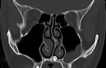
Computed tomography of the paranasal sinuses revealed the left-sided septum deviation and hypertrophic inferior conchae, as well as a mucocele on the floor of the left maxillary sinus and a radiopaque mass resembling an ectopic tooth in the left nasal floor (Figure 1). After consultation with the patient, and once informed consent had been obtained, an elective procedure under general anaesthesia was planned in order to remove the radiopaque mass.
Coronal computed tomography image of the paranasal sinuses, depicting a tooth in the left nasal cavity and left-sided septum deviation.

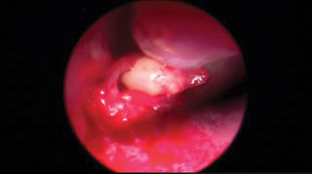
An incision was made over the mucosal thickening that covered the mass, thereby revealing the dental surface. The ectopic tooth was subsequently extracted from the palate using a little force (Figure 2). We then carried out a left-sided infundibulotomy, correction of the septum deviation and outfracture of the inferior nasal concha on both sides. Post-operatively, the patient made an uneventful recovery. There was no recurrence of symptoms during the one-year follow-up period.
Endoscopic photograph of the dental surface after incision of the nasal mucosa.

Discussion
An ectopic tooth in the nasal cavity is a rare phenomenon and the cause is unclear. Gupta and Shah (2001) reviewed the literature and found 49 well documented cases.Reference Gupta and Shah2 The age at which the intranasal teeth were detected ranged from 3 to 62 years. There appeared to be no site predilection (right or left), and more cases were found in males (60 per cent) than females.Reference Yeung and Lee1, Reference Gupta and Shah2
In most of the reported cases (as in ours), no aetiological explanation has been suggested or found. However, in a small number of cases some theories have been proposed, including tooth bud displacement due to trauma, osteomyelitis, exceptionally dense bone, or developmental disturbances such as cleft lip or palate.Reference Yeung and Lee1–Reference Gilbride and Smith3
Patients with intranasal teeth present with a variety of symptoms, including a feeling of a foreign body in the nose, nasal discharge, epistaxis, mild facial pain and external deviation of the nose.Reference Gupta and Shah2, Reference Smith, Gordon and De Luchi4 However, intranasal teeth may also be asymptomatic and revealed only by radiological investigation.Reference Kim, Kim, Chae, Hwang, Lee and Lee5
Other differential diagnoses of an intranasal dense radiopaque shadow include a foreign body, bony sequestrum, neoplasm, exostosis and rhinolith.
• Patients with an intranasal tooth may present with a diverse array of symptoms and complications
• Computed tomography or plain X-ray is helpful for diagnosis and operation planning
• Endoscopic removal of the intranasal tooth usually relieves the symptoms
• In most cases, the cause of the intranasal tooth remains unclear
The treatment of intranasal teeth entails surgical extraction, even though such teeth may remain asymptomatic; several cases have illustrated the potential significant morbidity associated with them.Reference Pracy, Williams and Montgomery6–Reference El-Sayed8 It is recommended that the tooth is extracted under endoscopic guidance. This recommendation is based on a review of 13 cases in which all patients had an uneventful recovery and the presenting symptoms disappeared after surgery.Reference Lee9 A recent publication has also reported an alternative transoral approach for impacted teeth next to the nasal floor.Reference Sammartino, Trosino, Perillo, Cioffi, Marenzi and Mortellaro10