Introduction
A Pott's puffy tumour is a subperiosteal abscess and osteomyelitis of the frontal bone which occurs as a suppurative complication of frontal sinusitis.Reference Babu, Todor and Kasoff1 This condition was first described by Sir Percival Pott in 1760. Owing to the introduction of antibiotics, this and other suppurative complications of sinusitis are now considered rare.Reference Pott2, Reference Bradley, Manning and Shaw3
In Pott's puffy tumour, the infection is spread anteriorly from the frontal sinus through the overlying bone, either by direct extension or via septic emboli, and manifests clinically as a soft tissue swelling over the forehead.Reference Dawes4, Reference Thomas and Nel5
Studies estimate that approximately 40 per cent of cases of Pott's puffy tumour present with an intracranial or orbital component.Reference Marshall and Jones6 Intracranial and orbital complications of sinusitis include epidural empyema, cerebral abscess, meningitis, dural and cavernous sinus thromboses, and orbital cellulitis and abscess, the development of all of which is strongly related to the venous drainage of the frontal sinus.Reference Kountakis, Senior and Draf7 The diploic veins of the frontal sinus travel through the anterior and posterior table foramina, communicating with the larger diploic veins and then with the dural venous sinus of the frontal bone. Infection can spread posteriorly from the frontal sinus, either through these valveless veins via a retrograde thrombophlebitis, or by direct extension across the bony limits of the orbit.Reference Osborn and Steinberg8 The mortality rate associated with intracranial complications of sinusitis ranges from 0 to 7 per cent, and is associated with delayed presentation and diagnosis.Reference Younis, Lazar and Anand9–Reference Gallagher, Gross and Phillips11
Pott's puffy tumour with intracranial complications is exceedingly rare in pregnancy. A search of the Medline database was performed using the search terms ‘Pott's puffy tumour and pregnancy’, ‘brain abscess and pregnancy’ and ‘complicated sinusitis in pregnancy’. Two cases of sinogenic brain abscess in pregnancy were identified.Reference Wax, Blackstone, Mancall, Cartin and Pinette12, Reference Kim, Kang, Huh, Yoo do, Cho and Kim13 A small number of spontaneous spinal epidural abscesses in pregnancy have also been reported.Reference Connealy, Lovgren, Tomich, Smith and Berg14–Reference Van Winter, Nielsen and Ogburn17
Our case represents the first report of Pott's puffy tumour in pregnancy, associated with both orbital and intracranial extension. This case also highlights some of the management dilemmas associated with intracranial complications of sinusitis in pregnancy.
Case report
A 21-year-old Indigenous Australian woman, primigravid at 35 weeks gestation presented with a 6-day history of right periorbital pain, swelling, erythema and associated persistent frontal headache. There was no nausea, vomiting or history of trauma. She had recently been discharged from hospital with threatened pre-term labour, but prior to this she had been well. She was a non-smoker and denied alcohol consumption or drug use.
On examination, the patient was febrile (38.1°C) with a right periorbital soft tissue swelling, chemosis and difficulty opening her right eye. She had normal pupillary reflexes and extra-ocular eye movements but reduced visual acuity in the ipsilateral eye (6/9).
Blood tests revealed a normal white blood cell count (11.6 109/L) but a raised C-reactive protein level (210 mg/L).
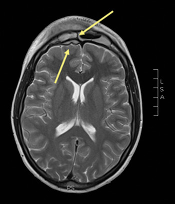
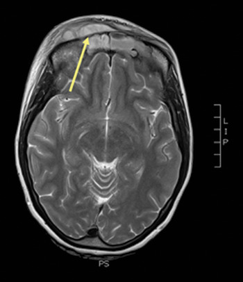
Magnetic resonance imaging (MRI) of the head was performed, rather than computed tomography, due to concerns regarding irradiation in pregnancy. This revealed a right frontal sinusitis with subperiosteal extension, lytic breakdown of the anterior table and a subgaleal collection (30 × 7 × 27 mm). A right-sided epidural empyema (20 × 5 × 18 mm) was noted posterior to the frontal sinus. A right-sided pre-septal orbital cellulitis could be seen, with no evidence of post-septal involvement (Figures 1 and 2).
Axial magnetic resonance imaging head scan showing the presence of right-sided infective frontal sinusitis and a right frontal pole empyema (arrows).

Axial magnetic resonance imaging head scan showing the frontal bone osteomyelitis and extracranial extension of the infection (arrow).

A multidisciplinary review was conducted involving infectious diseases, obstetric, neurosurgery and ophthalmology colleagues, and the patient was commenced on empirical broad spectrum intravenous (IV) antibiotics: meropenem 2 g 8 hourly and vancomycin 500 mg 8 hourly. Careful observation of the epidural collection and pre-septal cellulitis was recommended.
Intranasal saline douches and intranasal oxymetazoline and mometasone were commenced. Percutaneous aspiration of the frontal sinus was performed, draining 3 ml of frank pus. Daily fetal assessments, including fetal heart rate, revealed a normally progressing pregnancy.
Despite 72 hours of IV antibiotic therapy, the patient deteriorated clinically, becoming unable to open her right eye.
Thus, limited endoscopic sinus surgery was performed under general anaesthesia. This involved unilateral uncinectomy, middle meatal antrostomy, anterior ethmoidectomy and frontal recess dissection, which facilitated adequate drainage of the infected sinuses. Aspirates cultured Streptococcus milleri. A peripherally inserted central catheter was placed and the antibiotic regime was changed to IV ceftriaxone 2 g once daily and oral metronidazole 400 mg 8 hourly.
Post-operatively, the patient improved clinically, with a reduction in swelling and pain. On post-operative day three, the patient went into labour and an uneventful caesarean section was performed (at 36 weeks' gestation) at a high risk maternity hospital.
Five days post-delivery, after otolaryngology review, the patient was discharged back to her rural community with continued IV antibiotics through the peripherally inserted central catheter.
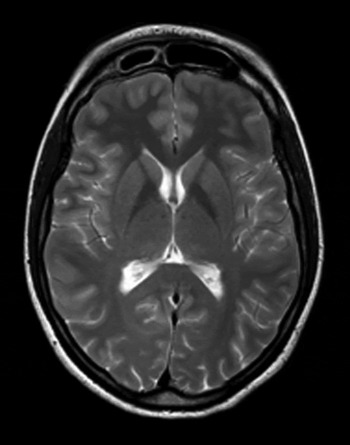
A follow-up MRI scan, performed three weeks post-discharge, showed a dramatic resolution of symptoms, with some minor induration of the subcutaneous tissue of the forehead and frontal sinus, but no collection or inflammatory changes present in the frontal sinus or epidural space (Figure 3). Intravenous antibiotics were ceased and oral amoxycillin and clavulanate (Augmentin Duo Forte; GSK, Uxbridge, UK) 875 mg/125 mg 12 hourly were commenced for four weeks.
Axial magnetic resonance imaging head scan taken three weeks after discharge, showing a dramatic improvement, with resolution of sinusitis and intracranial and extracranial components.

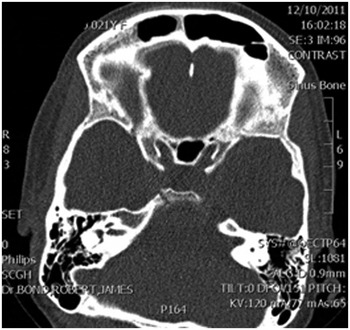
Six weeks after delivery, a computed tomography scan (Figure 4) confirmed complete resolution of sinusitis and complications.
Axial computed tomography head scan taken six weeks after delivery, confirming complete resolution of sinusitis and complications.

Discussion
Frontal sinusitis is common, and for the most part a fairly benign presentation. Pott's puffy tumour is an extracranial extension and subperiosteal abscess secondary to frontal osteomyelitis; a complication of frontal sinusitis. Clayman et al. found the condition in up to 0.5 per cent of cases of acute and chronic paranasal sinusitis.Reference Clayman, Adams, Paugh and Koopmann18 However, this study only included patients with sinusitis requiring hospital admission; its findings may therefore be an over-representation due to a selection bias for patients with severe sinus disease. Studies estimate that approximately 40 per cent of cases of Pott's puffy tumour present with an intracranial component, and an associated increased risk of mortality and morbidity.Reference Marshall and Jones6 The most common intracranial complication of sinusitis is subdural empyema.Reference Jones, Walker, Bassi, Jones and Punt10, Reference Singh, Van Dellen, Ramjettan and Maharaj19
The development of intracranial complications is significantly more frequent in males than females, and is most commonly seen in adolescence.Reference Clayman, Adams, Paugh and Koopmann18, Reference Small and Dale20, Reference Maniglia, Goodwin, Arnold and Ganz21 The hypothesis behind this difference relates again to vascular anatomy, with younger males having larger diploic veins allowing easier spread of infection.Reference Clayman, Adams, Paugh and Koopmann18
A relationship between pregnancy, mucosal oedema and rhinitis has been identified, but as yet no association has been found between pregnancy and sinusitis.Reference Ellegard, Hellgren, Toren and Karlsson22, Reference Ellegard23
In approximately half of all cases of Pott's puffy tumour and epidural abscess, a pathogen cannot be identified.Reference Gallagher, Gross and Phillips11 Where organisms are identified, multiple organisms are most commonly cultured, with anaerobes predominating.Reference Gallagher, Gross and Phillips11, Reference Giannoni, Stewart and Alford24 The most common organism is Streptococcus milleri, being detected in approximately 50 per cent of cases in which an organism is cultured.Reference Marshall and Jones6, Reference Gallagher, Gross and Phillips11, Reference Fenton, Smyth, Viani and Walsh25
The management of Pott's puffy tumour and associated intracranial complications involves IV antibiotics, nasal decongestants, sinus wash-outs, percutaneous aspiration and/or trephination, endoscopic sinus surgery, and craniotomy.Reference Fenton, Smyth, Viani and Walsh25
Cases of sinogenic intracranial complications occurring in pregnancy pose a number of management dilemmas. Firstly, information is very limited as there are so few reported cases. Two cases of sinogenic brain abscess in pregnancy have been reported. The first involved sphenoid sinusitis and a pituitary abscess in a 34-weeks' gestation woman, managed with a transsphenoidal evacuation and irrigation procedure alongside antibiotic therapy (cultures were positive for Streptococcus viridans). The following day, the patient went into labour and underwent an emergency caesarean section.Reference Kim, Kang, Huh, Yoo do, Cho and Kim13 The other case involved a maxillary and ethmoid sinusitis and temporal lobe abscess in a 36-weeks' gestation woman, managed by caesarean section under spinal anaesthesia and a 4-week course of antibiotics (with no neurosurgical procedure and no bacteria cultured).Reference Wax, Blackstone, Mancall, Cartin and Pinette12 Epidural collections in pregnancy occur most commonly as a complication of regional anaesthesia; however, there have been four reported cases of spontaneous spinal epidural abscess in pregnancy, presumably relating to transient bacteraemia and seeding, all of which required emergency laminectomy and decompression.Reference Connealy, Lovgren, Tomich, Smith and Berg14–Reference Van Winter, Nielsen and Ogburn17
A further management dilemma in pregnant patients with an intracranial collection involves the choice of treatment: whether to manage conservatively or surgically, which antibiotic regime to choose, and other issues of timing and induction.
Studies indicate that for large intracranial collections in non-pregnant patients (i.e. epidural and subdural abscesses larger than 1 cm in diameter), neurosurgical intervention with craniotomy is indicated.Reference DelGaudio, Evans, Sobol and Parikh26 In patients with smaller collections (i.e. smaller than 1 cm), the decision on whether to pursue a conservative or surgical approach becomes more difficult. In DelGaudio and colleagues' retrospective study, a treatment protocol of emergency endoscopic sinus surgery in conjunction with IV antibiotics for epidural collections smaller than 1 cm yielded a very low success rate, with 5 out of the 6 patients thus treated requiring further craniotomy for progressive symptomatology.Reference DelGaudio, Evans, Sobol and Parikh26
The use of craniotomy for sinogenic abscess in pregnancy has not been reported; however, there have been reported cases of brain abscesses in pregnancy in which neurosurgical interventions were safely performed. One such case involved a 23-year-old woman at 35 weeks' gestation who had an otogenic cerebellar abscess; she was successfully managed by caesarean section followed immediately by craniotomy and modified radical mastoidectomy.Reference Jacob, Kurien, Varghese, Aleyamma, Jasper and Prabu27 Craniotomy has also been efficacious in managing a pregnant woman with an occipital brain abscess secondary to a nocardial infection.Reference Braun, Kerson and Eisenberg28 Whilst there are limited reports of the management of brain abscesses in pregnancy, craniotomy has been shown to be safe and efficacious.
• Pott's puffy tumour is a suppurative complication of sinusitis
• The first report of Pott's puffy tumour in pregnancy is presented
• This 21-year-old woman at 35 weeks' gestation had complicated frontal sinusitis
• Multidisciplinary management was successful, with no short-term complications
The timing of delivery is also important in this setting, considering the risk posed by increased intracranial pressure. In a number of cases, clinicians have opted for an early caesarean section due to concerns regarding increased intracranial pressure during labour and spontaneous vaginal delivery.Reference Wax, Blackstone, Mancall, Cartin and Pinette12, Reference Jacob, Kurien, Varghese, Aleyamma, Jasper and Prabu27
Conclusion
This is the first reported case of a Pott's puffy tumour occurring in pregnancy. Management in a multidisciplinary setting can be successful, with no short-term complications. This case highlights the difficulties of managing complicated acute frontal sinusitis in pregnancy.