Introduction
The human capacity to represent space and to orient in an environment is manifested in the ability to create mental images and to talk about the environment from different perspectives. This ability is related to a navigation in various frames of reference. In the area of spatial cognition, the reference frame is considered as an orthogonal system with its origin (deixis center) in the retina, head, body or other points, objects, or array in space (Colby & Goldberg, Reference Colby and Goldberg1999; Behrmann, Reference Behrmann and Gazzaniga2000; McCloskey, Reference McCloskey and Rapp2001). In a series of psychological experiments, researchers concluded that reference frames are simultaneously activated (Carlson-Radvansky & Irwin, Reference Carlson-Radvansky and Irwin1993). The unanswered question is whether the simultaneous activation is expressed as a different activity in a single cortical area or whether different reference frames activate several brain centers.
The basic classification of reference frames in the area of neuroscience involves spatial coordinate systems based on egocentric and allocentric orientation (Howard & Templeton, Reference Howard and Templeton1966). In the egocentric frame of reference, the position of objects is encoded with a reference to the body of the observer or to the relevant body parts. Egocentric representations of objects may be used for goal-directed movements, such as reaching a target or avoiding a dangerous stimulus (Galati et al., Reference Galati, Lobel, Vallar, Berthoz, Pizzamiglio and Le Bihan2000). Spatial positions can also be coded in object-centered coordinates that are independent from the observer’s current position. This allocentric frame of reference is constituted of object-to-object relations. Knowing the allocentric position in the space, and having egocentric coordinates to other objects, navigators can form a map that allows object-to-object relations to be represented allocentrically (Klatzky, Reference Klatzky, Freksa, Habel and Wender1998).
Neurophysiological research in humans confirmed differences between the utilization of egocentric and allocentric frames of reference. Committeri et al. (Reference Committeri, Galati, Paradis, Pizzamiglio, Berthoz and LeBihan2004) attribute egocentric coding mainly to the dorsal stream (BA 7) and connected frontal areas, whereas allocentric coding is mediated by both dorsal and ventral regions. Fink et al. (Reference Fink, Marshall, Weiss, Stephan, Grefkes, Shah, Zilles and Dietrich2003) attribute allocentric processing to the right posterior parietal (BA 7) and ventral premotor cortex (BA 6). Wilson et al. (Reference Wilson, Woldorff and Mangun2005) reported greater activity in the left parietal cortex compared to the counterpart in the right hemisphere (hemispheric asymmetry) for the allocentric frame of reference.
There are also theories that attribute parallel processing of the egocentric and allocentric information to the parietal lobe (BA 7) and the hippocampal formation (BA 27, 28), respectively. Information from the parietal lobe is transferred to the hippocampus for a long-term storage in allocentric coordinates (Kesner, Reference Kesner2000; Save & Poucet, Reference Save and Poucet2000; Feigenbaum & Morris, Reference Feigenbaum and Morris2004). Other studies have associated the hippocampus with allocentric strategies and the striatum with egocentric strategies (Maguire et al., Reference Maguire, Burgess, Donnett, Frackowiak, Frith and O’Keefe1998; Hartley et al., Reference Hartley, Maguire, Spiers and Burgess2003; Iaria et al., Reference Iaria, Petrides, Dagher, Pike and Bohbot2003). The most recent study (Galati et al., Reference Galati, Pelle, Berthoz and Committeri2010) provides a summary of functional magnetic resonance imaging (fMRI) experiments with the static stimuli. According to this study, the posterior parietal and associated frontal regions are involved in processing the egocentric reference frame. The allocentric navigation is attributed to the specific parietal subregions and also to the hippocampal and retrosplenial region.
On the other hand, the way of stimuli presentation in these studies should be considered as insufficient (Gramann et al., Reference Gramann, Müller, Schönebeck and Debus2006). The experiments mentioned above are based on a static presentation of stimuli (there are various forms of the line bisection paradigm), which decreases their ecological validity. Schönebeck et al. (Reference Schönebeck, Thanhaüser and Debus2001) conducted a dynamic task built in a three-dimensional (3D) virtual reality environment consisting of a traverse through a virtual tunnel with straight and turned segments. In a subsequent study, Gramann et al. (Reference Gramann, Müller, Schönebeck and Debus2006) replicated this scenario and recorded an EEG signal to identify the neural correlates responsible for specific frames of reference processing. They employed the source reconstruction method (a combination of Loreta algorithms; Pascual-Marqui & Biscay-Lirio, Reference Pascual-Marqui and Biscay-Lirio1993, and temporal coupling; Darvas et al., Reference Darvas, Schmitt, Louis, Fuchs, Knoll and Buchner2001) that allows 3D reconstruction of signal sources. They localized higher mean source activation in the BA 7 (parietal cortex) for subjects who adopt an egocentric frame of reference similar to the previous studies and also BA 32 (anterior cingulate gyrus) for subjects who adopt an allocentric frame of reference. Lin et al. (Reference Lin, Chiou and Huang2008) concludes that allocentric strategy resulted in stronger activation in the occipital area (BA 17, 18, 19), and egocentric strategy was manifested by stronger activation in the parietal area (BA 7). In a collaborative study (Lin et al., Reference Lin, Chiou, Ko, Duann and Gramann2009), researchers adopted the tunnel task paradigm, the event-related spectral perturbations method for a signal analysis, and they did not find any performance-related brain activities for allocentric subjects. In the most recent research based on the tunnel task (Gramann et al., Reference Gramann, Onton, Riccobon, Müller, Bardins and Makeig2010), stronger alpha blocking was identified in or near to the right primary visual cortex (BA 17) in the turned segment of the tunnel for subjects who adopt egocentric reference frame. They also detected stronger alpha blocking of the occipitotemporal, bilateral inferior parietal (BA 7), and retrosplenial cortical areas (BA 26,29,30) in the first straight and turned segments for allocentric strategy. These results are consistent with the hypothesis of a continuous translation of egocentrically experienced visual flow into an allocentric model.
Since Gramann et al. (Reference Gramann, Müller, Schönebeck and Debus2006, Reference Gramann, Onton, Riccobon, Müller, Bardins and Makeig2010; Lin et al., Reference Lin, Chiou, Ko, Duann and Gramann2009) presented the tunnel task only in horizontal plane, we also administer upward and downward turned tunnels to reveal new findings related to the adoption of mentioned frames of reference in a vertical plane. A psychological study of frames of reference in a 3D environment was administered by Vidal et al. (Reference Vidal, Amorim and Berthoz2004). They concluded that the spatial updating process was more accurately performed for a terrestrial strategy (the head turns only in horizontal plane) and to some extent also for a subaquatic strategy (the head turns in both horizontal and vertical plane) than for a weightless (yaw and pitch turns) navigational style (Vidal et al., Reference Vidal, Amorim and Berthoz2004).
Although they have identified only slight differences in the behavioral response between terrestrial navigation (resembling the allocentric strategy in the tunnel task administered in vertical plane) and subaquatic navigation (resembling the egocentric strategy in vertical tunnels), we hypothesize that there are different brain areas involved in the processing of the egocentric and allocentric reference frame in vertical plane. Two research questions deal with this topic:
Are there differences in the EEG signal between participants who adopt egocentric and allocentric reference frames in a vertical plane?
Are there differences between the navigation in a horizontal and vertical plane?
Materials and methods
The experimental sample consisted of 38 participants (7 females and 31 males). The mean age was 28.8 years. All subjects had normal or corrected-to-normal vision. They were under no medication that affects the EEG signal and were neurologically intact. We recorded the EEG data in two laboratories (BioDat at Czech Technical University and the EEG and Sleep Lab at the Prague Psychiatric Center) with the same hardware and settings. The EEG signal was recorded from 19 unipolar sintered Ag/AgCl EEG electrodes, positioned under the 10-20 system, namely Fp1 and Fp2, F3, F4, C3, C4, P3, P4, O1, O2, F7, F8, T3, T4, T6, T6, FZ, CZ, PZ. The raw data were resampled from 250 (sampling frequency) to 128 Hz and the notch filter was applied to remove 50 Hz power supply interference.
Experimental procedure
The participants were informed prior to the experimental procedure that the task is focused on the spatial navigation but no further specification was given. After the introduction, each participant sat comfortably in front of a computer screen in a sound-attenuated room and the EEG cap was applied. The subjects were required to keep the track of their implied virtual 3D position with respect to their starting position within the tunnel traverse. The experimental instruction was translated from the study of Schönebeck et al. (Reference Schönebeck, Thanhaüser and Debus2001).
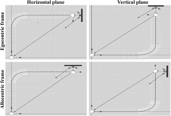
The tunnel task is based on a traverse through a virtual tunnel and subsequent identification of the origin of the tunnel (see Fig. 1). There are two 3D arrows displayed on the black screen at the end of each tunnel that represent the correct response within the egocentric and allocentric reference frame. The participants had to decide which of the displayed arrows pointed back to the origin of the traversed tunnel path. The subject’s choice corresponds to the reference frame he/she adopts as the navigation system. This method has several advantages. Dynamic stimuli are presented to the participants in a stable position that makes this method suitable for the EEG or fMRI studies. The same stimuli are presented to the whole experimental sample, which avoids the influence of intervening variables. Moreover, we administered this method without any pretest, which allows the experimental sample to be divided into the allocentric or egocentric group, independent of the experimenter’s intention or intervention. There are also some disadvantages in such an experimental design. The participants do not follow predefined strategies, so there should emerge new types of the navigation styles (e.g., they adopt different reference frames for specific planes or they choose strategy randomly).
Visualization of the tunnel task in a 3D environment. The head position is shown at the beginning and the end of the tunnel traverse for specific frames of reference (egocentric vs. allocentric) and specific planes (horizontal vs. vertical). The dark bar represents a computer screen with two arrows standing for the selection period.

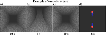
The animations of passages through a 3D virtual tunnel consisted of three segments: the first straight segment, the turned segment, and the second straight segment. The length of the two straight segments and the turned segment was constant. A fixation cross was present for 6 s prior to each trial. Each tunnel consisted of a 10-s traverse through the first straight segment, 6-s through the turned segment, and 10-s through the second straight segment (see Fig. 2). The bend of the turned passage varied between 30 and 90 degrees at intervals of 15 degrees. A total of 20 tunnels were presented to each subject, that is, 5 tunnels with variable curvature for each of four directions (up, down, left, and right). The tunnels were administered pseudorandomly to the participants, and there were no two tunnels in the same direction presented to the participant consecutively.
Example of a tunnel traverse in vertical plane. The figure shows the first straight (a), turned (b), second straight segment (c), and arrow selection period (d). The 6-s fixation period prior to each tunnel traverse is not visualized. Only the optical flow is presented to the participant.

The angular difference between the arrows (homing vectors) corresponds to the angle of the turned segment and represents the deviation of the egocentric frame in comparison with the allocentric frame (see Fig. 1). Each arrow stands for the correct answer for one type of the reference frame. The answers were evaluated to identify the subject’s preferred (native) reference frame. Participants who had selected the same frame of reference in above 80% of cases (16 of 20 answers) were considered as representative users of the particular reference frame. Each participant also undertook an inquiry after the experiment. This procedure helps to clarify subject’s strategy and it eliminates the errors caused by reinterpretation of the task.
EEG preprocessing
We performed a visual inspection of each EEG signal prior to the data analysis in order to detect obvious technical and biological artifacts and subsequently rejected these parts from further processing. Participants were instructed not to move during the tunnel traverse, so the amount of rejected period was less than 2% per participant. We also substituted the segments contaminated by eye blinking to improve the signal quality. Eye-blinking periods were manually marked and each eye-blinking segment was substituted with the signal of the same length recorded prior the segment. We did not measure either electrooculogram (EOG) or eye tracking to correct EEG signal in frontal electrodes.
Feature extraction
The signal was divided into the segments of constant length (1 s), and then, the following parameters were calculated: statistical parameters (minimum value, maximum value, mean value, standard deviation (s.d.), skewness, kurtosis), mean and maximum values of the first and second derivation of the samples in the segment. Further application of the Fourier transform resulted in the absolute/relative power for five EEG frequency bands, namely for delta (less than 3 Hz), theta (3–7 Hz), alpha (7–12 Hz), beta (12–30 Hz), and gamma (30–40 Hz) activities (the Fast Fourier Transformation [FFT] size was set to 128 sample points). The EEG coherences were computed from a 30-s window with a 5-s shifting interval.Footnote 1 We have also computed the correlation between the EEG electrodes (the signal was divided into segments 3 s in length) and the mean and maximum correlation values for each segment.
The wavelet transform was also applied to the signal segments. Daubechies 4 was used as the mother wavelet, and the signals were decomposed into four levels. These wavelet-based features included the statistical parameters of the wavelet coefficients corresponding to the different decomposition scales, zero crossings of wavelet coefficients, and Shannon’s entropy of the wavelet transform. We also calculated the mean and maximum values of the wavelet coefficients obtained after the application of the wavelet transform to the first and second derivative of the EEG signals. The data were processed in PSGLab Toolbox, which was developed in our laboratory (Gerla et al., Reference Gerla, Djordjevic, Lhotská and Krajča2010).
Feature selection
The input matrix for each subject consisted of 1916 features (93 features per electrode + correlations and coherences). We employed PRTools (van der Heijden et al., Reference van der Heijden, Duin, de Ridder and Tax2004) for the feature selection. First, we applied basic transforms to the data. We removed outliers and we also normalized the data. The further process of feature selection was divided into a number of steps. In the first stage, two algorithms were applied for the preselection. The features were individually evaluated by inter/intra distance and 1-nearest neighbor criterion to select 50 best features for each method. The inter/intra distance criterion (van der Heijden et al., Reference van der Heijden, Duin, de Ridder and Tax2004) is a distance-based class separability criterion that is a monotonically increasing function of the distance between the expectation vectors of different classes and a monotonically decreasing function of the scattering around the expectations. The 1-nearest neighbor method approximates the local density of the data patterns. These feature sets were used as the input for the subsequent processing.
In the next step, we applied forward, backward, and branch and bound feature selection algorithms to the preselected features. These algorithms were applied to both sets (inter–intra and 1-nearest neighbor), and the five best features that discriminate between egocentric and allocentric strategies were selected. We also applied optimization algorithms based on calculation of all possible combinations of 50 preselected features, and the best n-ary feature sets were evaluated (they varied from 1 to 15 features). The selected feature sets were tested by several classifiers to measure their discriminative quality. We employed a linear classifier, a quadratic classifier, and a naive Bayes classifier to calculate error rates.
Results
Administration of the tunnel task in vertical plane resulted in new navigation strategies compared to the task in horizontal plane (Schönebeck et al., Reference Schönebeck, Thanhaüser and Debus2001; Gramann et al., Reference Gramann, Müller, Schönebeck and Debus2006, Reference Gramann, Onton, Riccobon, Müller, Bardins and Makeig2010; Lin et al., Reference Lin, Chiou, Ko, Duann and Gramann2009). There were four participants (11%) who preferred the egocentric reference frame in horizontal plane and the allocentric frame in vertical plane navigation. They did not reflect the rotations in vertical plane, and they probably switched to the allocentric frame due to imagined gravity (see Discussion section for an explanation). We should not compare this strategy with the results of previous studies, so we excluded these participants from the EEG analysis. There were also 11 subjects (30%) who did not follow the instruction and they performed a mental U-turn at the end of the tunnel. They chose the arrow as if they were standing and looking back to the tunnel, although they had not been given such an instruction (see Materials and methods section). Although these subjects reported egocentric in the postexperiment inquiry, they were also excluded from EEG analysis because this change could have affected the results. A last group of five participants (13%) chose an egocentric or allocentric frame of reference randomly. It was not possible to analyze either behavioral or EEG data of this group because the participants were not able to describe the consistent navigation strategy in postexperiment inquiry. We consider the exclusion of the half experimental sample from the EEG analysis as a weak point of the adopted methodology; on the other hand, the subjects selected for the further EEG analysis adopted the egocentric or allocentric strategy without any instruction or pretest. This methodology was not presented in any previous study.
We did not identify gender differences in the reference frame adoption similar to Shelton and Gabrieli (Reference Shelton and Gabrieli2002, Reference Shelton and Gabrieli2004). Three women adopted the allocentric reference frame and two women adopted the egocentric frame in the selected sample. There was no correlation between the age of the participant and the preferred navigation strategy.
At the first stage, we calculated the error rates (percentage of trials with inconsistent reference frame) for mentioned navigation styles (except fully inconsistent “random selection” group). Participants who preferred egocentric strategy achieved mean error (M) 10% (s.d. 9.7) similar to the participants who adopt allocentric strategy (M 7.5%, s.d. 5.7) and mixed (egocentric frame in horizontal plane and allocentric frame in vertical plane) strategy (M 8.7%, s.d. 11.1). Surprisingly, the lowest mean error achieved participants who did mental turn at the end of each traverse (M 2.7%, s.d. 4.9). These results are counterintuitive because the mental imagery of self-motion increases the cognitive load during the task.
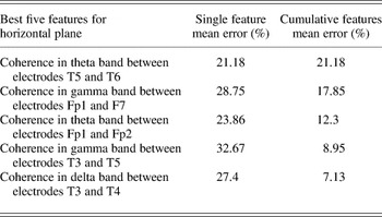
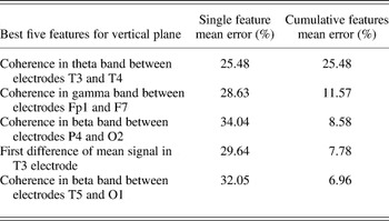
At the next stage, we processed the EEG data from 17 participants (9 participants preferred egocentric frame and 8 participants natively adopted allocentric frame in both planes). The data set for both planes was split to the two subsets (tunnels in horizontal and vertical plane). We analyzed the data from the whole tunnel traverse in horizontal and vertical plane (Tables 1 and 2). The coherences in theta and gamma bands were the most discriminative features in horizontal plane, namely theta and gamma intrahemispheric coherence in the right temporal lobe and gamma coherence in the right frontal lobe. There were also interhemispheric coherences between the orbitofrontal electrodes in the theta band and the temporal electrodes in the delta band. The interhemispheric coherence between the temporal lobes in the theta band and intrahemispheric coherence in the frontal lobe were the most discriminative features in vertical plane. Unlike horizontal plane, there were intrahemispheric coherences between the right parietal and occipital area and the left temporooccipital area in the beta band.
Best features for the whole tunnel in horizontal plane

There are mean errors for the best features. The error rates are calculated as mean values of three classifiers (Linear, Quadratic, and Naive). There are values for single best feature and cumulative error as the values of feature combinations.
Best features for the whole tunnel in vertical plane

We also tested these features with three mentioned classifiers and sevenfold cross-validation (Table 3). For the sake of objectivity, the values in Tables 1 and 2 were calculated as mean errors of all the three classifiers. We achieved the best results in both planes (M 7.55%) for forward search algorithm with a fixed number of features based on an inter–intra class search.
Feature selection and classifiers

The results of feature selection process evaluated by three classifiers (Linear, Quadratic, and Naive). There were three selection methods (Forward, Backward, and Branch and Bound Selection). We tested these methods with different mapping (inter–intra or nearest neighbor) with fixed number of parameters (five best features) but also as an optimization task.
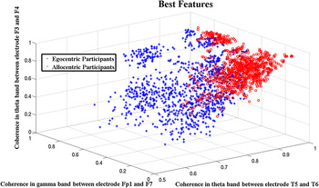
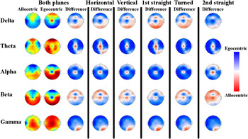
Finally, we visualized the best three features in a 3D graph (Fig. 3). The differences between the allocentric and egocentric groups were also visualized on scalp projections. Fig. 4 shows the mean activity in five spectral bands for allocentric and egocentric strategies (columns 1 and 2) and their difference maps (columns 3–8). The red and blue colors represent higher brain activity for the particular strategy, and white color stands for equivalent activity in both groups. There are no visible differences between horizontal and vertical planes (columns 4 and 5), but there is a change in the beta band activity for the second straight segment of the tunnel (column 8).
Feature space. The three best features for the whole tunnel in both planes are plotted in 3D space (each point stands for 1-s feature value). (+) represents egocentric participants and (o) represents allocentric participants.

Difference maps of tunnel task in horizontal plane, vertical plane, both planes, and specific parts of the tunnel (first straight segment, turned segment, second straight segment). The rows represent basic spectral bands. Average values for the egocentric and allocentric groups are given in columns 1 and 2. Columns 3–8 stand for difference maps. Higher activity in the specific area is represented by blue color for the egocentric group and by red color for the allocentric group. White color stands for similar activity for both groups. The values were calculated as the mean of all tunnel traverses.

Discussion
The analysis of the behavioral data in the vertical plane uncovers novel findings regarding the adoption of egocentric and allocentric reference frames in a 3D environment. The experimental sample consisted of participants who natively adopt egocentric reference frame in both planes (24%), participants who natively adopt the allocentric frame in both planes (21%), and participants who used an egocentric frame in horizontal navigation and an allocentric frame in vertical navigation (11%). We can interpret the behavior of the last group in terms of terrestrial navigation (Vidal et al., Reference Vidal, Amorim and Berthoz2004). Participants represented their body in the upright position at the end of the vertical tunnels, so there was no angular rotation of the head direction. Interestingly, none of the subjects adopted an allocentric frame in horizontal plane and an egocentric frame (resembling subaquatic navigation) in vertical plane. This finding leads us to the conclusion that people can (without any instruction) adopt two types of egocentric reference frame in a 3D environment. These findings require detailed analysis in further research.
The neurobehavioral results are partially consistent with the previous studies that attribute frames of reference processing to the parietal lobe, but we also identified other brain areas. We compared our results with fMRI and EEG studies based on a virtual reality simulation. A recent fMRI study based on navigation in a virtual environment (Wolbers et al., Reference Wolbers, Hegarty, Buüchel and Loomis2008) attributed egocentric navigation to the precuneus (BA 7) and the dorsal premotor cortex (BA 6). This result is consistent with the hypothesis that attributes allocentric and egocentric processing to the parietal cortex and nearby areas, but there are also fMRI studies based on a virtual reality simulation that localize allocentric activity to the inferior temporal (BA 20) and the posterior superior parietal cortex (BA 7) and egocentric activity to the anterior superior parietal (BA 7) and postcentral cortex (BA 1, 2, 3) (Shelton & Gabrieli Reference Shelton and Gabrieli2002, Reference Shelton and Gabrieli2004). These results are consistent with the hypothesis, which attributes the processing of reference frames to the brain areas other than parietal cortex.
In the area of EEG studies that employ a virtual simulation, Lin et al. (2008) have attributed egocentric processing to BA 7 and allocentric strategy to BA 17, 18, and 19. Gramann et al. (Reference Gramann, Müller, Schönebeck and Debus2006) localized higher mean source activity in BA 7 for the egocentric frame of reference, and the allocentric strategy induced higher activation in the anterior cingulate cortex (BA 32). A different type of analysis (Gramann et al., Reference Gramann, Onton, Riccobon, Müller, Bardins and Makeig2010) revealed stronger alpha blocking in BA 17 for subjects who adopt egocentric reference frame in the turned segment of the tunnel and stronger alpha blocking in BA 7 and BA 26, 29, 30 for allocentric strategy. When we compare our results with these studies, there is only a partial correspondence. The similar activity was detected only for vertical tunnels in our study. We detected differences in beta band coherence in the left occipital–parietal lobe. The beta frequency is interpreted as a conscious activity (Pfurtscheller & Lopes da Silva, Reference Pfurtscheller and Lopes da Silva1999). The coherences were higher for the allocentric strategy, but the detail analysis of beta activity in the separate electrodes (P4 and O2) revealed higher values in both electrodes for the egocentric group of participants (see Fig. 4 for basic visualization). We can interpret this as a coherent low beta activity in these electrodes for the allocentric frame of reference and noncoherent high beta activity for the egocentric strategy. There is a difference between P4 and O2 activity (it is higher in O2 electrode) in egocentric navigation compared to the similar activity between mentioned electrodes for egocentric strategy.
The data for the horizontal plane indicate interhemispheric theta coherences in both temporal lobes as the discriminative features. The problem is to interpret the interhemispheric coherences for the contralateral scalp locations because they can reflect indirect common/shared activity of the subcortical brain regions and also cortical activity between the two electrodes or hemispheric asymmetry. Although the results are consistent with the study of Wilson et al. (Reference Wilson, Woldorff and Mangun2005), we refrain from the interpretation of the interhemispheric coherences in this paper. It is necessary to replicate the scenario with more powerful EEG (at least 64 electrodes) to calculate the source reconstruction of the signal. This method should confirm or eliminate the hemispheric asymmetry hypothesis because there are no studies based on source reconstruction together with coherence calculation.
There was also the intrahemispheric coherence in the gamma band in the left temporal lobe as one of the most discriminative feature. It stands for higher gamma coherence in the egocentric group, but detail analysis revealed higher gamma activity (electrodes T3–T5) in the allocentric group (see Fig. 4). We can interpret this as a coherent low gamma activity for the egocentric frame of reference, while the allocentric strategy resulted in a noncoherent higher activity in these electrodes. Gamma activity is associated with cognitive functions and multimodal integration. These results are partially consistent with the fMRI study (even though these are different neuroimaging methods) of Shelton and Gabrieli (Reference Shelton and Gabrieli2004), which attributes allocentric processing to the temporal lobes and egocentric strategy to the parietal lobe; on the other hand, they differ from the EEG study (consider different methods of signal decomposition) that adopts the tunnel task (Gramann et al., Reference Gramann, Müller, Schönebeck and Debus2006, Reference Gramann, Onton, Riccobon, Müller, Bardins and Makeig2010). When we compare our results with the most recent tunnel task study (Gramann et al., Reference Gramann, Onton, Riccobon, Müller, Bardins and Makeig2010), there is hypothetically partial congruency. They detected alpha blocking and we localized higher gamma activity for the egocentric strategy. In contrast to all the studies mentioned here, we detected changes in the gamma band in the left frontal areas (electrodes Fp1–F7) in horizontal navigation and also in vertical plane navigation. There was higher coherence for the egocentric strategy but analysis of specific electrodes revealed coherent low gamma activity in the egocentric group and noncoherent higher activity for the allocentric strategy. The left frontopolar area is involved in memorizing tasks, and Fuster (Reference Fuster1989) detected large contingents of left prefrontal neurons that undergo sustained activation in delayed response experiments, for example, while a cue is being retained before the response is required. Thus, the difference in the left frontal gamma coherence that we have observed may be related to the different memory processing involved in the allocentric strategy. Summarizing the analysis of the whole tunnel in horizontal plane, we have identified differences between the navigation strategies in the brain areas other than the parietal lobe and nearby regions.
The differences among the results of the tunnel task experiments can be attributed to the signal analysis methods. Several types of analysis have been applied to the EEG signal, namely source reconstruction (Gramann et al., Reference Gramann, Müller, Schönebeck and Debus2006), event-related spectral perturbations (Lin et al., Reference Lin, Chiou, Ko, Duann and Gramann2009; Gramann et al., Reference Gramann, Onton, Riccobon, Müller, Bardins and Makeig2010; Plank et al., Reference Plank, Onton, Mueller, Makeig, Gramann and Holscher2010), and spectral analysis based on feature extraction and coherence calculation (presented study). Our results revealed coherences and signal derivations as the most discriminative features but they were not calculated in the previous studies. Hence, we excluded all the coherence, correlation, and derivation features from our data set, and we applied the same selection and classification algorithms as in previous analysis. This step resulted in 10% increase of error rate compared to the full-length data set but the results were more consistent with the mentioned studies. The best features were gamma activity in left temporal lobe similar to the fMRI results of Shelton and Gabrieli (Reference Shelton and Gabrieli2004), gamma and alpha activity in the medial frontal lobe similar to Gramann et al. (Reference Gramann, Müller, Schönebeck and Debus2006), beta activity in the left occipital lobe similar to Lin et al. (Reference Lin, Chiou, Ko, Duann and Gramann2009), but also gamma activity in the left prefrontal lobe. The differences among mentioned EEG studies should be clarified in a comparative study. The EEG data from all studies need to be analyzed by means of the methods described above, in order to identify the source of the variability in the results of the tunnel task studies. This meta-analysis should determine whether there is a variability in the data or in the analytical methods.
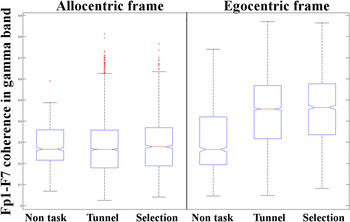
Finally, we compared the discriminative features in horizontal and vertical plane in order to answer the first study question. A basic visual comparison of the scalp projections for specific planes revealed some differences in the theta and beta bands (see Fig. 4; columns 4 and 5). When we compared numerical results in the separate planes (Tables 1 and 2), we have identified similar coherence between Fp1 and F7 as a discriminative feature for both planes. We plotted the mean feature values for the pretask condition (subject sits comfortably in front of a dark screen with his/her eyes open), the tunnel traverse, and arrow selection period (homing vector estimation) to exclude the hypothesis that the activity also differs in nonexperimental conditions. We observed similar values for both strategy groups in the pretask period, but the activity differed significantly in the tunnel traverse and the arrow selection period (Fig. 5). The coherence values for the allocentric group were stable in all conditions, but there was significant increase for the egocentric group in the tunnel task. We have tested differences between both navigation strategies using a nonparametric statistics (Kruskal–Wallis test) and the results confirmed our hypothesis. The feature values of the allocentric and egocentric participants in the pretask condition were not significantly different (H = 1, 1 df, P = 0.43) but the result for the tunnel traverse was statistically significant (H = 999, 1 df, P = 2.7 × 10−219). This feature represents a higher correlation in the gamma band between the electrodes on the left side of the orbitofrontal gyrus, but, as mentioned above, the results are not consistent with any of the studies in the area of spatial cognition. The scalp recordings of a frontal EEG activity in the gamma band are assumed to reflect memory encoding and retrieval (Gruber et al., Reference Gruber, Tsivilis, Montaldi and Muller2004). On the other hand, Yuval-Greenberg et al. (Reference Yuval-Greenberg, Tomer, Keren, Nelken and Deouell2008) attribute gamma activity to the onset of eye microsaccades and its time course is related to an increase in the rate of saccades. We should exclude this interpretation of gamma-related features in our research. Gramann et al. (Reference Gramann, El Sharkawy and Deubel2009) recorded the eye movements in horizontal tunnel task and there were no significant differences between allocentric and egocentric strategy related to saccades. This is indirect proof that the difference in the gamma activity between egocentric and allocentric strategy should not be attributed to the saccades. Moreover, a coherence between frontal electrodes as a discriminative feature does not correspond to the increase of gamma activity. The egocentric group in our research reveals coherent low gamma activity in both electrodes and there were high gamma activity in first electrode and low gamma in the second frontal electrode for the allocentric group. These results are not consistent with increase of gamma activity in frontal electrodes presented in Yuval-Greenberg et al. (2008). Unfortunately, we should not present any direct proof to the correlation between frontal gamma activity and the saccadic and microsaccadic activity of eyes as we did not record any data related to the eye movements.
Feature values within experimental conditions (tunnel traverses and arrow selection) and nonexperimental conditions (nontask) for allocentric and egocentric strategies. Boxplots summarize the statistical information about the selected epochs. The coherence between electrodes Fp1 and F7 in the tunnel task is considerably greater than in the pretask condition for the egocentric group but the coherence between the electrodes remains the same for the allocentric group.

There is also a shift in the theta coherence from T5–T6 electrodes in horizontal plane to T3–T4 electrodes in vertical plane. The absence of coherence in the beta band between P4 and O2 in horizontal plane is the most distinct observation compared to previous studies. The differences between the adopted strategies are also situated within the right side of the occipital–temporal area but in various spectral bands. There is higher intrahemispheric coherence for the egocentric strategy in the beta band (T5–O1) in vertical plane, and there is the same result for the gamma band (T3–T5) in horizontal plane (see Tables 1 and 2). The results in vertical plane cannot be compared with other EEG studies, as this is the first attempt to measure the frames of reference in a 3D environment.
Conclusion
The administration of the tunnel task in the vertical plane provided new insights into the area of spatial navigation. According to the results in Tables 1 and 2, 90% of the best features are interhemispheric and intrahemispheric coherences. This leads us to the conclusion that it is necessary to analyze not only specific electrodes and their characteristics but also the functional association or dissociation between the brain areas. The 10% difference in error rate between the coherences and electrode features confirms this hypothesis.
There is affirmative answer to the study question that there are differences in the EEG activity for the egocentric and allocentric navigation in vertical plane. Regarding the second study question, we have identified only one (of five) feature that discriminates reference frame processing both in horizontal plane and vertical plane.
In our future research, we would like to study the two types of egocentric reference frame adopted within the navigation in vertical plane in order to identify the neural correlates of these strategies. We would like to test a more complex scenario of the tunnel task and active navigation based on participant-free movement in a virtual environment to increase the ecological validity of our research. Our long-term goal was to localize the brain structures involved in the spatial information processing and to transfer this knowledge into the design of a neural model of this cognitive faculty.
Acknowledgments
This work has been supported by the Ministry of Education, Youth and Sports of the Czech Republic (under project No. MSM 6840770012 “Transdisciplinary Biomedical Engineering Research II”) and Grant Agency of Czech Republic (GACR) grant P407/11/P696.