INTRODUCTION
Accurate species diagnosis is fundamental to every biological investigation. Taxonomists face numerous challenges when assigning species-defining characteristics, especially when describing microscopic parasites with few appropriate and measurable morphological characters. This is particularly true for the parasite family Kudoidae Meglitsch, 1960.
Traditionally, species of the order Multivalvulida Shulman, 1959 were characterized by the number of spore valves and polar capsules, plus the presence of ornamentation or extensions of the valve margins (see Egusa, Reference Egusa1986). Under this scheme, kudoid species were described as having 4 spore valves and polar capsules. Recently, Whipps et al. (Reference Whipps, Grossel, Adlard, Yokoyama, Bryant, Munday and Kent2004) recognized that the classification based on polar capsule number rendered the genus Kudoa paraphyletic. So it appears that even major morphological characters (for example the number of spore valves) when taken alone, do not necessarily align with the degree of relatedness. Whipps et al. (Reference Whipps, Grossel, Adlard, Yokoyama, Bryant, Munday and Kent2004) proposed that members of the families Pentacapsulidae, Hexacapsulidae, and Septemcapsulidae become junior synonyms in the genus Kudoa within the Kudoidae, and that members of this genus are then characterized by having spores with 4 or more polar capsules.
Lom and Dyková (Reference Lom and Dyková2006) have also expressed the need for rigorous, detailed morphological descriptions, combined with molecular techniques, either prior to the proposal of new species or when revising existing descriptions within the phylum Myxozoa. As such, we favour a ‘whole evidence’ approach to species characterization. This article provides a mechanism for formally combining morphological, biological, and molecular characteristics for species descriptions of the myxozoan family, the Kudoidae.
MATERIALS AND METHODS
Sample collection
Fish from the families Apogonidae, Carangidae, and Pomacentridae were collected from Heron Island (23°27′S; 151°55′E), Lizard Island (14°39′S; 145°27′E), and Swains Reef (21°59.15′S, 150°20.71′E) on the Great Barrier Reef, Australia. Fish were caught using line, spear, or localized sprays of clove oil anaesthetic. The fish were usually euthanased by prolonged immersion in clove oil except for the larger fish, which were neurally concussed, then pithed.
The fish were examined for kudoid infection in the skeletal muscle by first excising a subsample (approximately 20×10×5 mm for fish less than 100 mm in length to caudal fork and 20×15×10 for larger fish) of muscle dorsal to the vertebral column in the posterior half of the fish. The muscle block was placed on a glass plate (76×50 mm), moistened with some vertebrate saline (0·85% salt), and macerated with scalpel blades. A second glass plate was placed on top and the liquid squeezed out into a 2 ml centrifuge tube. After allowing the spore suspension to settle, an aliquot (~15 μl) was pipetted from the bottom of the tube to a microscope slide and a cover-slip placed on top. The slide was then scanned for kudoid spores under an Olympus BH2 microscope at 400× magnification. When kudoid spores were detected, subsamples of skeletal muscle were preserved in 10% neutral buffered formalin for histological analysis and the whole fish (or for the large fish, a larger subsample of skeletal muscle) was preserved by freezing for morphological analysis of spores and for DNA extraction. Where material was limited, priority went to frozen samples from which both morphological information and DNA could be recovered. Additional spore suspensions were also air-dried and stained with Giemsa (Merck, Australia).
Prevalence in the field was recorded based on morphotype, where positive samples were labelled as having either stellate or quadrate spore shape, with or without spore projections. Approximate specific prevalence was determined by knowledge of whether the host species was found by subsequent morphometric and/or molecular analysis to be infected with only one, or more than one of the new kudoid species described in this report.
Morphometric analysis
Spore suspensions in saline were prepared as described in the sample collection methods and microphotographs were taken at 400× magnification with an Olympus BH2 microscope under phase-contrast lighting using a Nikon Digital Sight DS-L1 camera (Nikon Corporation, Japan). Spore measurements were taken from the microphotographs using the measuring tool in the Nikon Digital Sight DS-L1 that was calibrated using a stage micrometer. At least 30 spores from each parasite species were measured in accordance with a combination of schematics devised by Lom and Arthur (Reference Lom and Arthur1989), for spores with 4 polar capsules; by Whipps and Kent (Reference Whipps and Kent2006), for stellate spores; with additional measurements from Burger et al. (Reference Burger, Cribb and Adlard2007); and from the current work (see Figs 1 and 2).
Schematics for morphological characterization of kudoid spores in apical (left-hand column (A)) and side (right-hand column (B)) views. (1) For regular spores with an equal number of polar capsules. (2) For stellate spores with 1 polar capsule much larger than the others. (3) For spores with uneven valves and polar capsules, but with almost bilateral symmetry in the frontal and sagittal plane. (4) For spores with posterio-lateral projections. (5) For spores with an unequal number of polar capsules. Exclusive measurements are shown with upper case letters: A–B=Width (1A, 2A, 3A, 4A, 5A), C–D=Thickness (1A, 2A, 3A, 4B), E–F=Suture Radius (1A, 3A, 4A, 5A), B–F=Valve Width (measurement useful for species with an uneven number of polar capsules (5A), it is equal to half the width of radially symmetrical species with an even number of polar capsules), G–H=Apical Polar Capsule Length (1A, 4A, 5A), I–J=Apical Polar Capsule Width (1A, 4A, 5A), K–L=Length (1B, 2B, 3B, 4B, 5B), M–N=Side Polar Capsule Length (1B, 4B, 5B), O–P=Side Polar Capsule Width (1B, 4B, 5B), U–V=Projection Length (4B), W–X=Projection Breadth (4A), Y–Z=Anterior Extenion Height (5B). Variations dependent on certain morphotypes shown with lower case letters: a–b=Total Width (3A), g–h=Apical Large Polar Capsule Length (2A, 3A), i–j=Apical Large Polar Capsule Width (2A, 3A), m–n=Side Large Polar Capsule Length (2B, 3B), q–r=Apical Small Polar Capsule Width (2A, 3A), s–t=Side Small Polar Capsule Length (2B, 3B), w–x=Projection Width (4B).

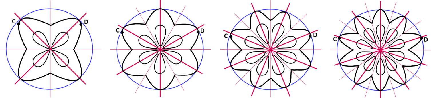
Schematics for measuring the thickness (C–D corresponding to Fig. 1) of radially symmetrical spores with an even number of polar capsules. Thickness is measured from the most distant points between 2 spore valves but does not go through the centre point of the spore. For 6-valved spores, the thickness measurement passes through 1 valve; for 8-valved spores, the thickness measurement passes through 2 valves; for 10-valved spores, the thickness measurement passes through 3 valves.

A full set of morphometric data was taken from multiple hosts for the characterization of each new species, except for K. kenti n. sp. where material was limited. Morphological characterization of K. kenti n. sp. was based on material collected from a single individual of Dischistodus perspicillatus (Pomacentridae).
Two samples of K. quadricornis (identity confirmed by morphology and DNA) from Carangoides fulvoguttatus (collected from Heron Island and Lizard Island) were used for morphological comparison with Kudoa paraquadricornis n. sp. using the above-mentioned schematics. A voucher specimen of air-dried spores stained with Giemsa of K. quadricornis ex Carangoides fulvoguttatus from Lizard Island was deposited in the collections of the Queensland Museum, Brisbane, Australia, registration number G465321.
The morphometric data were subjected to a principal component analysis using the software PAST version 1.82b (Hammer et al. Reference Hammer, Harper and Ryan2001) to determine the significance of morphological differences between host isolates of each new Kudoa species and for comparison of the new species with closely related ones for which raw data were available. Separate analyses were run on measurements from the apical and side view. Scatter plots with 95% confidence ellipses were generated using variant-covariant matrices. Each measurement was tested individually for significant difference between the means of host isolates and of the collective species measurements with closely related species using Stata™ 10.0 (StataCorp 2008); first by Bartlett's test for equal variance (one-way analysis of variance), followed by a 2-sample t-test (for either equal or unequal variance depending on the outcome of the Bartlett's test).
Histology
Sections (5 μm thick) were cut from the 10% formalin-fixed samples using standard histological techniques. Slides were stained with either haematoxylin and eosin (H & E) to observe the immunological response, or Giemsa and eosin to emphasize the polar capsules in contrast to the muscle fibres. Cover-slips were applied using DePeX (BDH, England). Digital, light microphotographs of the sections were taken at 400× magnification as described above.
DNA
DNA was extracted from spore suspensions that had been examined by microscopy for the morphological analysis mentioned in the morphometric methods. The suspensions were concentrated by centrifugation for 6 min at 4750 g and the supernatant discarded. The DNA from the pellet was extracted using a QIAgen DNeasy Kit (QIAGEN Inc., Valencia, California) according to the manufacturer's instructions. 18S rDNA was amplified using PCR primers KudMB1f 5′-GCC ATG GAT AAC TGT GGT AAA TCT AGA GC-3′ (Burger et al. Reference Burger, Barnes and Adlard2008) and universal 18R 5′-CTA CGG AAA CCT TGT TAC G-3′ (Whipps et al. Reference Whipps, Adlard, Bryant, Lester, Findlay and Kent2003b). 28S rDNA was amplified using the PCR primer combination of Kt28S1f 5′-CAA GAC TAC CTG CTG AAC-3′ and 28S1R 5′-GTG TTT CAA GAC GGG TCG-3′ (Whipps et al. Reference Whipps, Grossel, Adlard, Yokoyama, Bryant, Munday and Kent2004). PCR products were cloned and DNA sequence data were generated as described by Burger et al. (Reference Burger, Barnes and Adlard2008). DNA samples were extracted from spore suspensions of 2 isolates of K. quadricornis (the same suspensions used in the morphological analyses) to generate both 18S (GenBank Accession numbers: FJ792720 and FJ792721) and 28S rDNA sequence data (GenBank Accession numbers: FJ792753 and FJ792754). In addition, DNA from previously identified samples was used to generate 28S rDNA sequence data for a more comprehensive analysis: Kudoa amamiensis from Burger et al. (Reference Burger, Barnes and Adlard2008) (GenBank Accession numbers: FJ792726 to FJ792730), K. ciliatae from Burger et al. (Reference Burger, Cribb and Adlard2007) (GenBank Accession number: FJ792731), and K. monodactyli from Gunter et al. (Reference Gunter, Cribb, Whipps and Adlard2006) (GenBank Accession number: FJ792748). See Fig. 3 for GenBank specific isolate Accession numbers.
Neighbour-joining distance analysis of 18S rDNA (A) and 28S rDNA (B) from Kudoa spp. isolates. Bootstrap support based on 1000 replicates shown at nodes. Sequences from new species indicated in bold. Host name has been abbreviated, names in full are found in Tables 2, 4, and 5. Locality of new sequence isolate indicated prior to GenBank Accession number, HI, Heron Island; IH, Indian Head; LI, Lizard Island; SR, Swains Reef.

18S rDNA and 28S rDNA sequence data were aligned in BioEdit (Hall, Reference Hall1999) with other kudoid sequences available from GenBank using ClustalW (Thompson et al. Reference Thompson, Higgins and Gibson1994) and phylogenetic analyses were conducted using Mr Bayes 3.0 (Heulsenbeck and Ronquist, Reference Heulsenbeck and Ronquist2001) and PAUP* 4.0b10 (Swofford, Reference Swofford2002) as described in Burger et al. (Reference Burger, Cribb and Adlard2007) with the following modifications: in the Bayesian analysis, burnin was set to 5000 trees; for maximum likelihood analyses of both 18S rDNA and 28S rDNA sequence data, Modeltest 3.7 (Posada and Crandall, Reference Posada and Crandall1998) chose the best-fit model of (TrN+I+G) from the hierachical likelihood ratio tests and (GTR+I+G) from the Akaike Information Criterion. The bootstrap confidence values for maximum likelihood analyses were based on 100 fast-step heuristic search replicates (all other PAUP* 4.0b10 analyses used 1000 bootstrap replicates).
Pairwise differences were also analysed in PAUP* 4.0b10 to determine total nucleotide distance and percentage differences. The 18S and 28S rDNA genetic relationships among the kudoid species were represented in neighbour-joining trees (Fig. 3).
RESULTS
New species descriptions
Combined morphological and molecular analyses performed in the current study revealed 4 unique species of Kudoa. These are illustrated in Fig. 4.
Illustrations of apical (left hand column (A)) and side (right hand side column (B)) views from new species: Kudoa gunterae n. sp. (1); Kudoa whippsi n. sp. (2); Kudoa kenti n. sp. (3); and Kudoa paraquadricornis n. sp (4). Scale bar=10 μm.

Phylum Myxozoa
Class Myxosporea
Order Multivalvulida
Family Kudoidae Meglitsch, 1960
Genus KudoaMeglitsch, Reference Meglitsch1947
Kudoa gunterae n. sp
Type host: Abudefduf sexfasciatus (Lacepède, 1801), Scissortail sergeant (Teleostei, Pomacentridae) adult.
Type locality: Lizard Island (14°39′S; 145°27′E), The Great Barrier Reef, Queensland, Australia.
Location in the host: Histozoic, intracellular within myofibrils of skeletal muscle cells.
Prevalence: none of 11, Heron Island; 10 of 18, Lizard Island.
Other hosts: Others from the family Pomacentridae: Abudefduf septemfasciatus (Cuvier, 1830), Banded sergeant, 1/1 Lizard Island; Abudefduf sordidus (Forsskål, 1775) Blackspot sergeant, 1/1 Lizard Island; Chromis viridis (Cuvier, 1830), Blue green damselfish, 5/5 Lizard Island; Chrysiptera cyanae (Quoy and Gaimard, 1825), Sapphire devil, 2/11 Lizard Island; Dascyllus aruanus (Linnaeus, 1758), Whitetail dascyllus, 0/14 Heron Island, 23/65 Lizard Island; Dischistodus pseudochrysopoecilus (Allen and Robertson, 1974), Monarch damsel, 0/1 Heron Island, 1/4 Lizard Island; Neoglyphidodon melas (Cuvier, 1830), Bowtie damselfish, 8/9 Lizard Island; Plectroglyphidodon leucozonus (Bleeker, 1859), Singlebar devil, 5/7 Lizard Island; Pomacentrus chrysurus Cuvier, 1830, Whitetail damsel, 1/6 Lizard Island. From the family Apogonidae: Apogon properuptus (Whitley, 1964), Southern orange-lined cardinal fish, 1/4 Lizard Island.
Material deposited: Giemsa-stained air-dried spores (ex Ab. sordidus registration number: G465310), Giemsa and eosin-stained histological sections (ex Ab. sexfasciatus, Da. aruanus, and Pl. leucozonus registration numbers: G465311 to G465313), and haematoxylin and eosin-stained histological sections (ex Ab. sexfasciatus, Da. aruanus, and Pl. leucozonus registration numbers: G465314 to G465316); deposited in the collections of the Queensland Museum, Brisbane, Australia. 18S and 28S rDNA sequence data from various isolates were submitted to GenBank. 18S rDNA Accession numbers from FJ792707 to FJ792712 and 28S from FJ792732 to FJ792743. See Fig. 3 for GenBank specific isolate Accession numbers.
Etymology: specific name in honour of Dr Nicole Gunter, the University of Queensland, Australia for her continuous support in field collections and her comradeship.
Description. Plasmodia (Fig. 5I). Microscopic, polysporic. No inflammatory response evident. Spores of uniform development, extra-sporogonic stage not observed. Not found in other organs examined (brain, gall bladder, heart, kidney, or liver). Spores (Fig. 5A and B). Spore shape fitting the original description of the genus KudoaMeglitsch Reference Meglitsch1947. Spores with bilateral symmetry in the sagittal plane only. Stellate in apical view. No other morphotypes present. Polar capsules convergent, pyriform, with one much larger than the other three. In side view, spores are acute, scalene pyramidal. Turns of the polar filament not discernable. Spore measurements are shown in Table 1.
Comparison of Kudoa gunterae n. sp. and Kudoa whippsi n. sp. (A–D) Phase contrast micrographs of spores (Scale bar=10 μm). (A) Kudoa gunterae n. sp. ex Dascyllus aruanus in apical view and (B) side view; (C) Kudoa whippsi n. sp. ex Acanthochromis polyacanthus in apical view and (D) side view. (E–F) Principal component comparison of apical view (E) and side view (F) measurements with 95% confidence ellipses from Kudoa gunterae n. sp. (data points=crosses, left ellipse) and Kudoa whippsi n. sp. (data points=hollow squares, right ellipse) where component 1 is on the y-axis and component 2 on the x-axis. (G–H) Principal component comparison of apical view (G) and side view (H) measurements with 95% ellipses of host isolates. Ellipses from bottom to top, then right right, Kudoa gunterae ex Abudefduf sordidus (crosses) and ex Dascyllus aruanus (filled squares); Kudoa whippsi n. sp. ex Acanthochromis polyacanthus (filled inverted triangles), and ex Amphiprion akindynos (hollow squares). (I–J) Light micrographs of histological sections stained with Giemsa and eosin (Scale bar=20 μm). (I) Kudoa gunterae n. sp. and (J) Kudoa whippsi n. sp.

Spore measurements of Kudoa gunterae n. sp. and Kudoa whippsi n. sp. and related species with mean dimensions (μm), standard deviation, and range in parentheses

-, Not available; A, apical; S, side; LPCL, large polar capsule length; LPCW, large polar capsule width; SPCW, small polar capsule width; SPCL, small polar capsule length.
Morphological affinities: Intra-specific morphological variation was observed between the two host isolates measured (Abudefduf sordidus and Dascyllus aruanus); more so in apical view than in side-view measurements. However, there was still significant overlap of the measurements (Fig. 5G and H) from the 2 isolates.
Kudoa gunterae n. sp. closely resembles K. whippsi n. sp., and can be found infecting the same individual fish (i.e. co-infections of these 2 Kudoa species have been observed in Apogon properuptus, Chromis viridis, Neoglyphidodon melas, and Pomacentrus chrysurus). The most obvious morphological difference between these 2 species is that the 3 smaller polar capsules of K. gunterae n. sp. are smaller than those of K. whippsi n. sp., particularly when viewed in the apical view. Principal component analyses modelling the overall differences in morphometrics from apical and side view are shown in Fig. 5E and F. Kudoa gunterae n. sp. shows morphological similarity to other muscle-dwelling kudoids with a stellate spore shape and 1 polar capsule larger than the other 3 polar capsules (see Table 1) such as: K. cruciformum (Matsumoto, Reference Matsumoto1954), K. histolytica (Pérard, Reference Pérard1928) (possible junior synonym of K. thyrsites (see Whipps and Kent, Reference Whipps and Kent2006)), K. lateolabracisYokoyama et al. Reference Yokoyama, Whipps, Kent, Mizuno and Kawakami2004, K. megacapsulaYokoyama and Itoh, Reference Yokoyama and Itoh2005, K. minithyrsitiesWhipps et al. Reference Whipps, Adlard, Bryant, Lester, Findlay and Kent2003b, K. miriabilisNaidjenova and Gajevskaja Reference Naidjenova and Gajevskaja1991, and K. thyrsites (Gilchrist, Reference Gilchrist1924). Kudoa gunterae n. sp. is significantly smaller than K. megacapsula; smaller in width than K. cruciformum, K. histolytica, and K. thyrsites. Its width is closest to K. lateolabracis and K. miriablis, but it has a much smaller thickness and smaller polar capsule size, respectively. Other than K. whippsi n. sp., Kudoa gunterae n. sp. is morphologically closest to K. minithyrsites. There is overlap in most measurements except for the thickness and the apical large polar capsule width. Increased resolution between K. gunterae n. sp. and K. minithyrsites was obtained from DNA differences.
Molecular affinities: (Fig. 3, Table 2). Six 18S rDNA sequences of Kudoa gunterae n. sp. were generated from 6 individual fish (of 6 fish species) and 12 28S rDNA sequences from 12 individual fish (of 10 fish species). These isolates form a tight clade in all phylogenetic analyses with high bootstrap support (82–100%) and clade credibility (0·99–1·00). There is a maximum of 8 nucleotides of difference (99·4% sequence identity) in 18S rDNA and a maximum of 6 nucleotides difference (99·2% sequence identity) in 28S among the K. gunterae n. sp. isolates. The species most genetically similar to K. gunterae n. sp. is K. whippsi n. sp. The 18S sequences differ by 10–19 bp (99·2–98·7% sequence identity) between the 2 species. In 28S, there is a far more pronounced separation between K. gunterae n. sp. and K. whippsi n. sp., from 19 to 27 bp (97·1–96·1% sequence identity) between the 2 species.
18S rDNA nucleotide differences (lower triangle) and DNA percentage differences (upper triangle) of isolates from Kudoa gunterae n. sp. and Kudoa whippsi n. sp. and related species

Asep, ex Abudefduf septemfasciatus; Asex, ex Abudefduf sexfasciatus; Asor, ex Abudefduf sordidus; Ccya, ex Chrysiptera cyanae; Cvir, ex Chromis viridis; Nmel, ex Neoglyphidodon melas; Aaki, ex Amphiprion akindynos; Aben, ex Abudefduf bengalensis; Apol, ex Acanthochromis polyacanthus; Apro, ex Apogon properuptus; Sp, ex Sphyraena pinguis; Sq, ex Seriola quinqueradiata; Ch, ex Coryphaena hippurus; Po, ex Paralichthys olivaceus.
28S rDNA nucleotide differences (lower triangle) and DNA percentage differences (upper triangle) of isolates from Kudoa gunterae n. sp., Kudoa whippsi n. sp., and Kudoa thyrsties (closest geographical isolates)

In addition to the 18S rDNA comparison above, Daru, ex Dascyllus aruanus; Dpse, ex Dischistodus pseudochrysopoecilus; Pchr, ex Pomacentrus chrysurus; Pleu, ex Plectroglyphidodon leucozonus; Amel, ex Amphiprion melanopus; Awhi, ex Abudefduf whitleyi.
Kudoa gunterae n. sp. falls within a monophyletic clade of muscle-dwelling species with similar morphology. Specifically, a stellate spore shape with 1 of their 4 polar capsules much larger than the others. In 18S (Fig. 3A) this clade includes: K. lateolabracis (maximum 98·1% sequence identity), K. megacapsula (maximum 98·4% sequence identity), K. minithyrsites (maximum 97·8% sequence identity), and K. thyrsites (maximum 98·5% sequence identity). For 28S (Fig. 3B), there are only representative sequences from K. thyrsites, which has a maximum sequence identity with K. gunterae n. sp. of 91·6%.
Kudoa kenti n. sp
Type host: Amphiprion melanopus Bleeker, 1852, Fire clownfish (Teleostei, Pomacentridae) adult.
Type locality: Lizard Island (14°39′S; 145°27′E), The Great Barrier Reef, Queensland, Australia.
Location in the host: Histozoic, intercellular in skeletal muscle.
Prevalence: 3 of 4 from Lizard Island.
Other hosts: from the family Pomacentridae: Amphiprion akindynos Allen, 1972, Barrier reef anenomefish, 1/4 Lizard Island; Dischistodus perspicillatus (Cuvier, 1830), White damsel, 0/11 Heron Island, 1/3 Lizard Island; Plectroglyphidodon leucozonus (Bleeker, 1859), Singlebar devil, 1/7 Lizard Island.
Material deposited: Giemsa-stained air-dried spores (ex Di. perspicillatus registration number: G465317) deposited in the collections of the Queensland Museum, Brisbane, Australia. 18S and 28S rDNA sequence data from various isolates were submitted to GenBank. 18S rDNA Accession numbers from FJ792713 and FJ792714 and 28S from FJ792744 to FJ792747. See Fig. 3 for GenBank specific isolate Accession numbers.
Etymology: specific name in honour of Dr Michael Kent, Oregon State University, United States of America, for his contribution to myxozoan research.
Description: Plasmodia rare (1–2 per individual), round, approximately 1 mm diameter. Unfortunately, there was not enough material available for histological examination. Spores of uniform development, extra-sporogonic stage not observed. Not found in other organs examined (brain, gall bladder, heart, kidney, or liver). Spores (Figs 6A, B and 7). Spore shape fitting the original description of the genus KudoaMeglitsch, Reference Meglitsch1947. Spores quadrate with 4 equal rounded spore valves in apical view. Spores with tetramerous radial symmetry. Occasionally spores with 5 equal polar capsules and spore valves (see Fig. 7B) were observed. Polar capsules convergent, pyriform. In side view, spores are umbonate ellipsoid. Turns of the polar filament not discernable. Spore measurements are shown in Table 3.
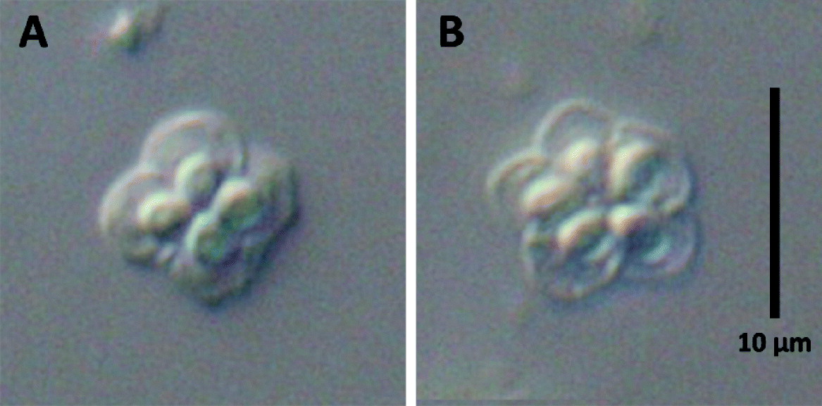
Comparison of Kudoa kenti n. sp. and Kudoa amamiensis (A–D) Phase contrast micrographs of spores (Scale bar=10 μm). (A) Kudoa kenti n. sp. ex Dischistodus perspicillatus in apical view and (B) side view; (C) Kudoa amamiensis ex Abudefduf bengalensis in apical view and (D) side view. (E–F) Principal component comparison of apical view (E) and side view (F) measurements with 95% confidence ellipses from Kudoa kenti n. sp. (data points=crosses, left ellipse) and Kudoa amamiensis (data points=hollow squares, right ellipse) where component 1 is on the y-axis and component 2 on the x-axis.

Polar capsule number variability in Kudoa kenti n. sp. (A) The most common morphotype with 4 polar capsules. (B) The minor morphotype with 5 polar capsules. Scale bar=10 μm.

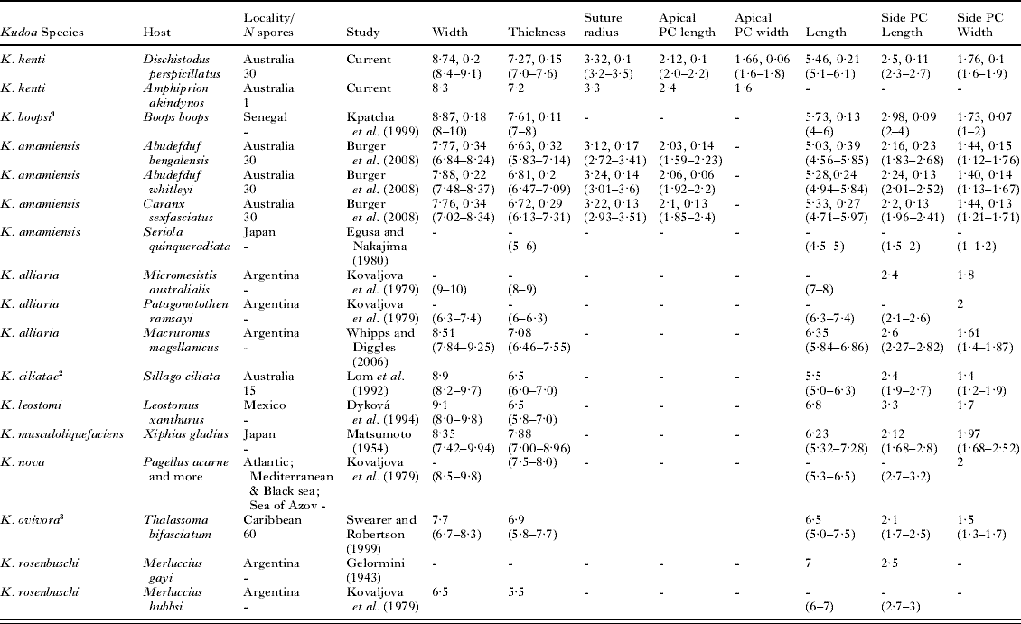
Morphological affinities: Kudoa kenti n. sp. has remarkably similar spore morphology to K. boopsiKpatcha et al. Reference Kpatcha, Diebakate, Faye and Toguebaye1999. However, K. kenti n. sp. differs by being found in skeletal muscle and infects reef fish of the Great Barrier Reef, rather than exclusively infecting than the gills of pelagic fish (Boops boops) of the Eastern Atlantic. Superficially, spores are similar to K. ciliataeLom et al. Reference Lom, Rohde and Dykova1992 and K. ovivoraSwearer and Robertson, Reference Swearer and Robertson1999, but K. ciliatae infects the smooth muscle of the intestine and produces macroscopic elongate white cysts and K. ovivora infects the ovaries.
Of the skeletal muscle-dwelling kudoids, K. amamiensis shows the most morphological similarity to K. kenti n. sp. In a comparison of spore morphology reported by Burger et al. (Reference Burger, Barnes and Adlard2008), K. amamiensis has a smaller width and thickness, but similar polar capsule length and suture radius, so that the polar capsules of K. amamiensis take up a larger proportion of the spore valve (53% vs 49%) and the spore valves of K. kenti n. sp. appear more lobate. Of the 20 other rounded quadrate, muscle-infecting kudoid species that have equal sized polar capsules, K. kenti n. sp. can be distinguished from K. aequidensCasal et al. Reference Casal, Matos, Matos and Azevedo2008, K. camarguensisPampoulie et al. Reference Pampoulie, Marques, Rosecchi, Crivelli and Bouchereau1999, and K. caudataKovaljova and Gajevskaja, Reference Kovaljova and Gajevskaja1983 by not having hair-like projections on the spore valves; K. azoniAseeva, Reference Aseeva2004 by the length at the central point being the longest axis in side view; K. bengalensisSarkar and Mazumder, Reference Sarkar and Mazumder1983, K. miniauriculataWhitaker et al. Reference Whitaker, Kent and Sakanari1996, and K. uncinataZhou and Zhao, Reference Zhou and Zhao2008 by not having apical extensions; Kudoa clupeidae (Hahn, Reference Hahn1917), K. crumena (Iversen and van Meter, Reference Iversen and Van Meter1967), K. funduli (Hahn, Reference Hahn1913), K. iwataiEgusa and Shiomitsu, Reference Egusa and Shiomitsu1983, and K. sciaenaeTeran et al. Reference Teran, Llicán and Luque1990 in having an undulate ellipsoid shaped spore in side view rather than pyriform; K. insolita (see Kovaljova et al. Reference Kovaljova, Shulman and Yakovlev1979) and K. paniformis (Kabata and Whitaker, Reference Kabata and Whitaker1981) by having even suture orientation; also, K. sebastea (Aseeva, Reference Aseeva2004), by not having ‘bolster thickening of its valves’. Kudoa kenti n. sp. shows most morphological affinity with K. alliaria 1979 (see Kovaljova et al. Reference Kovaljova, Shulman and Yakovlev1979), K. leiostomi (Dyková et al. Reference Dyková, Lom and Overstreet1994), K. musculoliquefasciens (Matsumoto, Reference Matsumoto1954), K. novaNaidjenova et al. Reference Naidjenova, Shulman, Donets, Greze, Delyamure and Nikolaeva1975, and K. rosenbuschi (Gelormini, Reference Gelormini1943). The width and thickness of K. kenti n. sp. both fall in the gap between the 2 different host isolates (Micromesistius australis and Patagonotothen ramzayi) from the original description of K. alliaria, but closer to the isolate identified recently from Macruronus magellanicus (see Whipps and Diggles, Reference Whipps and Diggles2006). Kudoa kenti n. sp. differs from K. alliaria in having a much smaller length and presenting as smaller macroscopic cysts. Kudoa leiostomi is larger in length and polar capsule length than K. kenti n. sp. Both K. nova and K. rosenbuschi have thicker spores (K. nova also has a larger polar capsule length) than K. kenti n. sp. There are overlapping spore measurement ranges with K. musculoliquefaciens, but it has more rounded polar capsules, smaller plasmodia, and causes substantial muscle liquefaction compared with K. kenti n. sp.
Molecular affinities: (Fig. 3, Table 4) Two 18S rDNA sequences of Kudoa kenti n. sp. were generated from 2 individual fish (of 2 fish species) and 4 28S rDNA sequences from 4 individual fish (of 3 fish species). These isolates cluster together in a tight clade in all phylogenetic analyses with high bootstrap support (100%) and clade credibility (1·00). There is only 1 nucleotide difference (99·9% sequence identity) between the two 18S rDNA sequences of K. kenti n. sp. and a maximum of 5 nucleotide differences (99·3% sequence identity) in 28S rDNA. The most genetically similar species, K. amamienis, differs from K. kenti n. sp. in 18S by 21–26 bp (98·6–98·2% sequence identity). The difference between the 2 species is far larger in 28S rDNA, being from 126 to 130 bp (80·2–79·4% sequence identity).
Spore measurements of Kudoa kenti n. sp. and morphologically similar species with mean dimensions (μm), standard deviation, and range in parentheses

-, Not available; PC, Polar Capsule.
1 Infects gills. 2 Infects smooth muscle of the intestine. 3 Infects ovaries.
18S rDNA nucleotide differences (lower triangle) and DNA percentage differences (upper triangle) of isolates from Kudoa kenti n. sp. and Kudoa amamiensis (from Burger et al. Reference Burger, Barnes and Adlard2008)

Abbreviations for Table 4A and B: Kk, Kudoa kenti; Ka, Kudoa amamiensis; Aa, ex Amphiprion akindynos; Dp, ex Dischistodus perspicillatus; Ab, ex Abudefduf bengalensis; Aw, ex Abudefduf whitleyi; Cs, ex Caranx sexfasciatus; Py, ex Pempheris ypsilichnus; Sq, ex Seriola quinqueradiata; Am, ex Amphiprion melanopus; Pl, ex Plectroglyphidodon leucozonus. After host isolate, numbers refer to the individual and letters refer to the clone number eg. Ka Ab 1a is the first clone of the first Kudoa amamiensis isolate sequenced from the first Abudefduf bengalensis.
28S rDNA nucleotide differences (lower triangle) and DNA percentage differences (upper triangle) of isolates from Kudoa kenti n. sp. and Kudoa amamiensi

Kudoa paraquadricornis n. sp
Type host: Caranx ignobilis (Forsskål, 1775), Giant trevally (Teleostei: Carangidae) adult.
Type locality: Heron Island (23°27′S; 151°55′E), The Great Barrier Reef, Queensland, Australia.
Location in the host: Histozoic, intracellular between myofibrils of skeletal muscle cells.
Prevalence: 1 of 3, Heron Island; 2 of 2, Lizard Island.
Other hosts: from the family Carangidae: Carangoides plagiotaenia Bleeker, 1857, Barcheek trevally, 3/3 Lizard Island; Caranx papuensis Alleyne and MacLeay, 1877, Brassy trevally, 2/6 Lizard Island; Caranx sexfasciatus Quoy and Gaimard, 1825, Bigeye trevally, 1/1 Lizard Island.
Material deposited: Giemsa-stained, air-dried spores (ex Caranx ignobilis registration number: G465318), Giemsa and eosin-stained histological section (ex Caranx ignobilis registration number: G465319), and haematoxylin and eosin-stained histological section (ex Caranx ignobilis registration number: G465320); deposited in the collections of the Queensland Museum, Brisbane, Australia. 18S and 28S rDNA sequence data from various isolates were submitted to GenBank. 18S rDNA Accession numbers from FJ792715 to FJ792719 and 28S from FJ792749 to FJ792752. See Fig. 3 for GenBank specific isolate Accession numbers.
Etymology: specific name refers to the similarity to Kudoa quadricornis.
Description. Plasmodia (Fig. 8J). Microscopic, polysporic. No inflammatory response evident. Spores of uniform development; extra-sporogonic stage not observed. Not found in other organs examined (brain, gall bladder, heart, kidney, or liver). Spores (Fig. 8A and B). Spore shape fitting the original description of the genus KudoaMeglitsch, Reference Meglitsch1947 and the emended diagnosis from Whipps et al. (Reference Whipps, Adlard, Bryant and Kent2003a). Spore body subquadrate in apical view with 4 equal rounded spore valves; stellate projections visible from each valve. Spore with tetromerous radial symmetry. No other morphotypes present. Polar capsules equal, convergent, pyriform. In side view, spore body pyriform with posterio-lateral projections from each valve, which appear distinct from the spore body. Turns of the polar filament not discernable. Spore measurements are shown in Table 5.
Comparison of Kudoa paraquadricornis n. sp. and Kudoa quadricornis. (A–D) Phase contrast micrographs of spores (Scale bar=10 μm). (A) Kudoa paraquadricornis n. sp. ex Caranx ignobilis in apical view and (B) side view; (C) Kudoa quadricornis ex Carangoides fulvoguttatus in apical view and (D) side view. (E–F) Principal component comparison of apical view (E) and side view (F) measurements with 95% confidence ellipses from Kudoa paraquadricornis n. sp. (hollow squares, right ellipse) and Kudoa quadricornis (crosses, left ellipse) where component 1 is on the y-axis and component 2 on the x-axis. (G–H) Principal component comparison of apical view (G) and side view (H) measurements with 95% ellipses of host isolates. Ellipses from left to right Kudoa quadricornis ex Carangoides fulvoguttatus from Lizard Island (filled squares) and Heron Island (crosses); Kudoa paraquadricornis n. sp. ex Carangoides plagiotaenia (filled inverted triangles), ex Caranx ignobilis from Heron Island (hollow triangles) and ex C. ignobilis from Lizard Island (hollow squares). (I) Principal component comparison of side-view measurements with 95% ellipses from Kudoa paraquadricornis n. sp., Kudoa quadricornis measurement from the current study, and the average measurements of Kudoa quadricornis from Whipps et al. (Reference Whipps, Adlard, Bryant and Kent2003a) without the thickness measurement. (J) Light micrograph of histological section stained with Giemsa and eosin (Scale bar=20 μm) of Kudoa paraquadricornis n. sp.

Morphological affinities: Minimal intra-specific morphological variation was observed between the 3 host isolates from Carangoides plagiotaenia and Caranx ignobilis collected at Heron Island; and Caranx ignobilis collected at Lizard Island (Fig. 8G and H).
Kudoa paraquadricornis n. sp. is the third Kudoa species known to have posterio-lateral projections. Kudoa haridasaeSarkar and Gosh, Reference Sarkar and Gosh1991 was the first; however, K. haridasae is much smaller (see Table 5) and differs in its host tissue location (coelozoic in the gall bladder). Kudoa paraquadricornis n. sp. closely resembles K. quadricornis but differs significantly in thickness and in the length of the projections. Figure 8E and F shows a separation in the principal component comparison of the morphological data from K. paraquadricornis n. sp. and K. quadricornis obtained in this study. A reduced analysis comparing the means of the side-view measurements of the original description of K. quadricornis from Whipps et al. (Reference Whipps, Adlard, Bryant and Kent2003a) (the thickness was not available) with the measurements of K. paraquadricornis n. sp., and K. quadricornis from this study placed the mean of the original description of K. quadricornis in the overlap between K. paraquadricornis n. sp. and K. quadricornis (Fig. 8I).
Molecular affinities: (Fig. 3) Five 18S rDNA sequences of Kudoa paraquadricornis n. sp. were generated from 5 individual fish (of 4 fish species) and 4 28S rDNA sequences from 4 individual fish (of 3 fish species). There are no nucleotide differences among these isolates in both 18S and 28S rDNA. K. paraquadricornis n. sp. clusters with the only other species of similar morphology for which there are genetic data, K. quadricornis, with relatively high bootstrap support (68–100%) and clade credibility (1·00). There is only 1 nucleotide difference (99·9% sequence identity) in 18S rDNA between the 2 species, but 25 nucleotides difference (96·4% sequence identity) in 28S rDNA.
Spore measurements of Kudoa paraquadricornis n. sp., Kudoa quadricornis and the morphologically similar species Kudoa haridasae, with mean dimensions (μm), standard deviation, and range in parentheses

-, Not available; PC, Polar Capsule.
* Infects the gall bladder.
Kudoa whippsi n. sp
Type host: Abudefduf bengalensis (Bloch, 1787), Bengal sergeant (Teleostei, Pomacentridae) adult.
Type locality: Heron Island (23°27′S; 151°55′E), The Great Barrier Reef, Queensland, Australia.
Location in the host: Histozoic, intracellular within myofibrils of skeletal muscle cells.
Prevalence: 3 of 12, Heron Island; none of 2, Lizard Island.
Other hosts: from the family Pomacentridae: Abudefduf whitleyi Allen and Robertson, 1974, Whitley's sergeant, 2/27 Heron Island, 1/1 Lizard Island; Acanthochromis polyacanthus (Bleeker, 1855), Spiny puller, 0/2 Heron Island, 6/15 Lizard Island; Amphiprion akindynos Allen, 1972, Barrier reef anenomefish, 4/4 Lizard Island; Amphiprion melanopus Bleeker, 1852, Fire clownfish, 1/4 Lizard Island; Chromis viridis (Cuvier, 1830), Blue green damselfish, 5/5 Lizard Island; Neoglyphidodon melas (Cuvier, 1830), Bowtie damselfish, 8/9 Lizard Island; Pomacentrus chrysurus Cuvier, 1830, Whitetail damsel, 1/6 Lizard Island. From the family Apoginidae: Apogon properuptus (Whitley, 1964), Southern orange-lined cardinal fish, 1/4 Lizard Island.
Material deposited: Giemsa-stained, air-dried spores (ex Am. akindynos registration number: G465322), Giemsa and eosin-stained histological sections (ex Ab. bengalensis, Am. akindynos, and Am. melanopus registration numbers: G465323 to G465325), and haematoxylin and eosin-stained histological sections (ex Ab. bengalensis, Am. akindynos, and Am. melanopus registration numbers: G465326 to G465328); deposited in the collections of the Queensland Museum, Brisbane, Australia. 18S and 28S rDNA sequence data from various isolates were submitted to GenBank. 18S rDNA Accession numbers from FJ792722 to FJ792725 and 28S from FJ792755 to FJ792763. See Fig. 3 for GenBank specific isolate Accession numbers.
Etymology: specific name in honour of Dr Christopher Whipps, State University of New York, United States of America for his contribution to kudoid research.
Description. Plasmodia (Fig. 5J). Microscopic, polysporic. No inflammatory response evident. Spores of uniform development, extra-sporogonic stage not observed. Not found in other organs examined (brain, gall bladder, heart, kidney, or liver). Spores (Fig. 5C and D). Spore shape fitting the original description of the genus KudoaMeglitsch, Reference Meglitsch1947. Stellate in apical view. No other morphotypes present. Spores with bilateral symmetry in the sagittal plane only. Polar capsules convergent, pyriform, with 1 much larger than the other 3. In side view spores are acute, scalene pyramidal. Turns of the polar filament not discernable. Spore measurements are shown in Table 1.
Morphological affinities: Moderate intra-specific morphological variation was observed between the host isolates (Amphiprion akindynos and Acanthochromis polyacanthus). However, there was still significant overlap of the measurements (Fig. 5G and H) from the 2 isolates.
Kudoa whippsi n. sp. closely resembles K. gunterae n. sp., and can infect the same individual fish (i.e. co-infections of these 2 Kudoa species have been observed in Apogon properuptus, Chromis viridis, Neoglyphidodon melas, and Pomacentrus chrysurus). The most obvious morphological difference between these species is that the 3 smaller polar capsules of K. whippsi n. sp. are larger than those of K. gunterae n. sp., particularly when viewed in the apical view. Principal component analyses modelling the overall differences in morphometrics from apical and side view are distinct, but overlap as shown in Fig 5E and F. Kudoa whippsi n. sp. shows similar morphological affinities as K. gunterae n. sp. (see the K. gunterae n. sp. description for specific details).
Molecular affinities: (Fig. 3, Table 2) Four 18S rDNA sequences of K. whippsi n. sp. were generated from 4 individual fish (of 4 fish species) and 9 28S rDNA sequences from 8 individual fish (of 6 fish species). These isolates form a clade in all phylogenetic analyses with moderate to high bootstrap support (56–99%) and high clade credibility (0·85–1·00). There is a maximum of 6 bp differences (99·6% sequence identity) among the 18S rDNA sequences of Kudoa whippsi n. sp. and 13 nucleotide differences (98·2% sequence identity) in 28S rDNA. The most genetically similar species is K. gunterae n. sp. Details on the differences between these 2 species are given in the K. gunterae n. sp. description.
Like K. gunterae n. sp., K. whippsi n. sp. falls within a monophyletic clade of muscle-dwelling species with a stellate spore shape, 1 polar capsule much larger than the other 3. In 18S (Fig. 3A) this clade includes: K. lateolabracis (maximum 97·6% sequence identity), K. megacapsula (maximum 98·1% sequence identity), K. minithyrsites (maximum 97·6% sequence identity), and K. thyrsites (maximum 98·5% sequence identity). For 28S (Fig. 3B), there are only representative sequences from K. thyrsites, which has a maximum sequence identity with K. gunterae n. sp. of 92·1%.
Morphological results
Morphological analyses conducted on species described here use 6 new measurements to characterize 4 novel kudoid species: Kudoa gunterae n. sp., K. kenti n. sp. K. paraquadricornis n. sp. and K. whippsi n. sp. These measurements are: Apical Polar Capsule Width, Apical Large Polar Capsule Length, Total Width (in addition to Spore Width for species with posterio-lateral projections), Projection Breadth, Projection Length and Projection Width (see Fig. 1) and a clear definition of thickness for radially symmetrical spores with an even number of polar capsules (see Fig. 2).
Intra-specific morphological variation was found between the host isolates from the new species as determined by principal component analyses (Figs 5G and H, 8G and H) and 2-sample t-tests. For the 2 host isolates of K. gunterae n. sp., there were statistically significant differences in the means (P-value <0·05) of thickness, apical large polar capsule length, apical large polar capsule width, side large polar capsule length and side small polar capsule length. The 2 host isolates of K. whippsi n. sp. showed statistically significant differences in the means (P-value <0·05) of width, thickness, apical large polar capsule length, apical small polar capsule width, side large polar capsule length and side small polar capsule length. Minimal intra-specific morphological variation was found between the 3 host isolates measured for K. paraquadricornis n. sp. Between the 2 Caranx ignobilis isolates, there were statistically significant differences in the means (P-value <0·05) from the thickness and the side polar capsule width. From the 2 isolates from Lizard Island, Carangoides plagiotaenia and Caranx ignobilis, there were statistically significant differences in the means (P-value <0·05) from the thickness, length, and side projection length. There were statistically significant differences in the means (P-value <0·05) between the isolates from different sampling sites recovered from Carangoides plagiotaenia from Lizard Island and Caranx ignobilis from Heron Island; in the apical polar capsule width, apical projection width, the length, and side projection length.
Nevertheless, in all instances, there was significant overlap of the data ranges found between the host isolates within each new species. The intra-specific variation between the isolates is less significant than the inter-specific morphological variation between closely related species that infect the same type of host tissue.
The measurements of the new species were compared with other kudoid species of similar morphology in Table 1, Table 3, and Table 5; and Figs 5E, 5F, 6E, 6F, 8E, 8F and 8G). From the combined raw data of the 2 host isolates of K. gunterae n. sp. and the 2 host isolates of K. whippsi n. sp., there were statistically significant differences (P-value <0·001) in all the mean measurements, except for thickness (P-value 0·221).
Kudoa kenti n. sp. was compared in the same manner specified above to K. amamiensis (raw data from Burger et al. (Reference Burger, Barnes and Adlard2008)). All mean measurements compared were statistically significantly different (P-value <0·001).
When measurements were compared between K. paraquadricornis n. sp. and K. quadricornis, the most significant differences were noted in the length of the posterio-lateral projections and to a lesser degree, the spore size in side view; but, no difference was observed in the size of the polar capsules. The mean measurements that were statistically significantly different (P-value <0·02) were: total width, suture radius, apical projection width, thickness, length, and side projection length. The additional thickness measurement resulted in greater separation between the two species as demonstrated in Fig. 8F and G, where Fig. 8I includes the average side view of K. quadricornis from Whipps et al. (Reference Whipps, Adlard, Bryant and Kent2003a). There was only 1 apical measurement supplied in Whipps et al. (Reference Whipps, Adlard, Bryant and Kent2003a), which has comparable ranges to the current K. quadricornis and K. paraquadricornis n. sp. measurements.
Representative spore microphotographs of these pairwise morphological comparisons are shown in Figs 5, 6 and 8A–D.
Phylogenetic analyses
Seventeen 18S rDNA sequences and 37 28S rDNA sequences were generated from the new species. Additional 28S rDNA sequences were generated from Kudoa amamiensis, K. ciliatae, K. monodactyli and K. quadricornis (2 isolates, 1 collected from Heron Island and 1 form Lizard Island) to allow a more comprehensive comparison of the genetic distances between species in both 18S and 28S rDNA regions.
The trimmed alignments of 18S and 28S rDNA sequences, including the new sequences (shown in bold in Fig. 3), were 1540 and 816 bp long, respectively. In 18S rDNA, the Kudoa sequences that spanned the entire alignment varied in size from 1444 (K. ciliatae) to 1510 bp (K. crumena). In 28S, the size of the Kudoa sequences varied from 667 (K. neurophila) to 759 bp (K. kenti n. sp.).
Trees of similar topology were obtained for each of the 18S and 28S rDNA sets of phylogenetic analyses (Bayesian, maximum likelihood, neighbour joining, and parsimony). Fig. 3 shows the molecular relationships between species of Kudoa, using neighbour-joining distance analyses.
There were several sequences and clades that varied in their position between phylogenetic analyses, but this variability relates to low bootstrap support (<50%) and clade credibilities (<0·5). In 18S, the species branch of K. megacapsula and the 2 species clades of: K. paraquadricornis n. sp. and K. quadricornis; and K. trifolia and K. unicapsula, did not maintain a fixed position in the trees. However, Kudoa megacapsula remained in the clade containing species with stellate spores and unequal sized polar capsules. Of the 85 Kudoa sequence replicates (a total of 36 species) in the 18S analysis, 7 other sequences did not have fixed positions: K. ciliatae, K. dianae, K. minithyrsites, K. paniformis, K. nova, and K. ovivora. There was a similar result in the 28S rDNA analysis except there were fewer species representatives that have corresponding sequence data available from GenBank with 58 Kudoa sequence replicates (a total of 18 species). In the 28S rDNA analysis, the phylogenetic position of 7 sequences; K. ciliatae, K. grammatorcyni, K. miniauriculata, K. monodactyli, K. ovivora, K. paniformis, and K. thyrsites ex Coryphaena hippurus, varied slightly due to their low bootstrap support and clade credibilities.
The 28S rDNA analyses of the new species isolates resulted in a similar intra-specific variation to 18S rDNA in total nucleotide differences (other than Fig. 2, see also Tables 2 and 4); however, 28S rDNA had a larger percentage intra-specific variation due to the relative size of the gene fragment – 816 bp from 28S vs 1540 bp from 18S. For instance, most species for which there were multiple replicates (K. amamiensis, K. gunterae n. sp., K. iwatai, K. kenti n. sp., Kudoa sp., K. whippsi n. sp.) vary by 0–8 bp in 18S, which equals 0–0·6% variation; and 0–13 bp in 28S, which equals 0–1·83% variation). The exception is that of K. thyrsites, which has a far greater degree of intra-specific variation, especially in 28S (in 28S 56 bp, which equals 8·6% variation; in 18S, 14 bp, which equals 1% variation).
The inter-specific variation is consistently much greater in 28S, for example there was 1 nucleotide difference between K. paraquadricornis n. sp and K. quadricornis in 18S, but 25 bp in 28S (a difference of 0·1% to 3·6%) and the difference between K. amamiensis and K. kenti n. sp. is a maximum of 26 bp in 18S, but up to more than 126 bp in 28S.
DISCUSSION
Molecular data are increasingly being applied to species characterization in the Kudoidae. This article aims to build on the morphological guidelines for species characterization of the Myxosporea devised by Lom and Arthur (Reference Lom and Arthur1989) to create a more specific structure for the definition of kudoid species, including DNA sequence data as an essential component.
Since the proposal to synonymize the Pentacapsulidae, Hexacapsulidae, and Septemcapsulidae into the Kudoidae (see Whipps et al. Reference Whipps, Grossel, Adlard, Yokoyama, Bryant, Munday and Kent2004), there are now 10 basic spore forms documented within this Family. Five distinct morphological forms are recognized from the apical view, and 5 are recognized from the side view. In apical view, spores present in the following forms: spores with radial symmetry, an even number of spore valves and equal sized polar capsules (including rounded quadrate, pointed quadrate, and spores of more than 4 polar capsules); stellate spores with unequal sized polar capsules; spores with uneven sized valves and polar capsules but with almost bilateral symmetry in the frontal and sagittal plane (eg. K. permulticapsula Whipps et al. 2003 a and K. tachysuraeSarkar and Mazumder, Reference Sarkar and Mazumder1983); spores with posterio-lateral projections; and spores with an unequal number of spore valves. Note that the thickness measurement has been removed from radially symmetrical spores with an uneven number of polar capsules as this measurement has frequently been interpreted in different ways and offers little for comparison with other species. For these spores the valve width, the suture radius, the polar capsule length and width are the important measurements in apical view. The 5 main spore forms in side view are: spores with equal sized polar capsules and no projections; spores with unequal sized polar capsules; spores with uneven valves and polar capsules but with bilateral symmetry in the frontal and sagittal plane; spores with lateral projections; and spores with apical projections.
The updated schematics for spore measurements are a compendium of several decades of morphological studies (Egusa, Reference Egusa1986; Lom and Arthur, Reference Lom and Arthur1989; Adlard et al. Reference Adlard, Bryant, Whipps and Kent2005; Whipps and Kent, Reference Whipps and Kent2006; Burger et al. Reference Burger, Cribb and Adlard2007). The new measurements included in this study allow for more comprehensive distinction between morphologically similar species. While measurement error due to indistinct character margins is often encountered using a light microscope at high magnification, errors are minimized by using replication from numerous spores.
Morphology is a fundamental aspect of species characterization and can be useful in the differentiation of closely related kudoid species. However, when multiple morphotypes with varying polar capsule numbers are encountered in a single plasmodium, these morphotypes do not indicate different species, as for K. chaetodoni, K. iwatai, K. lethrini, K. monodactyli, K. permulticapsula, K. shulmani, and K. yasunagai (see Naidjenova and Zaika, Reference Naidjenova and Zaika1970; Hsieh and Chen, Reference Hsieh and Chen1984; Whipps et al. 2003 a; Diamant et al. Reference Diamant, Ucko, Paperna, Colorni and Lipshitz2005; Gunter et al. Reference Gunter, Cribb, Whipps and Adlard2006; Burger et al. Reference Burger, Cribb and Adlard2007); but rather, the polar capsule number plasticity can be considered a characteristic feature of such species.
An overriding factor that differentiates similar morphological species can be the tissue that they infect. Tissue tropism has been shown by Burger et al. (Reference Burger, Cribb and Adlard2007) to be the most significant uniting biological factor that reflects genetic relationships among the Kudoidae. For example, the morphologically similar species with 4 polar capsules; K. alliaria (from the skeletal muscle), K. ciliatae (from the smooth muscle of the intestine), K. dianae (from the subepithelial connective tissue of the oesophagus and mesentery), and K. shiomitsui (from the pericardial cavity) are genetically closer to other species with less morphological similarity than those that infect the same tissue type rather than to each other. The ability to infect several different tissues is a characteristic of a few species such as K. dianae (see Dyková et al. Reference Dyková, Avila and Fiala2002) and K. iwatai (see Diamant et al. Reference Diamant, Ucko, Paperna, Colorni and Lipshitz2005). The relationships based on tissue tropism give support for the assignment of K. kenti n. sp. as a new species despite its close morphological resemblance to K. boopsi and to a lesser degree to K. ciliatae and K. ovivora. DNA sequence data for K. ciliatae and K. ovivora confirm the genetic distance between K. ciliatae and K. ovivora to K. kenti n. sp.
Even though kudoids are not highly host specific (Blaylock et al. Reference Blaylock, Bullard and Whipps2004; Diamant et al. Reference Diamant, Ucko, Paperna, Colorni and Lipshitz2005; Whipps and Kent, Reference Whipps and Kent2006; Burger et al. Reference Burger, Barnes and Adlard2008) and host differences are not reflected in genetic relatedness of Kudoa species (Burger et al. Reference Burger, Cribb and Adlard2007), the type host and host range are useful for subsequent diagnosis of infections. Also, an indication of the level of variation in morphology between host isolates is paramount, because host-induced factors can impact on specific spore size as seen with K. alliaria (see Whipps and Diggles, Reference Whipps and Diggles2006), K. hypoepicardialis (see Blaylock et al. Reference Blaylock, Bullard and Whipps2004), K. iwatai (see Diamant et al. Reference Diamant, Ucko, Paperna, Colorni and Lipshitz2005), K. thyrsites (see Whipps and Kent, Reference Whipps and Kent2006) and from the current study with K. gunterae n. sp.
In the presence of subtle differences in morphology in similar host individuals and sample collection sites, DNA nucleotide differences (especially those from 28S rDNA) can be useful for discriminating species. This is clearly shown in the differentiation between K. paraquadricornis n. sp. and K. quadricornis, which infect carangids on the Great Barrier Reef; and K. gunterae n. sp. and K. whippsi n. sp., which were occasionally found infecting the same host individual. This phenomenon is not unique to the Kudoidae. In a study conducted by Gunter and Adlard (Reference Gunter and Adlard2008) of bivalvulidan parasites on the Great Barrier Reef, 2 individuals of Pomacentrus chrysurus sampled from reefs off Lizard Island were infected with 2 morphologically similar species, Ceratomyxa cribbi and C. talboti, that were confirmed as separate species through reference to 18S rDNA nucleotide differences.
18S rDNA sequence data are important in species characterization of myxosporeans, to allow for comparisons of higher level phylogeny. 18S rDNA continues to be a useful genetic marker for species relationships among the order Bivalvulida (Gunter and Adlard, Reference Gunter and Adlard2009). However, for some morphologically similar species in the Kudoidae, more resolution can be obtained by the use of 28S rDNA, as seen with the differentiation of K. gunterae n. sp. and K. whippsi n. sp.; and K. paraquadricornis n. sp. and K. quadricornis.
Prior to this project, other gene regions have been evaluated for their efficiency to define kudoid species. Whipps and Kent (Reference Whipps and Kent2006) suggested that having a marker other than the small subunit rDNA cannot be underemphasized. They performed a comparison between isolates of K. thyrsites using 18S rDNA, 28S rDNA, internal transcribed spacer 1 (ITS-1), and heat shock protein 70 (hsp 70). The ITS-1 gene was considered too variable in the Kudoidae to be useful for species differentiation and the hsp 70 did not provide increased phylogenetic resolution. For the current study, using both 18S and 28S rDNA sequence data were adequate for species characterization and discrimination of small-scale inter-specific boundaries.
Clearly, it is not ideal to assign species boundaries simply on the level of nucleotide differences, but rather use these data as an indicator that points to existing, often subtle, morphological and/or biological differences. The 18S rDNA neighbour joining tree raises 2 significant taxonomic concerns: the relatively large genetic distance between different isolates of K. thyrsites; and the genetic similarity between K. alliaria, K. rosenbuschi, and the Kudoa sp. isolates. Whipps and Kent (Reference Whipps and Kent2006) demonstrated that genetic relationships among isolates of K. thyrsites correlated with geographical locality, but were reluctant to propose the erection of multiple species based solely on geography. Nonetheless, the evidence provided by these authors strongly suggests that K. thyrsites represents a species complex.
The Kudoa sp. isolates sequenced by Pascual and Abollo (Reference Pascual and Abollo2008), coupled with the K. alliaria and K. rosenbuschi sequences, show the same pattern of 18S rDNA genetic relationships (a maximum of 7 nucleotides variance, 0·5%) to the intra-specific variation of K. amamiensis isolates from Burger et al. (Reference Burger, Barnes and Adlard2008). Pascual and Abollo (Reference Pascual and Abollo2008) suggested that these species are either the same, or that 18S is too highly conserved to resolve them. Theoretically, the connectivity between a taxonomic binomen and a published genetic sequence must be completely without doubt. In reality the situation may be different. It is plausible for the following reasons that K. rosenbuschi in the study of Abollo et al. (Reference Abollo, Novoa and Figueras2005) may not refer to that taxon. The spores of Kudoa rosenbuschi are morphologically similar in size and shape to K. alliaria; however, muscle degradation is a characteristic feature of K. rosenbuschi (Gelormini, Reference Gelormini1943), which was not encountered in the Kudoa infection sequenced by Abollo et al. (Reference Abollo, Novoa and Figueras2005). Therefore, the K. rosenbuschi isolate and the Kudoa sp. sequences may actually be of K. alliaria, which produces macroscopic cysts, but does not degrade the muscle (Whipps and Diggles, Reference Whipps and Diggles2006).
From all available studies and the outcomes of this project, we have summarized schematics for morphological characterization and recommended the incorporation of 28S rDNA in addition to 18S rDNA sequence data for species descriptions in the Kudoidae. Four new species have been described here using these recommendations; Kudoa gunterae n. sp., K. kenti n. sp., K. paraquadricornis n. sp., and K. whippsi n. sp. In the future, we predict there may be more spore types that may not fit these suggestions; nevertheless, they are comprehensive in characterizing known kudoid infections.
ACKNOWLEDGEMENTS
The authors would like to thank Dr Nicole Gunter, Rick Gleeson, Tony Byrne, Associate Professor Tom Cribb, Abigail Downie, Dr Terrence Miller, Dr Matt Nolan, Chris Rohner, and Tane Sinclair-Taylor for assistance in fish collection and the staff and facilities of Heron and Lizard Island Research Stations. For access to molecular analysis equipment and assistance, we would like to thank Dr Andy Barnes and Dr Justice Baiano. For histology assistance, we would like to thank Chris Cazier from the Veterinary Pathology Laboratory, the University of Queensland.
FINANCIAL SUPPORT
This work was supported by research funds provided by the Queensland Museum Biodiversity Program and the School of Chemistry and Molecular Biosciences at the University of Queensland. We would also like to thank the ARC/NHMRC Research Network for Parasitology for travel funds.