INTRODUCTION
Spatial attention is a complex process that requires the engagement of multiple networks in the brain. Deficits in spatial attention can be dramatic and have dramatic effects on quality of life, as in the case of an acute left hemispatial neglect following damage to the right parietal cortex (Brain, Reference Brain1941; Heilman & Watson, Reference Heilman and Watson1977; Sobrinho et al., Reference Sobrinho, Santini, Marques, Gabriel, Neto, de Souza, Bazan and Luvizutto2018). In patients with left hemispatial neglect, the ability to attend to information in left egocentric space is severely impaired. These patients show a rightward bias when asked to bisect a horizontal line. This deficit is in attention and not attributable to a sensory defect, and has even been well demonstrated in mental imagery tasks (Bisiach & Luzzatti, Reference Bisiach and Luzzatti1978; Guariglia, Palermo, Piccardi, Iaria, & Incoccia, Reference Guariglia, Palermo, Piccardi, Iaria and Incoccia2013).
Hemispatial neglect is more frequent and severe with right than left hemisphere lesions (Ten Brink, Verwer, Biesbroek, Visser-Meily, & Nijboer, Reference Ten Brink, Verwer, Biesbroek, Visser-Meily and Nijboer2017). In most individuals, the right hemisphere is able to process information coming from the right and left hemispace and the left hemisphere is able to process information coming from the right (Heilman & Van Den Abell, Reference Heilman and Van Den Abell1980; Pardo, Fox, & Raichle, Reference Pardo, Fox and Raichle1991). This asymmetrical specialization is reflected in spatial biases evident in healthy individuals. For example, young adults have been reported to bisect lines to the left of midline, a phenomenon called “pseudoneglect” (Bowers & Heilman, Reference Bowers and Heilman1980; Jewell & McCourt, Reference Jewell and McCourt2000).
There have been reports that the leftward bias that healthy young adults often demonstrate on horizontal line bisection tests (pseudoneglect) diminishes with aging (Benwell, Thut, Grant, & Harvey, Reference Benwell, Thut, Grant and Harvey2014; Failla, Sheppard, & Bradshaw, Reference Failla, Sheppard and Bradshaw2003; Fukatsu, Fujii, Kimura, Saso, & Kogure, Reference Fukatsu, Fujii, Kimura, Saso and Kogure1990; Stam & Bakker, Reference Stam and Bakker1990). Some attribute this diminished pseudoneglect in older adults to a decline in the right hemisphere networks that mediate spatial attention, the “right hemi-aging” hypothesis (Barrett & Craver-Lemley, Reference Barrett and Craver-Lemley2008). In addition, with healthy aging, there is a decrease in asymmetrical brain activity during cognitive tasks (Cabeza, Reference Cabeza2002), called the hemispheric asymmetry reduction in older adults (HAROLD) phenomena. Thus, although it is not known what causes this age-related reduction in asymmetry in pseudoneglect, it is possible that this effect may be related to a reduced asymmetry in activation when performing spatial attention tasks (Benwell et al., Reference Benwell, Thut, Grant and Harvey2014). Support for this hemispheric activation asymmetry postulate comes from studies of patients who have experienced right hemisphere strokes that result in left hemispatial neglect. These patients show evidence of asymmetrical hemispheric (left greater than right) activation acutely (Corbetta, Kincade, Lewis, Snyder, & Sapir, Reference Corbetta, Kincade, Lewis, Snyder and Sapir2005; Umarova et al., Reference Umarova, Nitschke, Kaller, Kloppel, Beume, Mader, Bennet, Wilson, Glover and Weiller2016; Watson, Andriola, & Heilman, Reference Watson, Andriola and Heilman1977). Further, those patients who show improvement of neglect over time reveal an increase in symmetrical hemisphere activation (Corbetta et al., Reference Corbetta, Kincade, Lewis, Snyder and Sapir2005).
Neglect and pseudoneglect both appear to be related to right hemisphere dominance in mediating spatial attention. When healthy young adults that demonstrate pseudoneglect on line bisection experience interference in the functional activity of their right parietal lobe via transcranial magnetic stimulation (TMS), they demonstrate more severe left hemispatial neglect than those who do not demonstrate pseudoneglect (Varnava, Dervinis, & Chambers, Reference Varnava, Dervinis and Chambers2013). This may suggest that the degree of lateralization of attention is heterogenous and that the degree of pseudoneglect is related to the degree of right hemispheric attentional dominance.
If, with aging, there is a decrease in asymmetrical hemispheric activation during the performance of spatial bias tasks that recruit different brain regions, older participants may show a reduced bias in all spatial tasks, regardless of leftward or rightward biasing of a specific task. For example, solid line bisections and character line bisections produce divergent spatial biases in young adults. In character-letter line bisections, individual’s spatial bias on average deviates to the right of solid line bisections, reflecting left hemispheric lateralization for the processing of language content as well as focal attentional demands of the task (Lee, Kang, et al., Reference Lee, Kang, Park, Son, Lee, Adair, Heilman and Na2004; Lee, Kim, et al., Reference Lee, Kim, Kang, Park, Kim, Adair and Na2004). Solid line trisections also produce different biases, hypothetically due to engagement to different networks (Powers, Roth, & Heilman, Reference Powers, Roth and Heilman2005). Usage of trisection tasks has not been frequently reported in the literature and the methods of administration vary. However, common across all reported versions, individuals divide a line into 1/3 and 2/3 segments via a transection. Due to differences in attentional demands including hemispatial direction of attention, left and right trisections may elicit a diverging bias to each other and to solid line bisections.
With aging, right hemi-aging or HAROLD mechanisms would predict different performances on these tasks. Namely, with right hemi-aging, we would expect, relative to younger individuals, that older individuals would make transections to the right due to reduced influence of the right hemisphere relative to the left. If HAROLD is the dominant mechanism, we would expect, relative to younger individuals, that older individuals would make transections closer to the target regardless of direction of bias induced by each condition. In the current study, we compared older and younger individuals in four types of line bisection tasks previously shown to elicit biases to the left or right in neurologically healthy individuals including solid line bisection [leftward (Bowers & Heilman, Reference Bowers and Heilman1980; Jewell & McCourt, Reference Jewell and McCourt2000)], left trisection [leftward (Zivotofsky, Reference Zivotofsky2004)], right trisection [rightward (Zivotofsky, Reference Zivotofsky2004)], and character bisection [rightward (Lee, Kang, et al., Reference Lee, Kang, Park, Son, Lee, Adair, Heilman and Na2004)].
METHODS
Participants
Younger and older adults were recruited to participate in this study including 33 younger (age 27 ± 4.0, 15 women) and 37 older (age 68 ± 6.3, 14 women) neurologically and psychiatrically healthy individuals (see Table 1 for demographic information). Note, the first six of the older participants were not coded for age, only that they met the age inclusion criteria for the older group (age older than 60 and less than 80); thus, their average age is not included. Participants were right-handed as assessed by the Benton Handedness Questionnaire (degree of handedness indicated in Table 1) (Varney & Benton, Reference Varney and Benton1975), native speakers of English with at least 12 years of education. Sample size was determined based on prior research in the area and assumed a modest effect size.
Demographic attributes of younger and older participants

The participants were administered a structured interview, including questions about: age, education, occupation, history of learning disability, prescription medications, hypertension and diabetes, history of stroke occurrence, traumatic brain injury, other neurological conditions, alcohol use, tobacco use, illicit drug use, and psychiatric history. Older participants were administered a Mini-Mental Status Exam (MMSE) to decrease the likelihood of inclusion of individuals with a neurodegenerative disease.
The exclusionary criteria were (1) history of neurological dysfunction or injury (e.g., stroke, epilepsy, head injury with loss of consciousness); (2) learning disability, or DSM-IV Axis I psychiatric disorders or drug/alcohol abuse; (3) acute or chronic organ failure; (4) taking psychotropic medications that might influence alertness or arousal (e.g., benzodiazepines, barbiturates, anticonvulsants, neuroleptics, amphetamines, and dopaminergic agents); (5) history of significant brain developmental and/or birth disorders; (6) evidence for dementia or mild cognitive impairment of the older participants as indicated by scores <26 on the MMSE (Folstein, Folstein, & McHugh, Reference Folstein, Folstein and McHugh1975).
The University of Florida Institutional Review Board approved this study and the participants provided signed informed consent as approved by the Review Board.
Apparatus and Procedures
In addition to cognitive screening and interview as described above, a series of spatial bias assessments were performed on all participants. All stimuli were presented sequentially on a table and centered in front of participants. Presentation of stimuli was counterbalanced for order via a predetermined sequence for the entire sample. All participants completed solid line bisection, left trisection, right trisection, and character line bisection tasks. Each task required eight spatial judgments (e.g., eight solid line bisections). Each trial was presented on a separate 8.5 × 11 inch piece of unlined, white paper by a study coordinator. Lines were centered on the paper and were all presented in the landscape position. The coordinator placed the paper on a table in front of the participant. After each trial, the paper was removed and a new paper was placed. All transections were measured from the left end of the line.
Spatial Bias Tasks
Solid line bisections
For each trial, participants were presented with a 230-mm-long and 3-mm-thick solid line. The participants were asked to place a mark in the middle of the line or “bisect the line.” Participants marked the line with a pen, using their dominant hand, the center of each line. Displacement from the center was measured with a ruler in millimeters.
Solid line trisections
Participants were asked to use a pen to divide a 230-mm-long and 3-mm-thick solid line into thirds. For a right trisection, they were asked to place a mark on the line that would divide the line into two segments, with one-third of the line being on the right side and two-thirds of the line on the left side. For a left trisection, they were asked to divide the line into a left third and right two-thirds. Displacement from the actual portion of the line that made these two segments was measured with a ruler.
Character line bisections
Participants were asked to select the target letter closest to the center of a string of 26 letters. Participants were not told how many letters were in the line. Note, although it would be an insufficient approach (counter the instruction given), we did not attempt to prevent people from counting letters. Displacement from the center was measured in characters away from center.
Statistical Approach
Data were visually inspected and appeared approximately normally distributed. Further, metrics of skew and kurtosis did not suggest significant deviation from normal. Thus, to allow comparison between the type of tasks, a percentage deviation from accurate performance was calculated. The average transection across eight trials per condition was calculated, subtracted from the correct value (e.g., 115 mm for solid line bisection), and divided by the correct value. One-sample t tests were used to determine if overall participants’ bisections per condition deviated from zero and per group. A mixed design, repeated measures ANOVA was used for the two left biasing tasks and the two right biasing tasks. Multiple comparisons correction was implemented with Bonferroni.
Results
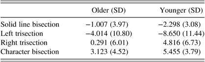
In all bisection and trisection conditions, the direction of deviation was the same in both age groups. Compared to a zero (correct) point (independent samples t test), overall, each bisection condition except right trisection was significantly different from zero; solid line bisection t(69) = −3.37, p < 0.001; left trisection t(69) = −4.60, p < 0.001; right trisection, t(69) = 1.93, p = 0.058; character bisection, t(69) = 8.17, p < 0.001. Within groups, younger participants in each spatial condition deviated from zero, whereas older participants were not different from zero in their estimation of right-sided trisections or solid line bisections, t(36) = 0.295, p = 0.770 and t(36) = −1.54, p = 0.132, respectively.
Overall, transections were different by condition (left and right trisection, sold line and character bisection), f(3, 100) = 25.07, p < 0.000, η 2 = 0.27. In pairwise comparisons, each bisection condition significantly differed from each other such that in solid line bisections and left trisections both groups deviated toward the left side of the line. In right trisections and character line bisections, both groups deviated toward the right side of the line. Further, there was an interaction effect such that older and younger participants deviated differently than each other across the four bisection conditions, f(3, 100) = 3.48, p = 0.048, η 2 = 0.05. In all tasks, the older participants deviated less from the target than did the younger participants (see Table 2 for mean values and standard deviations for each condition by age group).
Average spatial bias percent deviations from accurate in older and younger participants

“−” denotes leftward deviation
Discussion
The primary findings in this study are that older and younger individuals displayed expected biases in line bisection, left and right trisection, and character line bisection. Overall, participants deviated significantly to the left in both solid line and left trisection tasks and significantly to the right in right trisection and character line bisection tasks. Further, older participants were more accurate on all spatial tasks compared to younger participants. And, older participants’ spatial judgments did not differ from zero (essentially perfectly accurate) on solid line bisections and on right line trisections. The right hemi-aging hypothesis would predict that older participants would have deviated more to the right than the younger participants would have deviated. Rather, the results are consistent with the decreases in hemispheric asymmetry reduction with aging in older adults (HAROLD) (Cabeza, Reference Cabeza2002).
In the solid line bisection task, our younger population showed the characteristic leftward bias termed, pseudoneglect (Bowers & Heilman, Reference Bowers and Heilman1980). Replicating prior literature, in older adults, we found a reduced leftward deviation (Jewell & McCourt, Reference Jewell and McCourt2000). However, it is notable that the literature is not consistent on this issue with some studies showing reduction in pseudoneglect (or even rightward bias) and others showing increases in pseudoneglect with aging (Friedrich, Hunter, & Elias, Reference Friedrich, Hunter and Elias2018).
In younger participants, there is heterogeneity in the degree of pseudoneglect and this heterogeneity is associated with different spatial bias effects during interference of right parietal function with the use of TMS (Varnava et al., Reference Varnava, Dervinis and Chambers2013). There is also heterogeneity in performance in older individuals and, given different health trajectories in aging including vascular risk factors that contribute to divergent brain aging, one might anticipate greater variance in bisection performance with aging. However, in our sample, we do not see differences in variance across tasks as a function of age. The differences in bias, thus, represent a normalized shift in spatial judgments with aging.
It is unclear whether the age-related decrease in leftward bias on line bisection tasks represents a lateralized shift in the allocation of spatial attention produced by a reduction of right hemisphere activity (right hemi-aging) or a generalized difference in accuracy in spatial judgments associated with HAROLD. One means to disambiguate the two possibilities is to produce a task in which attention is directed to either the left or right side of space and to compare the changes between the two tasks as a function of aging. Thus, we administered a left and right trisection task and a character line bisection task, and found that older individuals were more accurate independent of the side of bias.
Compared with line bisection tasks, there are relatively little published data regarding the results of line trisection tests. In the few prior studies, the goals and administration differ from the current study. In these former studies, the goal was to learn the side of the line that the participant would first choose to perform their transection. In these studies, the participants were not directed to perform the trisections on one side of the other; they were allowed to choose (Zivotofsky, Reference Zivotofsky2004; Zivotofsky, Edelman, Green, Fostick, & Strous, Reference Zivotofsky, Edelman, Green, Fostick and Strous2007). However, in our paradigm, we directed participants to trisect the line to the right or to the left depending on the trial. The spatially directed trisection task may demand a greater degree of focal spatial attention to a segment of the line than the bisection task and thus, this task was expected to induce laterality biases. Further, the trisection task requires attention to be directed to the left or right side of the line. In the present study, with egocentric midline presentation of lines, that means that attention was directed to the left or right hemispace for left and right trisections, respectively. Given that the right hemisphere is able to allocate attention to the left and right hemispace, and the left hemisphere is able to allocate attention to the right hemispace, we expected that the left trisection task would produce a greater leftward bias than the right trisection task would produce a rightward bias.
In our trisection tasks, the younger adults showed a rightward bias on right trisections and a leftward bias on left trisections. As expected, the deviation of the leftward trisection was greater in magnitude than for the line bisection and greater than the relative bias in the right trisection task to the right. However, although in older adults, we found a similar pattern of change, in this group the deviation was smaller in magnitude than in the younger population in all conditions. It should also be noted that this task was apparently more difficult than the line bisection and the character bisection tasks, as indicated by greater variability in performance of both age groups. Although the reason for the difference in variability is unknown, in the line bisection task the participant has to select a point on the line where the right and left line segments appear to be of equal size, but in the trisection task the participant is required to select a point where one segment is twice the size of the other segment. This task would appear to require visuospatial computations and have greater demands on spatial working memory.
Similarly, on the character line bisection test, which has previously been shown to elicit a rightward bias in healthy young adults (Barrett, Reference Barrett2005), we not only replicated this result in both young and older adults but also demonstrated increased accuracy in older adults.
As mentioned, rather than the right hemisphere selectively aging (right hemi-aging), these accuracy shifts with age appear to reflect a reduction in functional hemispheric laterality. This reduction in asymmetry has been observed across domains, including working memory tasks (Reuter-Lorenz et al., Reference Reuter-Lorenz, Jonides, Smith, Hartley, Miller, Marshuetz and Koeppe2000), face-matching tasks (Grady et al., Reference Grady, Maisog, Horwitz, Ungerleider, Mentis, Salerno, Pietrini, Wagner and Haxby1994), episodic memory (Cabeza et al., Reference Cabeza, Grady, Nyberg, McIntosh, Tulving, Kapur, Jennings, Houle and Craik1997; Grady, Reference Grady2002), semantic retrieval tasks (Logan & Delheimer, Reference Logan and Delheimer2001; Stebbins et al., Reference Stebbins, Carrillo, Dorfman, Dirksen, Desmond, Turner, Martin, Hennig and Gabrieli2002), and inhibition tasks (Garavan, Ross, & Stein, Reference Garavan, Ross and Stein1999; Nielson, Langenecker, & Garavan, Reference Nielson, Langenecker and Garavan2002). It should be noted that these studies include tasks where younger adults tend to show left-lateralized activity (episodic memory encoding, semantic memory retrieval, verbal working memory) as well as tasks where they show right-lateralized activity (episodic memory retrieval, spatial working memory, and response inhibition). Regardless of the domain or the pattern of activity in younger adults, older adults appear to have a reduction of asymmetry.
Hemispatial neglect is more likely to occur following a brain injury in older individuals (Gottesman et al., Reference Gottesman, Kleinman, Davis, Heidler-Gary, Newhart, Kannan and Hillis2008). Based on the reduction of pseudoneglect observed with aging, hemispatial neglect should be less common and less severe with aging. These contradictory findings suggest that other factors may be important in the prevalence and severity of neglect associated with aging. In patients who have experienced right hemisphere strokes that result in left hemispatial neglect acutely, studies with FDG PET have shown that there is a relative decrease in metabolic activity around the site of injury. The degree to which neglect resolves in these patients is associated with an increase in metabolic activity within the right hemisphere, resulting in a relative balance of activity between the left and right hemispheres (Corbetta et al., Reference Corbetta, Kincade, Lewis, Snyder and Sapir2005). Further, in another study, whether or not patients with stroke experienced acute recovery was predicted by increases in functional activity between right parietal and left prefrontal/parietal regions (Umarova et al., Reference Umarova, Nitschke, Kaller, Kloppel, Beume, Mader, Bennet, Wilson, Glover and Weiller2016).
Recovery from stroke often depends on the recovery of functionally impaired perilesional areas (Furlan, Marchal, Viader, Derlon, & Baron, Reference Furlan, Marchal, Viader, Derlon and Baron1996; Yi et al., Reference Yi, Kim, Shim, Oh, Kim and Jung2019). Even with a decrease in right hemispheric attentional dominance, the incidence of left hemispatial neglect is more common with aging, and this age difference may be related to decreases in the ability of the perilesional areas to mediate the functions of the injured hemisphere.
More symmetrical brain activity is associated with line bisection performances that are closer to midline. However, this activity likely does not reflect compensatory activation or any decrease in functional localization as a function of age. To that point, older adults tend to be more susceptible to distraction or have more difficulty multitasking (Backman, Laukka, Wahlin, Small, & Fratiglioni, Reference Backman, Laukka, Wahlin, Small and Fratiglioni2002). The increased diffusivity of activation with age may increase susceptibility to interference effects in the performance of functionally proximal tasks (Hicks, Bradshaw, & Kinsbourne, Reference Hicks, Bradshaw and Kinsbourne1978). Though unknown, this could have implications for the performance of daily activities, for instance, fall risk with increased attentional demands could interact with changes in automaticity of walking tasks (Clark, Christou, Ring, Williamson, & Doty, Reference Clark, Christou, Ring, Williamson and Doty2014).
An ideal spatial attention task produces functional asymmetries linked to spatial attention processes involved in the task and should not be driven by other parameters known to produce asymmetries, such as motoric responses, eye movement, and visual attention shifting (Petit et al., Reference Petit, Zago, Vigneau, Andersson, Crivello, Mazoyer, Mellet and Tzourio-Mazoyer2009; Reference Petit, Zago, Mellet, Jobard, Crivello, Joliot, Mazoyer and Tzourio-Mazoyer2015; Shulman et al., Reference Shulman, Pope, Astafiev, McAvoy, Snyder and Corbetta2010). Many other factors have been shown to influence line bisection performance. Despite not explicitly controlling for some of these variables, reduction in hemispheric asymmetry with aging remains the most efficacious explanation of the effects that we see. However, these are factors which should be considered when designing future experiments.
Overall, the current investigation demonstrates aging effects in spatial biases such that older adults are more accurate in spatial judgments regardless of direction of bias. Further, the direction of biases induced by different tasks remains with aging, suggesting similar functional specificity in terms of system-network recruitment. These results are consistent with aging-related decreases in hemispheric asymmetry in activation regardless of spatial task. In addition, we have introduced a new method of measuring lateralized spatial bias, a modification of trisection approaches previously published by others. Further research on shifts in attention should consider the inclusion of spatial tasks that elicit different directions of biases in evaluating the mechanisms of such changes as well as potential effects on ecological performance such as in driving or in the context of rehabilitation.
ACKNOWLEDGEMENTS
This work was supported by the Department of Veterans Affairs (VA I01CX000744).
Michał Harciarek contribution to this study was supported by the National Science Center, Poland (grant number 2015/17/B/HS6/03951), PI: Michał Harciarek.
CONFLICTS OF INTEREST
The authors have nothing to disclose.