The use of an implantable cardioverter-defibrillator is the gold-standard therapy for the prevention of sudden cardiac death in several situations despite implantation challenges. Reference Carro, Cereda, Annoni, Annoni and Marianeschi1 A dearth of dedicated devices for small children forces physicians to implant the device in individualised manners.
Case report
The case was an 8-kg, 15-month-old boy diagnosed with high-risk long QT syndrome, which was refractory to appropriate medical therapy. A detailed and complex ethical process was exhausted, and the patient was transferred to the operating room. After conventional general anaesthesia, a median sternotomy was made, and the pericardium was opened longitudinally. An endocardial implantable cardioverter-defibrillator lead (Sprint Quattro Secure 6935; Medtronic Inc, Minneapolis, MN, USA) was placed around the heart with its shock coil attached to the posterior surface of the heart. The lead tip was fixed with a suture. Subsequently, an epicardial pacemaker lead (CapSure SenseTM EPI 4968–25cm; Medtronic Inc, Minneapolis, MN, USA) was fixed on the epicard for sensing purposes. The defibrillator end of the implantable cardioverter-defibrillator lead was connected to the shock socket of the defibrillator device (Protecta XT VR; Medtronic Inc, Minneapolis, MN, USA) while the sensing-pacing end was capped. The end of the pacemaker lead was connected to the sensing-pacing socket. The implantable cardioverter-defibrillator device was placed in an abdominally created pocket and secured after fixation. The sternum and all superficial tissues were closed in conventional manners, and the patient was transferred to the ICU. The defibrillation threshold was checked in the ICU. An arrhythmia was detected accurately, and effective therapy was achieved with a 20 J DC shock.
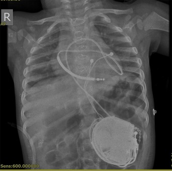
The patient had no pleural effusions or pneumothorax after the procedure (Fig 1). A few days after the procedure, the patient was discharged on propranolol therapy. During a follow-up of 8 months, implantable cardioverter-defibrillator analyses revealed stable lead impedances, appropriate capture thresholds, suitable sensing, stable pacemaker function, no malfunction, and no adverse events. One episode of ventricular fibrillation was detected, and it was converted with an implantable cardioverter-defibrillator shock.
Chest X-ray shows the position of implantable cardioverter-defibrillator epicardial leads and generator and no pneumothorax or pleural effusions.

Discussion
Children with a history of sudden cardiac death or ventricular tachycardia refractory to medical treatment may benefit from implantable cardioverter-defibrillator implantation. Reference Gradaus, Wollmann and Köbe2 Small body size has resulted in individualised and novel implantation techniques in this age group. Reference Stephenson, Batra and Knilans3
Sughimoto et al Reference Sughimoto, Tsuchida and Hayashi4 described a 6.0-kg, 2-month-old boy who underwent implantable cardioverter-defibrillator implantation, during which the coil lead was placed posterior to the left atrium and the generator was positioned in a space between the internal and external abdominal oblique muscles. Bryant et al Reference Bryant, Aboutalebi, Kim, Kertesz and Morales5 reported epicardial implantable cardioverter-defibrillator implantation via a median sternotomy in a 5-week-old infant boy weighing 4.9 kg with cardiopulmonary arrest. They placed a bipolar pace lead on the right ventricle, the shock coil in the transverse sinus, and the generator pocket below the left costal margin. Silver et al Reference Silver, Liberman and Chung6 described a 3.6-kg, 1-month-old infant who underwent implantable cardioverter-defibrillator placement, during which a median sternotomy was followed by the placement of the sensing leads on the right atrium and the right ventricle, while the shocking coil was placed posterior to the heart in the left pleural space and the generator was positioned in the abdominal space. Kriebel et al Reference Kriebel, Ruschewski and Gonzalez7 reported implantable cardioverter-defibrillator implantation by a partial sternotomy in 8 patients via the extracardiac technique. Czapla et al Reference Czapla, La Meir, Verbessern, Brugada and Wellens8 reported 3 cases with a small midline subxiphoid incision. The shock lead position was intrapericardial, and the generator was placed in a submuscular pocket in the upper abdomen.
In this study, we reported a case of epicardial implantable cardioverter-defibrillator implantation via a median sternotomy. In our experience, this technique can be used as a safe alternative for very small children. We will report the long-term results in due course for use in further such attempts.
Financial support
This manuscript received no grants and contracts or other forms of financial support.
Conflicts of interest
None.