INTRODUCTION
The Sepiolidae is one of the least known groups among the cephalopods. The members of this family are relatively small (with mantle length (ML) up to 100 mm), have lateral fins on the mantle and can be found in all oceans. Most of them are benthic, only the Heteroteuthinae are pelagic (Naef, Reference Naef1923). The genus Heteroteuthis Gray, 1849 is the most pelagic taxon in the family Sepiolidae and is adapted to an oceanic lifestyle. The big photophore, the ventral shield and the capability to eject bioluminescent bacteria characterize this genus (Naef, Reference Naef1923; Young, Reference Young1977). Heteroteuthis includes five species, three of them belonging to the subgenus Heteroteuthis Gray, 1849 and two to the subgenus Stephanoteuthis Berry, Reference Berry1909 (Nesis, Reference Nesis and Levitov1987). The type species of the genus is Heteroteuthis dispar (Rüppel, 1845), described from the Mediterranean Sea, which is distributed in the North Atlantic Ocean as well.
The other species of this genus are: Heteroteuthis weberi Joubin, Reference Joubin1902, described from the sea around Indonesia (Mers des Colonies, Indo Neerlandaises), H. hawaiiensis Berry, Reference Berry1909 from the Pacific Ocean, the sea around Hawaii and the Great Australian Bight, H. dagamensis Robson, Reference Robson1924, reported from South Africa, coast of Natal, and H. serventii Allan, Reference Allan1945, described from New South Wales and eastern Australia. Relatively little information can be found about these four members of the genus.
Although Heteroteuthis dispar is a very common cephalopod, little information about it is available. It has a two year life cycle. Spawning occurs on the seabed and the hatchlings ascend to epipelagic layers. Immature individuals travel within the water column in depths between 25 and 1000 m, whereas mature animals are distributed in the near-bottom layers (Boletzky, Reference Boletzky1978; Hoving et al., Reference Hoving, Laptikhovsky, Piatkowski and Önsoy2008).
As part of the cataloguing and revision of the Mollusca Collection in the Zoological Museum Hamburg (ZMH), several small cephalopods from the Atlantic Ocean belonging to the genus Heteroteuthis were found. Based on these animals this study provides information about the taxonomy and distribution of the species from the genus Heteroteuthis in the Atlantic Ocean and a full description for both species.
MATERIALS AND METHODS
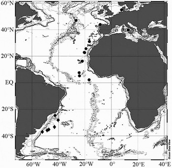
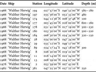
The specimens were captured during four expeditions with the research vessel ‘Walther Herwig’ (WH) to the North and South Atlantic Oceans in the years 1966, 1968, 1976 and 1982. Some of the expeditions were part of the German exploration fisheries programmes for the South-West Atlantic Ocean and the West African coast. The sampling gears were an Engel mid-water trawl (MT 1600) and an otter trawl. Sampling depths ranged between 150 and 600 m on open water with ground depths up to 4300 m. The specimens were collected at the sites depicted in Figure 1, the station list is given in Table 1. The material was fixed in 10% formalin and preserved in 70% ethanol. It is deposited in the Mollusca Collection of the Zoological Museum Hamburg (ZMH) and consists of 58 individuals, 23 from the North Atlantic Ocean, and 35 from the South Atlantic Ocean.
Map showing study area with capture locations: (•) Heteroteuthis dispar; (♦) Heteroteuthis dagamensis; (■) type locality H. dispar; (*) type locality H. dagamensis.

Station data.

The syntype material of the species Herteroteuthis dispar (male) was loaned from the Senckenberg Naturgeschichtliches Museum (SNG) and of Heteroteuthis dagamensis (female) from the Natural History Museum London (NHM). Dissections were made after Bello (Reference Bello1995), using a Wild M5A dissecting microscope. Four males and three females from the North Atlantic Ocean as well as five males and five females from the South Atlantic Ocean were dissected. Radulae, suckers (enlarged and normal ones) and tentacular clubs were taken from one female and one male of each species and examined using a LEO 1525 electronic scanning microscope (SEM) after shading with carbon evaporation using a carbon vaporizer PD770AZw from Leybolo-Heraus.
Eggs, spermatophores and club suckers were counted and measured under a Leica MZ 12 dissecting microscope. The measurements and indices follow Jereb & Roper (Reference Jereb, Roper, Vecchione, Jereb and Roper2005) and Roper & Voss (Reference Roper and Voss1983). The classification followed in this paper is the one proposed by Sweeney & Roper (Reference Sweeney and Roper1998).
The abbreviations and definitions are as follows: ML, mantle length; MW, mantle width; MWI, mantle width index; Fl R, fin insertion right side; FL R, fin length right side; FWS R, fin width right side; FP R, fin position right side; FLI, fin length index; FWI, fin width index; Fl L, fin insertion left side; FL L, fin length left side; FWS L, fin width left side; FP L, fin position right side; HW, head width; HWI, head width index; HL, head length; HLI, head length index; ED, eye diameter; EDI, eye diameter index; AF, arm formula; AL I R, arm length arm I right side; ALI I R, arm length index arm I right side; AL II R, arm length arm II right side; ALI II R, arm length index arm II right side; AL III R, arm length arm III right side; ALI III R, arm length index arm III right side; AL IV R, arm length arm IV right side; ALI IV R, arm length index arm IV right side; AL I L, arm length arm III left side; ALI I L, arm length index arm I left side; AL II L, arm length arm II left side; ALI II L, arm length index arm II left side; AL III L, arm length arm III left side; ALI III L, arm length index arm III left side; AL IV L, arm length arm IV left side; ALI IV L, arm length index arm IV left side; Keel I R, keel existing on arm I right side; Keel II R, keel existing on arm II right side; Keel III R, keel existing on arm III right side; Keel IV R, keel existing on arm IV right side; Keel I L, keel existing on arm I left side; Keel II L, keel existing on arm II left side; Keel III L, keel existing on arm III left side; Keel IV L, keel existing on arm IV left side; HcLI I R, hectocotylized arm index arm I right side; HcLI II R, hectocotylized arm index arm II right side; TL L, tentacle length left side; TL R, tentacle length right side; CIL R, club length right side; CIL L, club length left side; CILI, club length index; SC I R, sucker count arm I right side; SC II R, sucker count arm II right side; SC III R, sucker count arm III right side; SC IV R, sucker count arm IV right side; SC I L, sucker count arm I left side; SC II L, sucker count arm II left side; SC III L, sucker count arm III left side; SC IV L, sucker count arm IV left side; SC Club, club sucker count; CIRC, club sucker row count; ASD, arm sucker diameter (diameter of largest sucker on the arm); ASIn I, arm sucker index arm I; ASIn II, arm sucker index arm II; ASIe (III), arm sucker index arm III; ASIn IV, arm sucker index arm IV; Web A, web length between arm I right and arm I left side; Web B, web length between arm I and II; Web C, web length between arm II and III; Web D, web length between arm III and IV; Web E, web length between arm right IV and arm IV left; SpL, spermatophore length; FuL, funnel length; GiL, gill lamellae count (number of outer gill lamellae); SpLI, spermatophore length index; EgLI, egg length index.
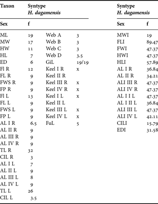
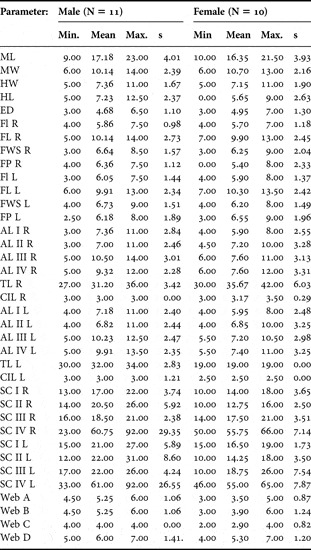
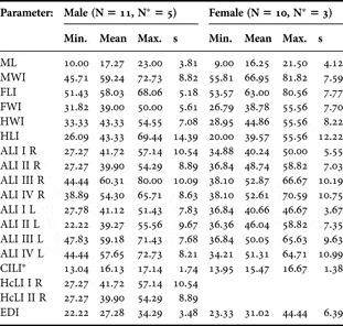
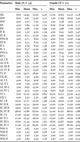
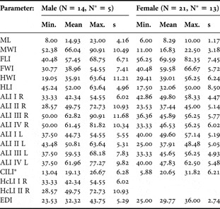
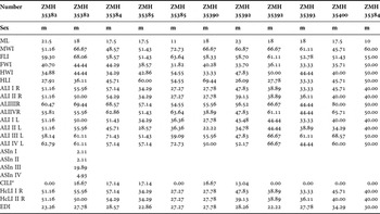
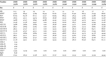
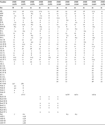
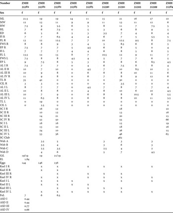
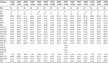
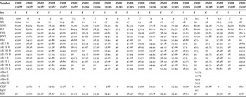
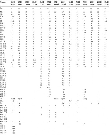
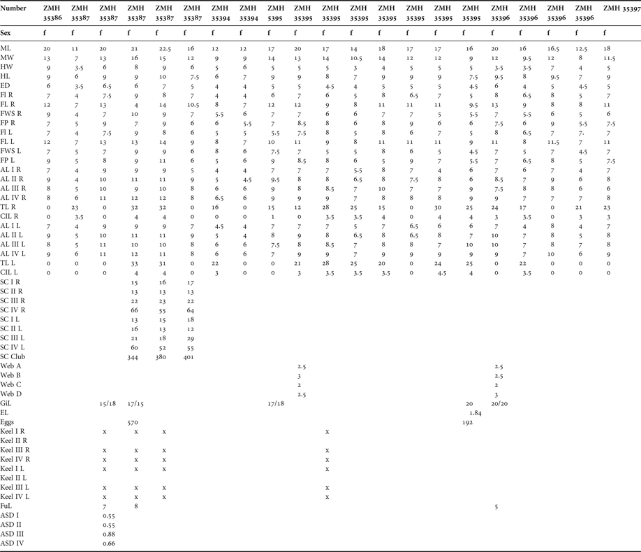
Indices and measurements for the species are given in Tables 2 to 5 and morphometrics and indices of all studied individuals are given in Appendices 1 to 10.
Measurements (in mm) of Heteroteuthis dispar.

Indices of Heteroteuthis dispar.

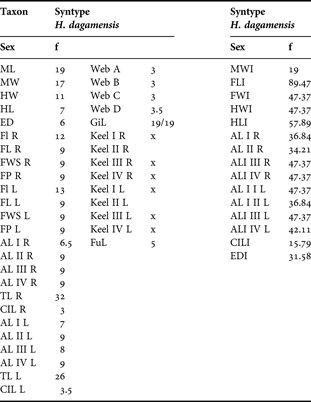
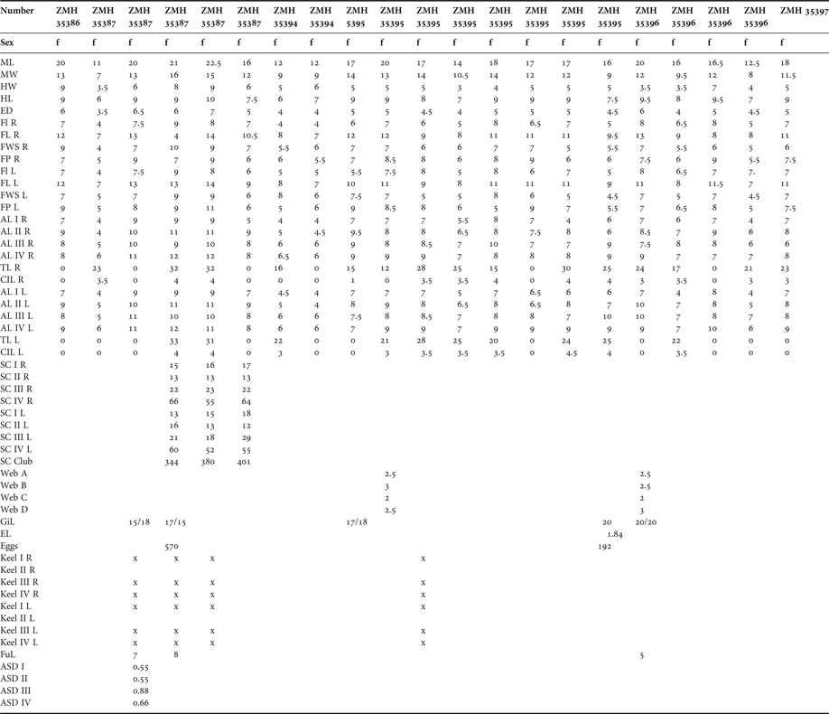
Measurements (in mm) of Heteroteuthis dagamensis.

Indices of Heteroteuthis dagamensi s.

The capture locality map was made using Ocean Data View (Schlitzer, Reference Schlitzer2002).
SYSTEMATICS
DIAGNOSIS
(Modified after Naef, Reference Naef1923): small Sepiolidae (ML < 30 mm), mantle free from head, projected forward ventrally forming a ventral shield. Large fins attached at the posterior dorsal half of the mantle. Visceral photophores fused, forming a large structure covering the ink sac. A silvery glance covers the ventral and lateral side and the periocular region. Males have greatly enlarged suckers (about five to nine times as large as normal ones) on arm pair III. The hectocotylized right arms I and II are usually connected with a muscular band. Females have bare tips on arm pairs I and II and transverse ridges on arm pair II.
TYPE SPECIES
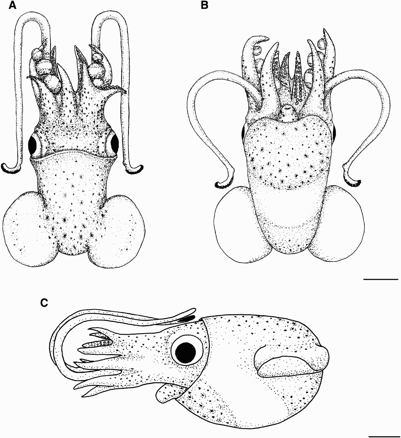
Habitus of Heteroteuthis dispar (Rüppel, Reference Rüppel1844). ZMH 35390, male: (A) dorsal view; (B) ventral view. ZMH 35391, female: (C) lateral view. Scale bar: 5 mm.

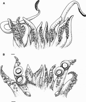
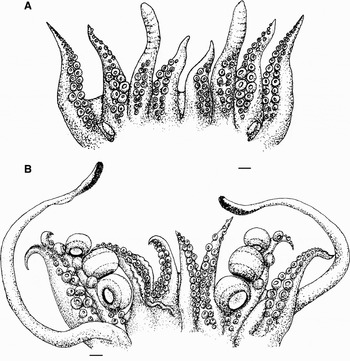
Arm crown of Heteroteuthis dispar (Rüppel, Reference Rüppel1844) ZMH 35382 (A) female and (B) male. Scale bar: 1 mm.

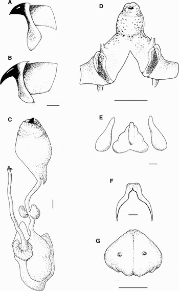
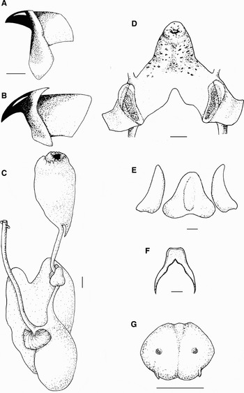
Heteroteuthis dispar (Rüppel, Reference Rüppel1844). ZMH 35392, male: (A) lower beak; (B) upper beak; (C) digestive tract. ZMH 35400, male: (D) funnel and funnel locking apparatus; (E) funnel organ; (F) nuchal cartilage. ZMH 35382, female: (G) photophore. Scale bars: A–C, E–G, 1 mm; D, 5 mm.

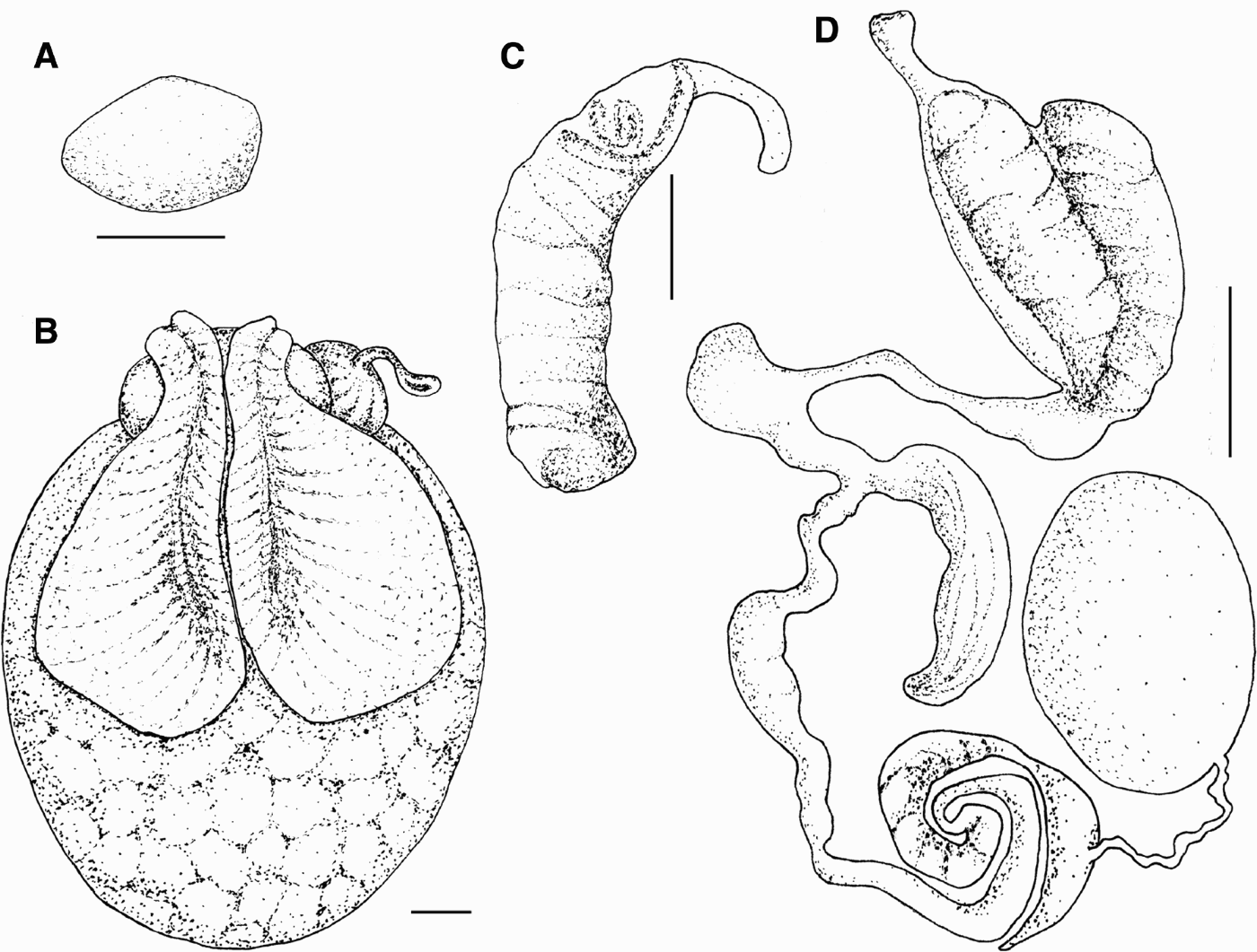
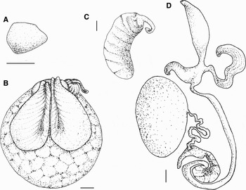
Heteroteuthis dispar (Rüppel, Reference Rüppel1844) ZMH 35382: (A) egg; (B) female genitalia; (C) spermatophore; (D) male genitalia. Scale bars: B, 1 mm; C, 5 mm; D, 3 mm.
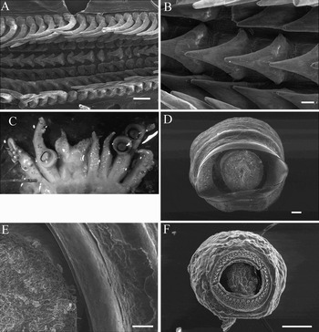
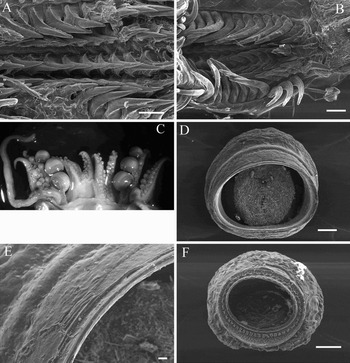
Heteroteuthis dispar (Rüppel, Reference Rüppel1844). ZMH 35382, female: (A) radula; (B) detail of radula. ZMH 35382, male: (C) arm crown. MH 35392, male: (D) enlarged arm sucker; (E) detail of enlarged sucker. ZMH 35382, female: (F) normal arm sucker. Scale bars: A, E, F, 100 µm; B, 20 µm; C, D, 200 µm.

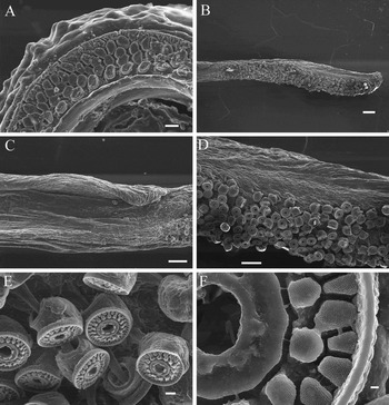
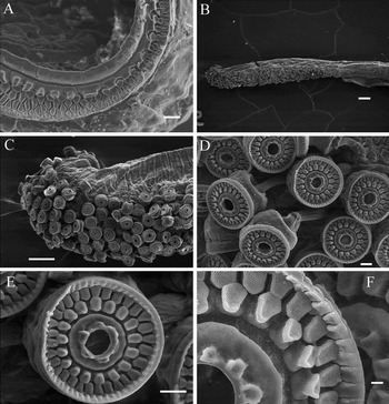
Heteroteuthis dispar (Rüppel, Reference Rüppel1844). ZMH 35392, male: (A) detail of normal sucker. ZMH 35382, male: (B) tentacle club; (C) detail of club keel; (D) detail of club; (E) club suckers. ZMH 35392, female: (F) detail of club suckers. Scale bars: A, E, 10 µm; B, 200 µm; C, D, 100 µm; F, 1 µm.

SYNONYMY
Sepiola dispar Rüppel, Reference Rüppel1844
Rossia (Heteroteuthis) dispar Gray, 1849
Rossia dispar Verany, Reference Verany1851
Heteroteuthis dispar Troschel, Reference Troschel1857
Cirroteuthis meangensis Lo Bianco, Reference Lo Bianco1903
TYPE MATERIAL
Syntype SNG 26796: male, ML 13 mm; type locality: Messina, Mediterranean Sea.
MATERIAL EXAMINED
Syntype SNG 26796: ♂, ML 13 mm; additional material: ZMH 35382, WH Station 174: 2 ♂, ML 21.5 mm, ML 18 mm, 3 ♀, ML 21.5 mm, ML 19 mm, ML 19 mm; ZMH 35384, WH Station 178: 2 ♂, ML 17.5 mm, ML 9 mm, 1 ♀, ML 10 mm; ZMH 35385, WH Station 180: 2 ♂, ML 17.5 mm, ML 11 mm, 2 ♀, ML 14 mm, ML 11 mm; ZMH 35390, WH Station 8: 1 ♂, ML 18 mm, 1 ♀, ML 15 mm; ZMH 35391, WH Station 11: 1 ♀, ML 21 mm; ZMH 35392, WH Station 13: 2 ♂, ML 23 mm, ML 18 mm; ZMH 35393, WH Station 13: 1 ♂, ML 18 mm, 1 ♀, ML 16 mm; ZMH 35399, WH Station 2: 1 ♀, ML 17 mm; ZMH 35400, WH Station 381: 1 ♂, ML 17.5 mm; (ZMH 35388, WH Station 184 and ZMH 35383, WH Station 177: two small individuals, dried out and not used for the examinations).
DIAGNOSIS
Males have at least two greatly enlarged suckers (about nine times as large as normal ones) on the distal half of arm pair III.
DESCRIPTION
Mantle (Figure 2): animals small, with maximum mantle length of 23 mm for males and 21.5 mm for females. Mantle firm and muscular, oval, longer than wide, in females rounded ventrally (especially in mature females, Figure 2C), in males sometimes pointed V-shaped. Mantle wide (MWI 43–73 in males, 56–88 in females). Mantle sometimes covering the distal part of the eyes, sometimes leaving a short free neck, ventrally extended anteriorly but not covering the funnel. Fins: oval, placed dorsally on the mantle, about half as long as mantle (FLI 58 in males, 63 in females), reaching the dorsal midpoint of the mantle with their frontal end and slightly overlapping the distal part of the mantle.
Head: narrower than mantle, length about 40% of ML in both sexes. Eyes large, round, located mediolaterally on the head (EDI 27 in males, 31 in females). Funnel (Figures 2B & 4D): tubular, always free, at least at the distal part. About 1/3 of ML. Funnel organ (Figure 4E): consisting of three parts. One large central triangle with basal rounded apices and one lateral clubbed pad on each side. Funnel locking apparatus (Figure 4D): well defined, large, oblong oval, deeply curved distally and with pronounced edges medially, proximally shallow and flat. Nuchal cartilage (Figure 4F) saddle-shaped, anterior part broad, posterior part divided and pointed.
Arms (Figure 3): male (Figure 3B): arm formula: III: IV: I: II. First arm pair generally slightly longer than second arm pair, which is the shortest. Third arm pair longest (ALI 44–80), fourth pair second longest (ALI 39–66). First and second right arms hectocotylized (HcLI I and HcLI II about 40), fused and connected with a muscular band (Figures 3B & 6C). Second right arm with thick glandular tissue at base. Suckers pedunculated, arranged in two rows, attached ventrolaterally. Suckers on right arms I and II only appear on distal third to the tips. Suckers on left arms I and II are normal-sized (ASD 0.38), reaching from basis to tip. Arm III (Figure 6C) has two greatly enlarged suckers (ASD 3.58) on its second half, relatively close to tips, eventually followed by some smaller suckers. Enlarged suckers with a smooth chitinous sucker ring, infundibulum smooth (Figure 6D, E). Basis mostly with nine normal suckers. Suckers of fourth arm slightly larger in central section (ASD 0.88), followed by normal suckers reaching tips of the arm and there becoming very small. Suckers at the end of arm IV in several rows of three to six. Relatively inconspicuous keel on arms I, II and IV.
Normal suckers with a chitinous ring, infundibulum with three to six rows of blunt polygonal projections (Figure 7A). Web formula: D: B: A: C. Web A nearly as long as B, reaching slightly more than half of arm length. Web C shortest and web D longest, reaching about 2/3 of arm length. Web E not developed.
Female (Figure 3A): arm formula: IV = III: II: I. First arm pair shortest (ALI 35–50), followed by second arm pair (ALI 37–59). Third arm pair longer (ALI 36–66), fourth arm pair almost equal (ALI 34–65). Generally arms on right side a bit longer. Suckers on arm pair I normal-sized (ASD 0.4), not reaching tip. Arm pair II with ridges on anterior half, suckers covering only first third of arm (Figure 3A). On arm pair III suckers reaching tip and are larger at the midsection and basis (ASD 0.7). On fourth arm pair suckers larger in the midsection (ASD 0.6), followed by normal suckers and very small suckers close to tips in three to six rows. Keels inconspicuous and not spread regularly, but mostly appearing on arm pair I and IV. Normal suckers appearance like in males (Figure 6F). Web-like in males.
Tentacles in both sexes (Figure 3A): long, at least twice as long as body length, as wide as arms, width decreasing towards the end. Small keels observable at clubs basis (Figure 7C). Clubs (Figure 7B) moderately thickened and covered by very small suckers in about 16 rows crosswise and 55 rows lengthwise with more than 462 suckers per club (Figure 7D). Club suckers of tentacles attached ventrolaterally, rim composed of two rows of polygonal pads, central ring thicker at inner edge with up to seven blunt projections (Figure 7E, F).
Gills with 17 to 21 lamellae on outer demibranch, in males mostly 17 to 19 and in females 19.
Digestive tract (Figure 4C): buccal mass oval, compact, large with a small flag at its caudal end. Lower beak (Figure 4A): hood strong and short, wings slender and almost translucent. Lateral walls almost translucent. Upper beak (Figure 4B): with long curved pointed rostrum, hood slender. Lateral walls thin, almost translucent. Radula (Figure 6A, B): rachidian pointed, margins serrated, smoothly rounded. First lateral with one proximal point, margins serrated, basis wide. Second lateral large, with a keel expanding towards basis, margins smooth without teeth. Third lateral very large, twice as long as rachidian, strongly curved without sharp margins and blunt keels on frontal and posterior edge. No lateral plates.
Oesophagus slender, tubular. Posterior salivary glands bean-shaped. Stomach sac-like with no special features. Caecum kidney-shaped, covered with striae. Digestive gland large, with two lobes enclosing stomach and genitalia, frontal margin covered by large ink sac. Intestine slender, about 1/3 of total length of digestive tract. Anal flaps and one anal pod present. Ink sac cushion-shaped, covering photophore dorsally. Photophore (Figure 4G) rounded to lobe-shaped in different expressions, with two papillae and two openings.
Male reproductive system (Figure 5D): testis located on posterior dorsal mantle cavity, covering spermatophoric complex. Testis' shape from ovoid to elongated. Vas deferens coiled and short. Spermatophoric gland horn-like. Seminal vesicle coiled. Accessory gland lobe-like, appendage of accessory gland bubble-like. Needham sac spindle-shaped to oval, depending on the amount of spermatophores inside. Penis short and without special features. Spermatophores (Figure 5C) large (SpLI 27–54), compact, about four times as long as wide. Sperm cord with few wide coils (9–11), light brown coloured. Cement body small. Ejaculatory apparatus short, about one-fifth of spermatophore length and translucent.
Female reproductive system (Figure 5B): ovary ovoid, placed in the posterior part of mantle cavity, in mature females large, displacing other organs. Nidamental glands paired, tear-shaped, located ventrally, covering two-thirds of ovary. Accessory nidamental glands fused, oval, located above the distal portion of the nidamental glands. Oviductal gland resembling a small pumpkin. Only one oviduct on the left side, opening short and doughnut-shaped. Mature oocytes (Figure 5A) ovoid, largest 1.84 mm (EgLI 8.6). Ovary with about 156 mature and many immature oocytes. Seminal receptacle large, located on the posterior end of visceral mass, expanding half of ovary and part of nidamental glands when filled. Spermatangium consists of one long coiled tube with one pointed and one rounded end. Up to two spermatangia were found in one female.
Preserved animals of reddish-brown colour at mantle and at basis of arms. Dorsally between eyes dark-brown coloured. Several chromatophores appear in these coloured parts. Ventral region of body and periocular region have a silvery metallic glance.
DISTRIBUTION
North Atlantic Ocean and Mediterranean Sea (Reid, Reference Reid, Jereb, Jereb and Roper2005).
REMARKS
In living animals, tips of the arms and fins are translucent except for some brownish chromatophores placed distally on the fins (Bello & Biagi, Reference Bello and Biagi1995). Animals are able to illuminate their ventral side in a blue-green glow. The maximum wave-length of the photophore lies between 485 and 490 nm (Nixon et al., Reference Nixon and Young2003).
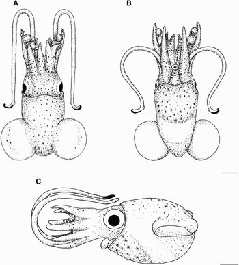
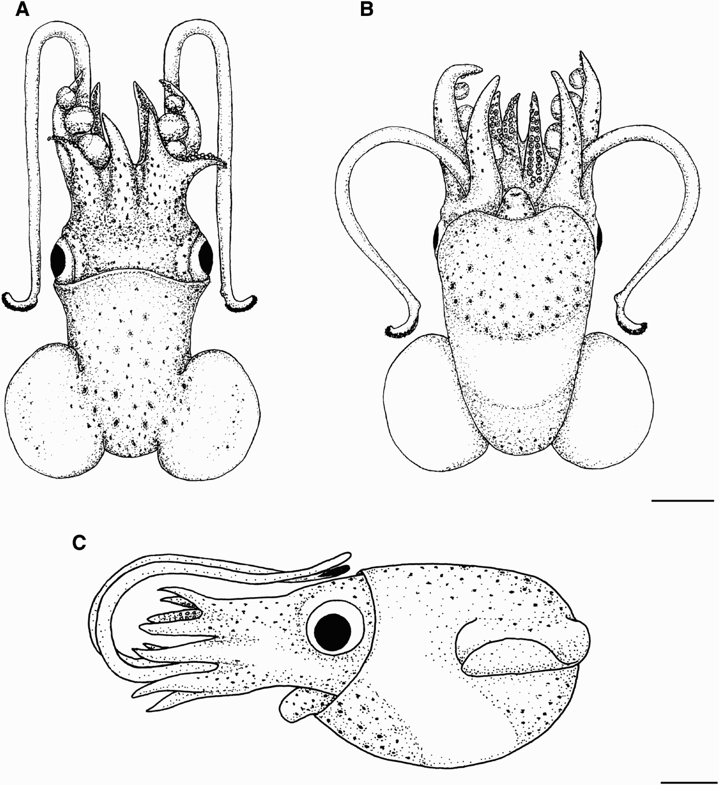
Habitus of Heteroteuthis dagamensis (Robson, Reference Robson1924). ZMH 35395, male: (A) dorsal view; (B) ventral view. ZMH 35396, female: (C) lateral view. Scale bar: 5 mm.
Arm crown of Heteroteuthis dagamensis (Robson, Reference Robson1924). (A) ZMH 35396 female and (B) ZMH 35397 male. Scale bar: 1 mm.

Heteroteuthis dagamensis (Robson, Reference Robson1924). ZMH 35397, male: (A) lower beak; (B) upper beak; (C) digestive tract. ZMH 35387, male: (D) funnel and funnel locking apparatus; (E) funnel organ; (F) nuchal cartilage. ZMH 35387, female: (G) photophore. Scale bars: A–C, E–G, 1 mm; D, 2 mm.

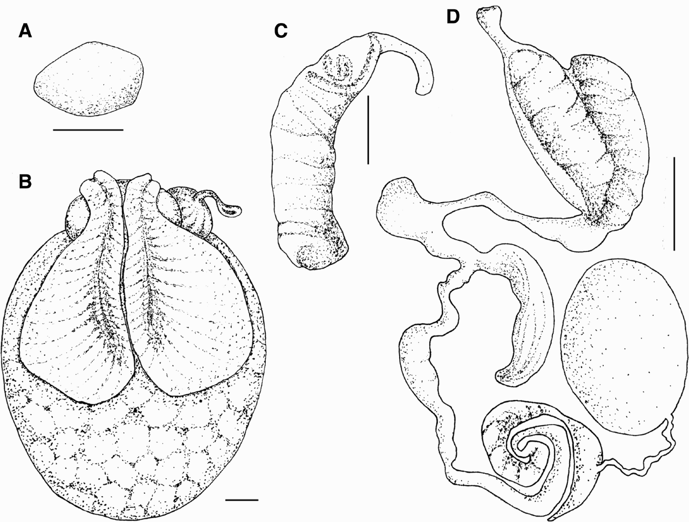
Heteroteuthis dagamensis (Robson, Reference Robson1924). ZMH 35387, female: (A) egg; (B) female genitalia. ZMH 35397, male: (C) spermatophore; (D) male genitalia. Scale bar: 1 mm.

Heteroteuthis dagamensis (Robson, Reference Robson1924). ZMH 35387, female: (A) radula. ZMH 35397, male: (B) detail of radula; (C) arm crown. ZMH 35395, male: (D) enlarged arm sucker; (E) detail of enlarged sucker. ZMH 35387, female: (F) normal arm sucker. Scale bars: A, B, F, 100 µm; C, D, 200 µm; E, 20 µm.

Heteroteuthis dagamensis (Robson, Reference Robson1924). ZMH 35387, female: (A) detail of normal arm sucker. ZMH 35395, male: (B) tentacle club; (C) detail of club tip; (D) club suckers; (E) sucker club; (F) detail of club sucker. Scale bars: A, B, 200 µm; C, 100 µm; D, E, 10 µm; F, 2 µm.

SYNONYMY
Stephanoteuthis hawaiiensis dagamensis Robson, Reference Robson1924.
TYPE MATERIAL
Syntype BMNH 19249949-50: one female, ML 19 mm, type locality: Natal coast, South Africa.
MATERIAL EXAMINED
Syntype BMNH 19249949-50: 1 ♀, ML 19 mm; additional material: ZMH 35386, WH Station 194: 1 ♀, ML 20 mm; ZMH 35387, WH Station 197: 2 ♂, ML 15 mm, ML 15 mm, 5 ♀, ML 11 mm, ML 20 mm, ML 21 mm, ML 22.5 mm, ML 16 mm; ZMH 35394, WH Station 28: 2 ♂, ML 18 mm, ML 11 mm, 2 ♀, ML 12 mm, ML 12 mm; ZMH 35395, WH Station 89: 2 ♂, ML 21 mm, Ml 16 mm, 8 ♀, ML 17 mm, ML 20 mm, ML 17 mm, ML 14 mm, ML 18 mm, ML 17 mm, ML 17 mm, ML 16 mm; ZMH 35396, WH Station 89: 4 ♂, ML 13 mm, ML 17 mm, ML 17 mm, ML 10 mm, 4 ♀, ML 20 mm, ML 16 mm, ML 16.5 mm, ML 12.5 mm; ZMH 35397, WH Station 29: 4 ♂, ML 23 mm, ML 13 mm, ML 12 mm, ML 8 mm, 1 ♀, ML 18 mm.
DIAGNOSIS
Males with three greatly enlarged suckers (about five times as large as normal ones) on arm pair III, always on inner side, alternating with three enlarged suckers (at least one and a half times as large as normal ones) at outer side of arm pair III.
DESCRIPTION
Mantle (Figure 8): animals small, with maximal mantle length 23 mm in males and 22.5 mm in females. Mantle firm and muscular, oval, longer than wide, in females rounded ventrally (especially in mature females), in males sometimes V-shaped, (MWI 52–90 in males, 56–82 in females), sometimes covering eyes distally, sometimes leaving a short free neck, ventrally extended anteriorly but not covering funnel. Fins: oval, placed dorsally on the mantle, about half as long as mantle (FLI 57 in males, 59 in females) reaching dorsal midpoint of mantle with frontal ends, slightly overlapping distal part of mantle.
Head: narrower than mantle, length about 50% of ML in both sexes. Eyes large, round (EDI 32 in males, 30 in females), located mediolaterally on head. Funnel (Figures 8B & 10D): tubular always free, at least at distal part. About 1/3 as long as ML. Funnel organ (Figure 10E): consisting of three parts. One large central triangle with basally rounded apices and one lateral clubbed pad on each side. Funnel locking apparatus (Figure 10D): well defined, large, oblong oval, distally deeply curved, medially with pronounced edges, especially at outer margin, proximally shallow and flat. Nuchal cartilage (Figure 10F) saddle-shaped, posterior part divided and pointed.
Arms (Figure 9): male (Figure 9B): arm formula: III: IV: I: II. First arm pair generally slightly longer than second arm pair, which is shortest. Third arm pair longest (ALI 37–90), fourth one second longest (ALI 37–80). First and second right arms hectocotylized (HcLI I 42 and HcLI II about 49) fused and connected with a muscular band (Figures 9B & 12C), second right arm with thick gland tissue at basis. Suckers pedunculated in two rows, attached ventrolaterally. Suckers on right arms I and II only appear on distal third to tips. Suckers on left arms I and II have a normal size (ASD 0.44) and reach from basis to tip. Arm pair III (Figures 9B & 12C) has three greatly enlarged suckers (ASD 2.36) on inner side, alternating with three medium-sized suckers (about one and a half times as big as normal ones) at outer side. Basis with about nine normal suckers. Fourth arm pair, suckers slightly larger in midsection (ASD 0.66), followed by normal suckers reaching tips of arm and becoming very small, standing in several rows of three to six. Relatively inconspicuous keel mostly on arms I, III and IV. Enlarged suckers with a smooth chitinous ring with no lateral pads (Figure 12D, E). Normal suckers with a chitinous ring, infundibulum with three to five rows of blunt polygonal projections. Web formula: A = D: B: C. Web C shortest, web B second longest. Web A as long as web D. Web E not developed.
Female (Figure 9A): arm formula: IV: III: II: I. First arm pair shortest (ALI 24–48), followed by second arm pair (ALI 33–56). Generally third arm pair (ALI 33–56) slightly longer than fourth arm pair (ALI 44–60). Arms on right side in mean a bit longer. Arm pair II with ridges on anterior half (Figure 9A). Suckers on arm pair I of normal size (ASD 0.5) not reaching tip. On arm pair II suckers only cover first third of arm. On arm pair III reaching tip and are larger at middle and basis (ASD 0.8). At fourth arm pair, suckers larger in mid-section (ASD 0.6), becoming smaller to the tips, standing in three to six rows. Relatively inconspicuous keel on arms I, III and IV. Normal sucker appearance as in males (Figures 12F & 13A). Web-like in males.
Tentacles in both sexes: long, at least twice as long as body length, as wide as arms, width decreasing towards the ends, with small keels at club basis (Figure 13B). Clubs (Figure 13B, C) moderately thickened and covered by very small suckers in about 16 rows crosswise and 55 rows lengthwise, more than 401 suckers per club (Figure 13B). Club suckers of tentacles attached ventrolaterally, rim composed of two rows of polygonal pads, central ring thicker at inner edge with up to six blunt projections (Figure 13D–F).
Gills lamellae: modal for males is 19, for females 20.
Digestive tract (Figure 10C): buccal mass oval, compact, large with a small flag at its caudal end. Lower beak (Figure 10A): hood strong and short, wings slender and almost translucent. Lateral walls almost translucent. Upper beak (Figure 10B) with long, curved pointed rostrum, hood slender. Lateral walls thin, almost translucent. Radula (Figure 12A, B): rachidian sharp pointed, margins serrated and rounded with sharp step at basis. First lateral with one proximal point, margins serrated, basis wide. Second lateral large, with a keel expanding towards basis, margins smooth without teeth. Third lateral very large, twice as long as rachidian, strongly curved without sharp margins and blunt keels on frontal and posterior edges. No lateral plates. Oesophagus slender, tubular. Posterior salivary glands bean-shaped. Stomach sac-like without special features. Caecum sac-like and kidney-shaped, covered with striae. Digestive gland large with two lobes enclosing stomach and genitalia, frontal margin covered by large ink sac. Intestine slender, about 1/3 of total length of digestive tract. Anal flaps and one anal pod present. Ink sac cushion-shaped covering photophore dorsally. Photophore (Figure 10G) rounded to lobe-shaped in different expressions, with two papillae and two openings.
Male reproductive system (Figure 11D): testis located on the posterior dorsal mantle cavity, covering spermatophoric complex. Testis' shape from ovoid to elongated. Vas deferens coiled and mid-long. Spermatophoric gland horn-like. Seminal vesicle coiled. Accessory gland lobe-like, appendage of accessory gland bubble-like. Needham sac spindle-shaped to oval, depending on the amount of spermatophores inside. Penis short without special features. Spermatophores (Figure 10C) large (SpLI 39–45), compact, about four times as long as wide. Sperm cord with few wide coils (7–9), light brown coloured. Cement body small. Ejaculatory apparatus short, about one-fifth of spermathophore length and translucent.
Female reproductive system (Figure 10B): ovary ovoid, placed in the posterior part of mantle cavity, in mature females large, displacing other organs. Nidamental glands paired, tear-shaped, located ventrally and covering 2/3 of ovary. Accessory nidamental glands fused, oval, located above distal portion of nidamental glands. Oviductal gland resembling a small pumpkin. Only one oviduct on left side, opening short and doughnut-shaped. Mature oocytes (Figure 11A) round, largest 1.84 mm (EgLI 9.2). Between 192 and 570 mature and many immature oocytes in ovary. Seminal receptacle large, located on posterior end of visceral mass, expanding half of the ovary and part of the nidamental glands when filled.
Spermatangium consists of one long coiled tube with one pointed and one rounded end.
Coloration same as in H. dispar.
DISTRIBUTION
South Atlantic Ocean and South African coast.
DISCUSSION
The examination of the material has demonstrated that the separation of the genus Heteroteuthis into the subgenera Heteroteuthis and Stephanoteuthis cannot be supported. Until now Heteroteuthis dispar has been placed in the subgenus Heteroteuthis and H. dagamensis in the subgenus Stephanoteuthis. According to Nesis (Reference Nesis and Levitov1987) the following attributes separate both subgenera: Stephanoteuthis: females' second arm pair orally flattened, transversally striated and bent outwards, males having a deep web A at about 90% of arm length, second and third arms being equal in length with bare tips and hectocotylized arms (first and second right arm) having no muscular band between them. However, this does not apply to both of the examined populations. The characters that should differ have proven to be within the range of variation of both species. Males of Heteroteuthis dagamensis have a muscular band between both hectocotylized arms. The variation of this character is high, ranging from a clear band to a nearly invisible connection in both species. The intraspecific variability is generally high in both species and sexes, and most characters lie within the range of variation of the species. The females of both species are morphologically identical and cannot be separated at all. Significant differences could not be found by comparing the syntype of H. dagamensis with the examined female material.
The valid description of H. dagamensis from Robson (Reference Robson1924) is very poor, consisting of only ten lines and no figures. The description was made with only one individual, a female, and given the fact that females are not useful to separate species of Heteroteuthis, is very problematic, and therefore Heteroteuthis dagamensis is considered a doubtful species.
Young et al. (Reference Young, Roper, Mangold and 2008) examined Heteroteuthis from South Africa including males and identified them as H. dagamensis, because they were found in the waters close to the Locus Typicus of H. dagamensis. The comparative examination with male data by Young et al. (2008) and the animals examined here showed the same significant character: three greatly enlarged suckers on the inner side of arm pair III with the alternating moderately enlarged suckers on the outer side of these arms. Based on these data it was possible to clearly determine the examined specimens from the South Atlantic Ocean as H. dagamensis, and not as a new species, and to confirm the existence of this species in the area. Although a partial description of H. dagamensis is given in the Tree of Life web site (Vecchione et al., Reference Vecchione, Young and Roper2007), the synonymization of H. serventii with H. dagamensis is doubtful. Also changing the Locus Typicus of H. dagamensis and replacing it with the Locus Typicus of H. serventii is not correct and changes of nomenclature status of species within the internet are not accepted by the ICZN, chapter 3 Article 8 and chapter 6 Article 23.3. In order to clarify the taxonomic position of Heteroteuthis dagamensis, a full description of this species is given.
The proposal of Nesis (Reference Nesis and Levitov1987) that Heteroteuthis atlantis Voss, Reference Voss1955 is a synonym of H. dispar can be confirmed. The morphology of the photophore and other characters are very variable, giving no support to sustain the species H. atlantis. This variation can be seen for example in the body shape and the photophore, comparing the published figures from Voss (Reference Voss1955) with Figures 2 & 7 and Figures 4G & 10G from the present work. Generally the variation of most characters is very high in the genus Heteroteuthis.
The assumption of Hoving (2008) that Heteroteuthis is more common near seamounts and submarine ridges and seldom in open water cannot be supported, because almost all studied specimens came from the open sea with ground depths up to 4300 m.
The distribution of Heteroteuthis dispar can be enlarged to the north: for the first time a specimen was caught in the Northern Sea, close to the Elbe Estuary.
This is the first confirmed report for H. dagamensis for the South-Western Atlantic Ocean near the coast of Brazil. Concerning the distribution of Heteroteuthis the results show that the Atlantic Ocean can clearly be separated into the northern hemisphere, where H. dispar occurs and the southern hemisphere, where H. dagamensis occurs. There seems to be a border of distribution in the tropical Atlantic Ocean between both species.
The distribution of H. dagamensis comprises the South Atlantic Ocean and can be related to the South Atlantic Gyre current that describes a circle covering the whole South Atlantic Ocean, reaching from the southern tip of South Africa to the north of Mar de la Plata of South America. Heteroteuthis dispar is the most abundant cephalopod species in the Ligurian Sea (Relini, Reference Relini1995) and the second most abundant cephalopod species in the Aegean Sea (Hoving et al., Reference Hoving, Laptikhovsky, Piatkowski and Önsoy2008). Heteroteuthis dispar is a key species in the Mediterranean Sea (Bello & Pipitone, Reference Bello and Pipitone2002; Hoving et al., Reference Hoving, Laptikhovsky, Piatkowski and Önsoy2008). Although Heteroteuthis dispar only has a very minor commercial importance (Belcari & Sartor, Reference Belcari and Sartor1993) it is commercially significant indirectly as it is an important prey for many animals, like Thunnus alalunga and Aristaemorpha foliacea (Bello, Reference Bello1998; Bello & Pipitone, Reference Bello and Pipitone2002), which are the targets of commercial fisheries. In this respect it could be economically interesting to know more about the distribution, abundance, behaviour and taxonomy of Heteroteuthis dispar and the other species of the genus. Knowledge of distribution and abundance of Heteroteuthis dispar in the Mediterranean Sea is improving, but there is still only little information about H. dispar in the Atlantic Ocean or about the other four species of this genus. A revision of the genus is needed to clarify the taxonomic status of H. serventii because the descriptions of males are indispensable to distinguish the species. The designation of neotypes based on males for species described only by females should be considered.
ACKNOWLEDGEMENTS
The authors are grateful to Angelika Brandt for her support during the work, to Bernhard Hausdorf and Ramona Timon for providing the material, to Uwe Piatkowski for help with references, to Renate Walter for pictures with the SEM, to Henk-Jan Hoving for information about reproduction of Heteroteuthis, to Ute Mühlenhardt-Siegel for help with the map and to Cornelia Warneke-Cremer, Marion Stössel, Gerd Edel, Walter Foltin, Simon Weigmann and Caryl Rotermund for comments on this study.
Indices of Heterotetuhis dispar male (N = 11, N* = 5).

Indices of Heteroteuthis dispar female (N = 9, N* = 10, N** = 3).

Morphometric measurements of Heteroteuthis dispar male (in mm).

Morphometric measurements of Heteroteuthis dispar female (in mm).

Indices of Heteroteuthis dagamensis male (N = 14, N* = 5).

Indices of Heteroteuthis dagamensis female (N = 21, N* = 13).

Morphometric measurements of Heteroteuthis dagamensis male (in mm).

Morphometric measurements of Heteroteuthis dagamensis female (in mm).
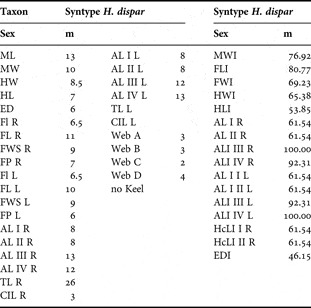
Morphometric measurements (in mm) and indices of Heteroteuthis dispar SNG 26796 male.

Morphometric measurements (in mm) and indices of Heteroteuthis dagamensis BMNH 19249949-50 female.