Introduction
Bacidia rosella (Pers.) De Not. is the type species of the genus Bacidia and has been reported from Europe, northernmost Africa, Macaronesia, Asia and North America during the last century (Almborn Reference Almborn1948; Awasthi & Mathur Reference Awasthi and Mathur1987; Hafellner Reference Hafellner1995; Egea Reference Egea1996; Seaward Reference Seaward1996; Yıldız & John Reference Yıldız and John2002; Kovaleva Reference Kovaleva2004). In my treatment of the North American corticolous species of Bacidia and Bacidina (Ekman Reference Ekman1996), I recognized a few specimens from the Great Lakes area of the USA and Canada as B. rosella. These specimens, however, were found to deviate in some important respects from European B. rosella, but the few and small specimens from North America did not at that time permit any further taxonomic conclusions. In 2002, I was presented with a few well-developed specimens from central Sweden that agreed perfectly with the North American ones. This made me realize that these specimens were not accidental deviants of B. rosella, which is found in southern Sweden (Almborn Reference Almborn1948; Arup et al. Reference Arup, Ekman, Kärnefelt and Mattsson1997), but instead belong to a separate species. Subsequently, I came across specimens from the Komi Republic of Russia, on the west side of the Ural Mountains. A search for a described name for this species proved unsuccessful, and it is consequently described here as new.
Materials and Methods
Eight specimens of Bacidia rosellizans from Sweden and five from Russia were available for study (deposited in hb. Hermansson, hb. Jonsson & Nordin, LD and UPS), eight of which were large enough to allow measurements of most or all morphological characters under study. These measurements were pooled with the information from three out of the four specimens of B. rosellizans available to Ekman (Reference Ekman1996; as B. rosella). Morphological information concerning B. rosella was taken either from Llop et al. (Reference Llop, Ekman and Hladun2007) or from measurements made on Swedish specimens in UPS. A syntype of Lecidea alabastrina Ach., a name claimed to be a synonym of B. rosella, was made available to me by herbarium H.
Morphological and chemical methods follow those of Ekman (Reference Ekman1996). Measurements are recorded as ‘(minimum –) minimum average of n observations in a specimens – average – maximum average of n observations in a specimen (– maximum) (SD = standard deviation of the sample, N = number of specimens studied, n = number of observations per specimen).’ Exceptions from this rule are conidial sizes (given as ‘minimum – maximum’) and sizes of excipular cells (difficult to measure but with little variation; given simply as lumen width or as ‘maximum length × maximum width’).
The following abbreviations are used: ‘K’ for a 10 % aqueous solution of potassium hydroxide and ‘I’ for a 0·3 % plus 0·6 % aqueous solution of I2 and KI, respectively.
Characters that are not mentioned in the species description fit the general generic description provided by Ekman (Reference Ekman1996: 60–61).
The Species
Bacidia rosellizans S. Ekman sp. nov
MycoBank no.: MB513356
Bacidiae rosellae similis sed thallus tenuior pallidior, apothecia minora margine tenuiore, ascosporae breviores angustiores septis paucioribus, conidia longiora septata.
Typus: Russia, Komi Republic, Troitsko-Pechorskii, Pechoro-Ilich Zapovednik, Hret Shuka Eliz, western slope of the mountain W of Ichet-Parus-Yol’, 140 km ENE Troitsko-Pechorsky. 62°52′N 58°50′E, alt 300 m, corticolous on Populus in wet aspen stand of Vaccinium-grass type in mixed forest. 15 July 2006, J. Hermansson 15110 & T. N. Pystina (UPS—holotypus).
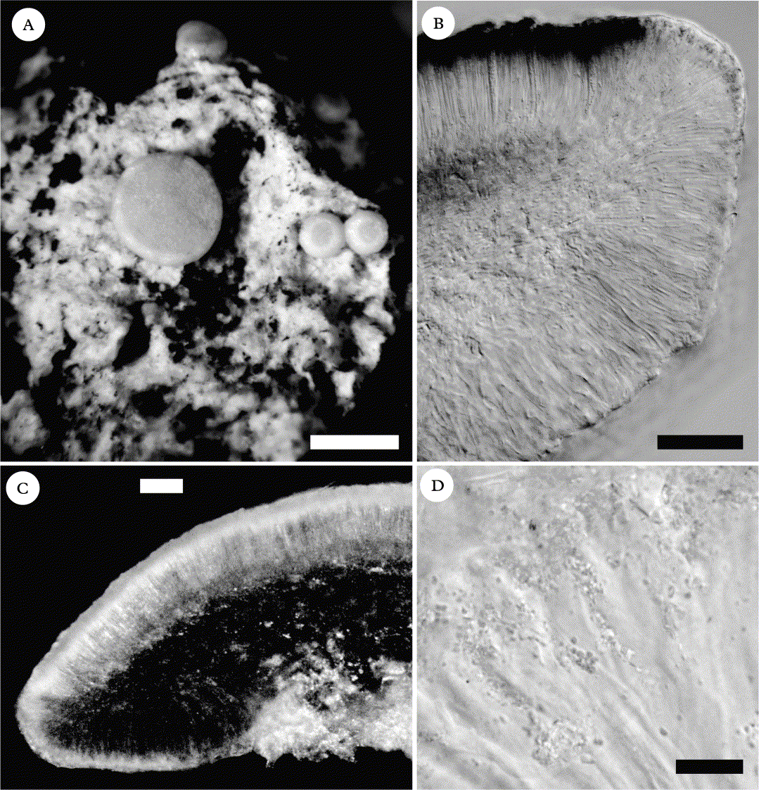
(Fig. 1A–D)
The morphology of Bacidia rosellizans. A, thallus with apothecia (holotype, Hermansson 15110 & Pystina); B, section through apothecium in differential interference contrast (Jonsson 3689), showing most of the proper exciple and parts of the hypothecium and hymenium (the dark layer in the upper part of the hymenium is caused by a thick layer of crystals blocking the light, not by a pigment); C, section through apothecium between two polarizing filters (Jonsson 3689), displaying the distribution of crystals (in white); D, crystals in upper part of hymenium in an ordinary bright field (Hermansson 3461b). Scales: A = 0·5 mm; B & C = 50 μm; D = 10 μm.

Thallus crustose, indeterminate, thin, almost white to light grey, ranging from continuous and of thin warted areoles, slightly or not at all cracked, to discontinuous and of discrete, wart-like areoles. Prothallus lacking or present between discrete areoles, whitish to light grey.
Apothecia biatorine, (0·3–)0·4–0·6–0·7(–1·1) mm diam. (SD = 0·2, N = 9, n = 10), at first flat, remaining so or becoming more or less convex with age. Disc pale pink, pale yellowish pink, or pale brownish pink. Margin usually slightly paler than the disc, distinct, slightly raised above the disc in young apothecia, soon level with the disc, persistent or occasionally becoming excluded in convex apothecia, (33–)45–53–63(–82) μm thick (SD= 13, N= 6, n= 10) in flat apothecia. Proper exciple with minute colourless crystals (less than 1 μm long) that form a dense opaque layer along the upper part of the rim but often decrease gradually in frequency in the lower part, dissolving in K; interior excipular hyphal cells dichotomously branched, with long and narrow lumina (c. 1·5 μm wide) and very thick and gelatinized walls; terminal excipular hyphae forming an irregular single-celled layer of cells with enlarged, globose or ellipsoid lumina, up to 9 × 6 μm. Hypothecium of dense textura intricata, colourless or very pale yellowish, K+ intensifying when pigmented (probably corresponding to Rubella Orange in the pigment nomenclature of Ekman Reference Ekman1996). Hymenium (58–)62–71–85(–87) μm thick (SD = 8, N = 10, n = 5), colourless, inspersed with crystals between the paraphyses apices in the upper 10–15 μm; crystals mostly less than 1 μm long, colourless, dissolving in K. Paraphyses (1·3–)1·5–1·6–1·7(–2·0) μm wide in mid-hymenium (SD = 0·1, N = 8, n = 5), unbranched or moderately branched in the upper part, infrequently anastomosed; apices ± narrowly clavate, (1·6–)2·2–2·4–2·7(–3·2) μm wide (SD = 0·4, N = 10, n = 10), without pigment. Asci clavate; young spore mass with a pointed ocular chamber; tholus staining dark blue in I with a paler blue, narrowly conical axial body, the zone closest to the axial body concolorous with the rest of the tholus or slightly darker. Ascospores 8 per ascus, acicular, straight, curved, or sigmoid, coiled in young asci, (34–)41–49–58(–73) μm long (SD = 7, N = 10, n = 10), (1·6–)1·9–2·1–2·4(–2·6) μm wide (SD = 0·3, N = 10, n = 10), (16·5–)20·7–21·8–23·5(–30·0) times as long as wide (SD = 3·8, N = 10, n = 10), with (3–)5·6–8·2–11·5(–17) septa (SD = 2·5, N = 10, n = 10).
Pycnidia half-immersed in the thallus, colourless, 100–175 μm diam. Conidia filiform, slightly curved, distinctly 5–9-septate, 27–35 × 1·2–1·6 μm.
Chemistry. Thallus without acetone-soluble lichen substances or (usually) with atranorin in trace amounts. Apothecia with atranorin in fairly high concentrations (forming at least part of the characteristic crystals in the hymenium and proper exciple).
Etymology. The epithet rosellizans means ‘approaching rosella’, referring to the similarity with the close relative B. rosella.
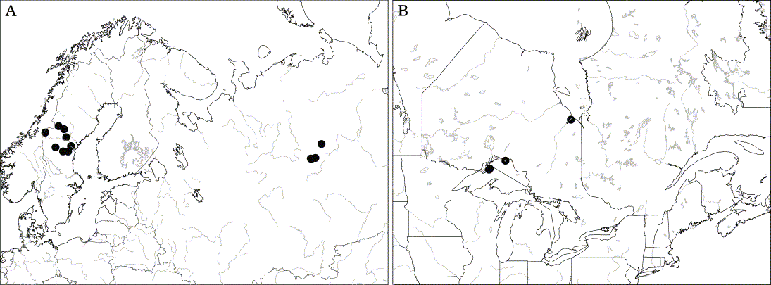
Distribution. Currently known from scattered sites in the provinces of Medelpad, Hälsingland, and Jämtland in central and northern Sweden, a few sites in the Pechora river delta in the Komi Republic on the west side of the Ural Mountains in Russia, a single site near James Bay in northern Ontario, Canada, and on the Slate Islands in Ontario and Isle Royale in Michigan, USA., both islands situated in Lake Superior (Fig. 2). The species is probably overlooked, although rare, and is likely to occur also in Norway and Finland and to be more widely distributed in Russia. Remarkably, the earliest known European specimen was collected as late as in 1993. I have come across no older European specimens of B. rosellizans in three major Scandinavian herbaria (LD, S, UPS).
Known world distribution of Bacidia rosellizans. A, Europe; B, North America.

Substratum and habitat. Bacidia rosellizans has been found on Populus tremula and Salix caprea in Sweden, on Populus tremula and once on a snag of Betula in Russia, and on Populus tremuloides and once on Acer spicatum in North America. The habitat seems to be open taiga with a humid local climate, often in the vicinity of mires or water bodies, at low altitude (near sea level to 480 m a.s.l). Forest stands with B. rosellizans usually consist of a mixture of deciduous trees (Populus tremula, Salix caprea, and Betula in Europe, Populus tremuloides, P. balsamifera and Betula papyrifera in North America) and conifers (Picea abies and Pinus sylvestris in Europe and at least Abies balsamea and Pinus banksiana in North America). Some of the Russian collections were made from pure stands of Populus, surrounded by coniferous forest, on soils that had possibly been disturbed by landslides (J. Hermansson, pers. comm.).
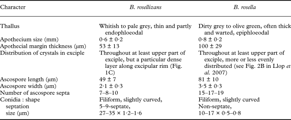
Similar species. In habit, the new species has the stature of a poorly developed B. rosella or something in between B. rosella and a pale-fruited Cliostomum griffithii (Sm.) Coppins without the dark pycnidia. However, B. rosellizans and B. rosella differ in a number of important respects, which are summarized in Table 1. The two species differ also in their distribution and habitat. In Sweden, which is currently the only country from which both species are known, their distributions do not overlap, with B. rosella on broad-leaved trees in forests or open tree-stands in the south and B. rosellizans on Populus and Salix in coniferous forests further north.
Characters separating Bacidia rosellizans from the similar B. rosella. Measurements for B. rosella referring to the ascospores were taken directly from Llop et al. (Reference Llop, Ekman and Hladun2007), whereas measurements of apothecium size and apothecial margin thickness are based on original measurements on Swedish specimens in UPS (N = 10, n = 10). Quantitative information is given as ‘average ± SD’, except number of ascospore septa (‘first quartile – median – third quartile’) and conidium size (‘minimum – maximum’).

Additional specimens examined. Sweden: Hälsingland: Ramsjö par., Ensjölokarna nature reserve, c. 10 km NW of Ramsjö, 2002, Jonsson 2265 (hb. Jonsson & Nordin). Medelpad: Stöde par., 15 km SW of Stöde, NW of Ulvsjön, 2002, Nordin 2266 (hb. Jonsson & Nordin); Liden par., between Backbodarna and Dacksjön, 4·3 km ENE of Liden church, 2007, Jonsson & Nordin 3664 (hb. Jonsson & Nordin). Jämtland: Åsane par., SW slope of Kronskogsberget, NE of Äldåmagasinet, 4·5 km SW of Åsarna, 2006 Nordin 3067 (hb. Jonsson & Nordin); Borgvattnet par., Duvknösen, 13 km SE of Borgvattnet church, 2008, Jonsson 3690 (hb. Jonsson & Nordin); Hotagen par., at Småtjärnarna S of Långvattenhöjden, 50 km NW of Strömsund, 2003, Nordin 2342 (hb. Jonsson & Nordin); Kall par., Skäckerfjällen nature reserve, at the river from Nedre Ottsjön to Anjan, just N of the road, E of the river, 2008, Arup L08094 (LD); Ström par., c. 1 km W of Kattögeltjärnen, 6·5 km W of Lövberga, 15 km NE of Strömsund, 2005, Jonsson 3689 (hb. Jonsson & Nordin).—Russia: Komi Republic: Troitsko-Pechorski, Pechoro-Ilych Zapovednik, Yaksha, 18·5 km SE of Yaksha, riverbank of Pechora, 1993, Hermansson 3593a (UPS); Troitsko-Pechorski, Pechoro-Ilych Zapovednik, Yaksha, Volosnitskaya Staziza, 15 km SE of Yaksha, 1993, Hermansson 3361b, 3367b (hb. Hermansson); Troitsko-Pechorski, Vozchennost Andjunga Parma, N-facing slope of “hill 227.3” at Pechora, 1·5 km WSW of Ust-Un'ja, 50 km ESE Yaksha, 1993, Hermansson 3461b (hb. Hermansson).—Canada: Ontario: Slate Islands, Patterson Island, in cove and stream from High Dam Lake, in conifer woods along shore, 1977, Wetmore 29024 (MIN); Cochrane District, near Moose Factory, on SE shore of Moose River opposite Moose Factory Island, 1969, Brodo 14717c (CANL; in specimen of Bacidia subincompta, under which it is filed).—USA: Michigan: Keweenaw Co., Isle Royale Nat. Park, on point N of Tallman Isl. in Tobin Harbor, 1983, Wetmore 49341 (MIN); Keweenaw Co., Isle Royale Nat. Park, on S side of Tonkin Bay, 1980, Wetmore 41319B (MIN).
Discussion
A search through the literature did not reveal any existing names for B. rosellizans. Particular attention was paid to six names referring to candidates for corticolous species of Bacidia that were described from Finland, never synonymized with a species name in current use, nor reported from anywhere else (Vainio Reference Vainio1922; Santesson et al. Reference Santesson, Moberg, Nordin, Tønsberg and Vitikainen2004), viz. Bacidia atrolivida (Vain.) Zahlbr., B. dolera (Nyl.) Blomb. & Forssell, B. invertens Vain., B. sparsa Kullh., B. tenella Kullh., and B. vegeta Vain. Detailed descriptions by Vainio (Reference Vainio1922), however, mention black or brown apothecia in all except B. vegeta, a brown, reddish, or purplish hypothecium in B. dolera, B. vegeta, and B. sparsa, or bacilliform to fusiform ascospores with blunt ends and a maximum of seven septa in B. invertens, B. atrolivida, B. vegeta and B. sparsa. Syntypes of B. sparsa and B. tenella in UPS reveal that the former is a synonym of B. subincompta (Nyl.) Arnold, whereas the latter is an unidentified lichen that cannot be referred to Bacidia or any other genus in the Ramalinaceae sensu Ekman (Reference Ekman2001). Furthermore, two names listed as synonyms of B. rosella by Zahlbruckner (Reference Zahlbruckner1927), Lichen anceps Rutstr. and Lecidea alabastrina Ach., were checked. Lichen anceps was described by Rutström (Reference Rutström1794) and soon reduced to a variety under Lecidea rosella (Pers.) Ach. (= Bacidia rosella) by Acharius (Reference Acharius1810), which may be the reason for Zahlbruckner's treatment. No one has been able to trace original material of any of the lichens described by Rutström (Reference Rutström1794). He collected corticolous lichens exclusively in the province of Halland in south-westernmost Sweden (Rutström Reference Rutström1794: 2; Krok Reference Krok1925), which is outside the range of B. rosellizans, but well within the range of B. rosella. However, I consider it unlikely that Lichen anceps is a synonym of Bacidia rosella, as mature apothecia were described as dark reddish (“scutellis… adultioribus obscure rufis”). Lecidea alabastrina was briefly described by Acharius (Reference Acharius1808) but the nominal variety was simply declared not to occur in Sweden. In a more detailed treatment (Acharius Reference Acharius1810), he explained that Lecidea alabastrina var. alabastrina was found in Switzerland, which is clearly outside the range of Bacidia rosellizans. The only syntype of Lecidea alabastrina Ach. in herbarium H (H-ACH 427) belongs to an unidentified species of Biatora Fr.
Bacidia rosella and B. rosellizans are not only morphologically similar, they are likely to be closely related. They share many traits that are likely to be plesiomorphic within the genus (given the phylogenetic trees in Ekman Reference Ekman2001), such as ascus apex characteristics, acicular ascospores, filiform and more or less curved conidia, and the very thin excipular cell lumina inside very thick and gelatinized cell walls. However, B. rosella and B. rosellizans also share some character states that are likely to be apomorphic, viz. the almost complete lack of pigments as well as the crystals in the hymenium and proper exciple that consist at least partly of atranorin.
With the description of the two Bacidia rosella look-alikes, B. rosellizans and B. thyrrenica Llop (Llop et al. Reference Llop, Ekman and Hladun2007), the distribution of B. rosella has proven to be much more restricted than previously thought. The distribution is mainly (possibly even exclusively) European, extending approximately as far north as the oaks (Quercus) and as far south as some montane areas of the Mediterranean region. Reports from Russia (Kovaleva Reference Kovaleva2004), northern India (Awasthi & Mathur Reference Awasthi and Mathur1987), and northern Africa (Egea Reference Egea1996; Seaward Reference Seaward1996) need critical re-examination. There is currently no indication that B. rosella occurs in North America, despite repeated reports. Its rarity, combined with a distribution mainly restricted to the parts of lowland Europe strongly affected by human development, should make this species a good candidate for future international red-listing.
I am grateful to Janolof Hermansson, Fredrik Jonsson, Ulrika Nordin and Ulf Arup for sharing with me most of the specimens on which this paper is based. I thank Anders Nordin for providing me with the Latin diagnosis and the curators of herbarium H for sending Acharian type material on loan. This study was financially supported by a grant from the Swedish Species Information Centre through the Swedish Taxonomy Initiative. This paper is dedicated to Ingvar Kärnefelt on his 65th birthday. Ingvar was a generous supporter of my early interest in lichens during my time as a student. I am very grateful to him for putting the faith in me to start a PhD project aiming at a taxonomic revision of the North American corticolous species of Bacidia and Bacidina, an idea that he enthusiastically endorsed from the start in 1988. This paper is a natural continuation of that PhD project, which Ingvar supervised until its completion in 1996.