Introduction
The Lifetech Cera™ PDA Occluder is a ceramic-coated device similar in shape to the Amplatzer duct occluder (ADO I). It is popular because of its flexibility, low profile and lower cost. We present two cases of spontaneous embolisation and successful retrieval of ceramic-coated PDA devices from the descending thoracic aorta despite perfect initial angiographic result.
Case reports
Case 1
A 15-month-old girl (weighing 8.2 kg) with a Krichenko Type E patent arterial duct (PDA) measuring 2.2 mm at the pulmonary end underwent duct closure with a Lifetech Cera 4/6 mm PDA Occluder using the standard techniqueReference Boehm, Emmel and Sreeram 1 with a satisfactory echocardiographic and angiographic result (Fig 1).
Case 1. (a) Baseline aortogram demonstrating a slightly tubular PDA. (b) Lifetech Cera Occluder 4/6 mm deployed: aortogram prior to release. (c) Aortogram after release: PDA device waist appears slightly reduced and the distal splay is not seen. (d) Attempts at snaring the embolised device in the descending aorta from the venous side with a 10 mm Gooseneck snare through a 4F JR catheter. (e) Successful retrieval of the embolised PDA device. (f) Deployment of a 6/8 mm Lifetech Duct occluder: the waist and splaying of the pulmonary end are satisfactory.

There was spontaneous embolisation to the descending aorta after 2 hours. The device was retrieved using a 10 mm Amplatz Gooseneck snare (Medtronic Inc., Minneapolis, Minnesota, United States of America) passed through a 7F Cook sheath, which was introduced through the femoral vein and passed across the duct. The duct was closed using 6/8 Cera PDA Occluder in the same sitting. The patient’s lower limb perfusion post-embolisation and retrieval was normal.
Case 2
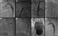
A 6-month-old girl (weight 5.5 kg) with a Krichenko Type A duct measuring 2.1 mm at the pulmonary end underwent successful transcatheter closure using a Lifetech 4/6 mm PDA Occluder (Fig 2a–c). Despite appropriate deployment, the device embolised to the descending aorta after 12 hours. Attempts at snaring the device from the venous end across the duct were unsuccessful. The body of the device was captured with a 10 mm Amplatz Gooseneck snare, but the device could not be slenderised within the sheath due to device-sheath malalignment and the screw at the pulmonary end could not be snared in view of its location deep down. Thereafter, the device was successfully retrieved from the arterial side by holding the metallic hub at the aortic end using a 5.5 Fr Cook Bioptome (Cook Medical, Bloomington, Indiana, United States of America) and slenderising the device into a 6Fr short sheath.
Case 2. ( a ) Descending aortogram demonstrating a tubular PDA. ( b ) Post-deployment, the Lifetech Cera device has a good waist with a distal splaying at the pulmonary end. ( c ) Post release, the waist appears reduced and the distal splaying at the pulmonary end is absent. ( d ) Device in descending aorta successfully captured with a 10 mm Gooseneck snare. ( e ) Attempts at collapsing the device into the sheath were unsatisfactory. ( f ) Snared device captured from arterial side with a Bioptome. ( g ) Captured device slenderised and removed from right femoral artery sheath. ( h ) Successful PDA occlusion with a 6/6 ADO2.

Post-procedure, the patient had loss of right-sided pulse. Moreover, with the device in the descending aorta for more than 2 hours prior to successful recapture and retrieval, the distal perfusion was affected, and she had a stormy post-retrieval course requiring peritoneal dialysis for 3 days. Fortunately, the renal function recovered completely. The duct was closed at a later date with a 6/6 mm ADO II device (St Jude Medical, Abbott) with a good result. (Fig 2h) During the PDA occlusion at the second sitting, a descending thoracic aortogram was done, which revealed right iliofemoral block with excellent collateral circulation. (supplementary Fig 1)
Discussion
The Amplatzer Duct Occluder (St Jude Medical, Abbott, St. Paul, Minnesota,United States of America) has been in clinical use for over two decades. The standard sizing dictum for the ADO-shaped devices is that the pulmonary end of the device should be 2 mm larger than the narrowest diameter of the duct.Reference Boehm, Emmel and Sreeram 1
The Lifetech Cera PDA occluder (Lifetech Scientific Co. Ltd, Shenzhen, China) is a similar shaped self-expanding device made of nitinol mesh with titanium nitride ceramic surface coating.Reference Chamié, Simões, Chamié de Queiroz and Mattos 2 This device is softer and more flexible than the ADO, and generally allows a smaller delivery sheath size and easy deployment. Chamie et al also reported increased softness and flexibility and recommended that oversizing the Cera device to form a tighter waist was safe.Reference Chamié, Simões, Chamié de Queiroz and Mattos 2
Complications with the ADO are rare and include device embolisation or device protrusion producing coarctation or branch pulmonary artery obstruction. Late device embolisation into the pulmonary arteries is known to occur if the retention disc has prolapsed into the ductal ampulla prior to release.Reference Boehm, Emmel and Sreeram 1 , Reference Bilkis, Alwi and Hasri 3 Of the device embolisations reported, the majority of dislodgements are to either branch pulmonary artery due to duct anatomy, undersizing the device or improper deployment. Bilkis et al reported three embolisations (1.5%) in 209 patients, all to the pulmonary arteries.Reference Bilkis, Alwi and Hasri 3 In a prospective multi-center study, El-Said et al evaluated data from eight centers and 496 transcatheter PDA closures (338 device closures, rest coils). They described three device embolisations (<1%) (two ADO, one AVPII), all to the pulmonary arteries and all in type C ducts. In addition, there were four device malpositions with overall device dislodgement/malposition rate of 2%.Reference El-Said, Bratincsak and Foerster 4 In a recent meta-analysis of infants treated with transcatheter PDA occlusion,Reference Backes, Rivera and Bridge 5 Backes et al reported an overall device embolisation rate of 2.6%.
PDA device embolisation to the aorta is often associated with pulmonary arterial hypertension, or is secondary to improper device position with the pulmonary end of the device remaining within the body of the duct and not extending into the pulmonary artery.Reference McMullan, Moulick and Jonas 6 – Reference Khan, Almashham, Almoukirish and Momenah 8 Vijalalaxmi et al have reported embolisation in eight of 1325 patients (3.3%) of PDA with pulmonary hypertension.Reference Vijayalakshmi, Setty, Narasimhan, Singla and Manjunath 7 Khan et al reported device embolisation to the aorta (6/4 ADO) following improper positioning of the device prior to release.Reference Khan, Almashham, Almoukirish and Momenah 8
In contrast to cases of PDA device embolisation reported in literature, both our cases were unique in that pulmonary arterial pressures were normal and initial device position on angiography appeared optimal. Our hypothesis of embolisation in these patients is that in tubular ducts, the pulmonary arterial end of these softer devices did not splay adequately. Consequently, the device got squeezed out of its initial position towards the aortic side during recoil at the time of release. (Figs 1b and c and 2b and c). Second, we also feel that there was inadequate stenting function of the cylindrical body of the softer Cera device against the ductal wall, resulting in device displacement into the descending aorta secondary to the downward force of the aortic pressure wave on the retention skirt of the device.
Embolised PDA devices have been successfully retrieved from the descending aorta with a snare or bioptome.Reference Vijayalakshmi, Setty, Narasimhan, Singla and Manjunath 7 , Reference Koneti, Bakhru, Penumatsa and Lalukota 9 In our first case, the device was successfully retrieved within the sheath with a snare. However, in the second case, despite snaring, it was not possible to slenderise the device within the sheath due to malalignment between the device and sheath. Although all retrievals described in the literature are by snaring or holding the pulmonary end, in this case we grasped the knob at the aortic end of the device with a five Fr Bioptome, which offered a grip of sufficient strength enabling easy slenderisation of the device into a 6Fr sheath.
Conclusion
We conclude that for tubular ducts, the softer ceramic-coated devices may need to be oversized to form a tighter waist with a pulmonary flare as in our first case during second deployment, or else it may be prudent to use a vascular plug or ADOII as was done in our second case.
Supplementary materials
To view supplementary material for this article, please visit https://doi.org/10.1017/S104795111900091X
Acknowledgements
None.
Financial Support
This research received no specific grant from any funding agency, commercial or not for profit sectors.
Conflicts of Interest
Dr Bharat Dalvi, Supratim Sen and Sneha Jain have no conflict of interest related to this manuscript and have no financial dealings with any device company. Drs Sen and Jain use both Lifetech and Amplatzer devices in their regular pediatric interventional practice. They do not have any preference for one device manufacturer over the other, and the choice of device is always based on cost, patient preference and device availability. The authors have attended conferences and workshops organised or sponsored by both device companies.
Ethical Standards
This research does not include human experimentation and describes cases from routine clinical practice.