Introduction
Rhinoscleroma is a chronic granulomatous infectious disease of the respiratory tract. The disease mainly affects the nose, but the upper air passages, trachea and bronchi may also be involved either with or without nasal granuloma.Reference Gonzalez-Angulo, Marques-Monter, Greenberg and Cerbon1
In the history of the disease, three dates are important. In 1870, Ferdinand Ritter von Hebra was the first physician to make a clinical description of this disease, naming it rhinoscleroma (hard nose). In 1876, Johann von Mikulicz described for the first time the histology of the disease and the characteristic foam cells that bear his name. In 1882, Anton von Frisch isolated the encapsulated bacillus (Klebsiella rhinoscleromatis), which he considered to be the aetiological agent of rhinoscleroma.Reference Hoffman, Loose and Harkin2
Although rhinoscleroma has been sporadically observed in all parts of the world, it is endemic in certain areas. It is most prevalent in Poland, Hungary, Southern Russia, Egypt, Indonesia, and Central and South America.Reference Gadomski3–Reference Fisher and Dimling5
The disease occurs almost equally in both sexes and at almost any age but with the greatest incidence between 15–35 years.Reference Hara4 The disease is contagious and often reported in more than one member of the same household, family or vicinity. Poverty and a lack of cleanliness are important factors.Reference Gadomski3 Scleroma is a disease affecting mainly the nose; however, other sites may be affected including the pharynx, larynx, trachea and bronchi. Other rare sites may include the Eustachian tube (otoscleroma), maxillary sinus (antroscleroma) and ethmoid sinus (ethmoidal scleroma).Reference Gamea and El-Tatawi6
Treatment of rhinoscleroma has been attempted for many decades without much success. Many antibiotics, including streptomycin,Reference Hara, Pratt, Levine and Hoyt7 aureomycin,Reference Botros, Hamilton, Floyd, Mufti and Imam8 rifampicin (both systemicReference Gamea and El-Tatawi9 and localReference Gamea10) and ciprofloxacin,Reference Eltatawi11 have been tried as a treatment, but it is unclear if antibiotics can lead to complete cure.
Botros et al. (1954) stated that the incidence of scleroma is decreasing in Egypt. This study mentioned that the huge characteristic saddle nose, the involvement of the upper lip, and the perforation of the septum and palate was seldom encountered at that time.Reference Botros, Hamilton, Floyd, Mufti and Imam8
Those observations motivated us to review cases of rhinoscleroma presented to Department of Otolaryngology at Tanta University Hospitals during the last five years in order to detect recent changes that may have been encountered in the clinical and histopathological patterns of rhinoscleroma. Tanta University lies in the middle of Nile Delta in Egypt, and its Department of Otolaryngology receives thousands of patients every year from all Nile Delta governorates. Data obtained from our patient series study might be used to show changes in the pattern of rhinoscleroma in Egypt.
Materials and methods
Twenty-eight rhinoscleroma patients were included in this study. Patients attending the Department of Otolaryngology at Tanta University Hospitals from January 2014 to January 2019 were included. Patients had not received medication for their nasal symptoms for at least three months at the time of their attendance. The cases were diagnosed by clinical, bacteriological and histopathological data.
A detailed history was taken for each patient and a thorough clinical otorhinolaryngological examination with a special emphasis on nasal examination was performed to diagnose and determine the stage of the disease.
Nasal biopsies were taken from the granuloma or from the lower border of the inferior turbinate under surface anaesthesia without adrenalisation. Each specimen was divided into two parts: one part was processed and stained with haematoxylin and eosin stain for histopathological study, and the other part was processed for electron microscopic study.
All patients received medical treatment in the form of 500 mg ciprofloxacin tablets to be taken twice daily for twelve weeks. Follow up by bacteriological and histopathological examination was performed. If the results were still positive after twelve weeks of treatment, another twelve-week course of ciprofloxacin was given until the results were negative on three successive occasions at three-month intervals. Cured patients were followed up every three months in order to detect any recurrences.
Results
Demographic findings
Twenty-eight patients with rhinoscleroma were included in this study. There were 13 males (46.5 per cent) and 15 females (53.5 per cent). Patient age ranged from 16 to 62 years. A positive family history was encountered in seven cases (25 per cent). Twenty-two patients (78.5 per cent) came from rural areas.
Clinical findings
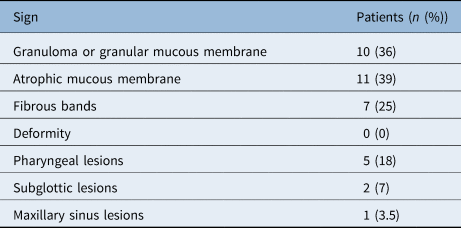
The nose was found to be affected in all 28 cases (100 per cent). Pharyngeal lesions were seen in 5 cases (18 per cent), and subglottic lesions were observed in 2 cases (7 per cent). One case showed involvement of the right maxillary sinus (3.57 per cent).
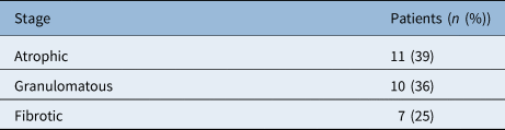
The incidence of various clinical stages of rhinoscleroma in our series are shown in Table 1. The atrophic and fibrotic stages together comprised 18 patients (64 per cent), and the granulomatous stage was recorded in 10 patients (36 per cent).
Incidence of the various stages of rhinoscleroma

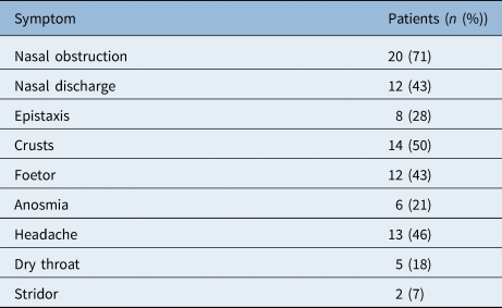
Different symptoms and signs of rhinoscleroma in our series are represented in Tables 2 and 3, respectively.
Incidence of rhinoscleroma symptoms

Incidence of rhinoscleroma signs

Histopathological findings
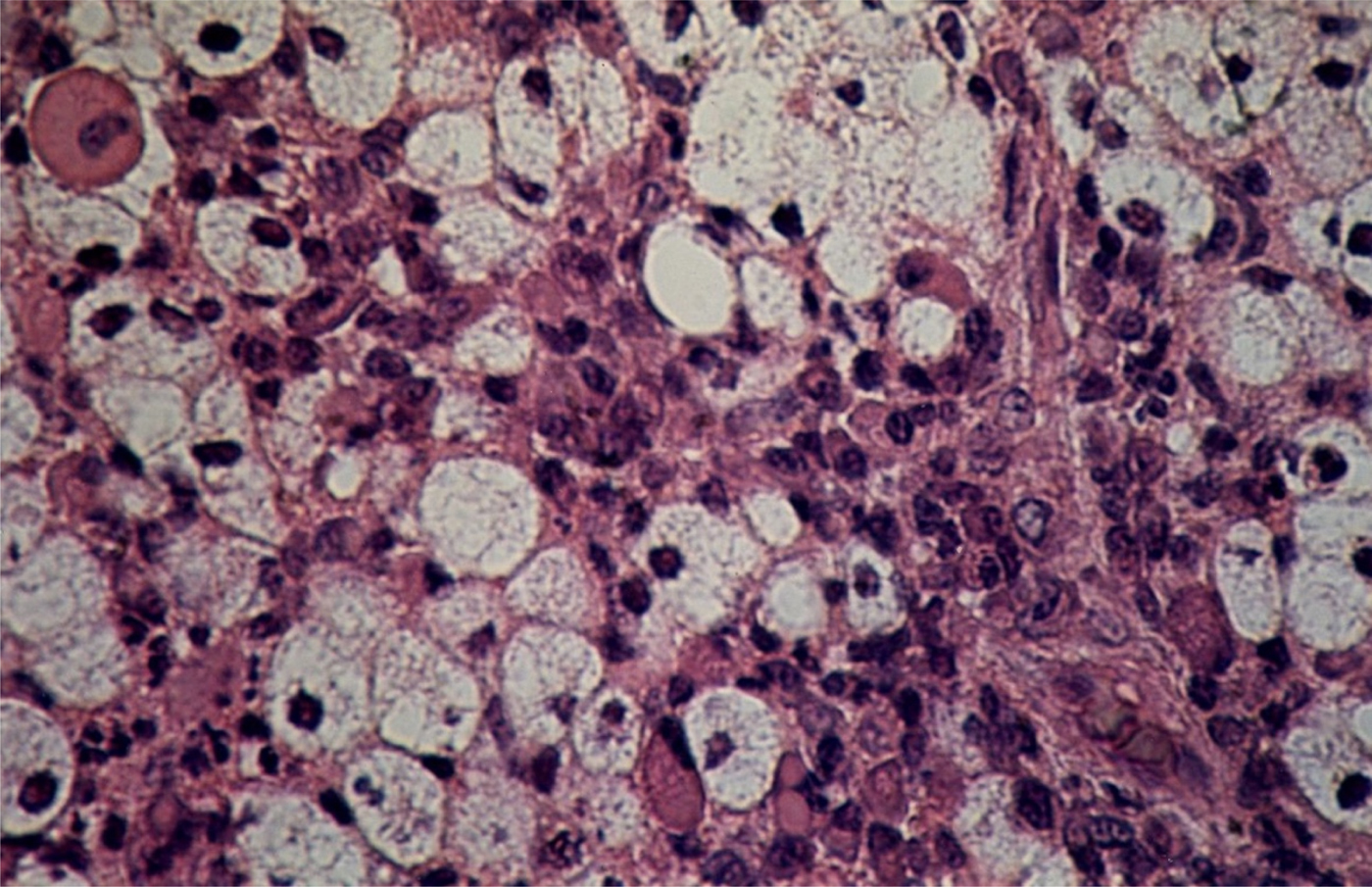
In all patients, histopathological findings were specific to different degrees of rhinoscleroma. Squamous metaplasia of the epithelium was seen in all cases. The lamina propria showed granulomatous infiltration with typical Mikulicz cells, plasma cells, Russell bodies, lymphocytes and few neutrophils (Figure 1). Although the histopathological features of the different stages of the disease varied, most of the elements of the lesion could be seen in every stage but with varying abundancy.
Mikulicz cells in rhinoscleroma appearing as large, pale vacuolated cells with round or pyknotic nuclei. Some Russell bodies appear with central or peripheral nuclei. (H&E; × 400)

Electron microscopy findings
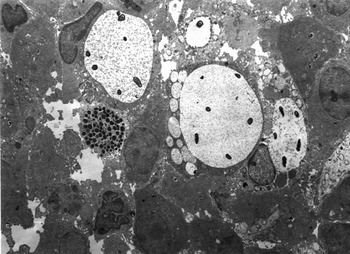
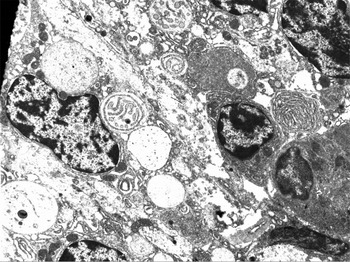
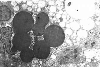
Metaplasia of the surface epithelium was a constant feature in rhinoscleroma. However, the degree of stratification was variable in different areas. Mikulicz cells were not numerous. They appeared large and irregular in shape. The cytoplasm appeared to be almost exclusively formed of large vacuoles with the organelles located in the narrow intervesicular bridges. Some large vacuoles showed variable numbers of klebsiella bacilli, but bacilli were rarely seen extracellularly (Figure 2). Plasma cells were numerous and had their typical appearance (Figure 3). Their endoplasmic reticula were dilated with small spheres of electron dense granules gradually forming Mott cells and typical Russell bodies (Figure 4). Fibroblasts and fibrous bundles were encountered in some cases.
Electron micrograph showing Mikulicz cells. The vacuoles containing bacilli appear larger than the others. The vacuoles contain fine granular material. A mast cell is seen with its characteristic secretory granules. (×4600)

Electron micrograph showing plasma cells with compressed nucleus, abundant mitochondria and whorls of rough endoplasmic reticula. (×8600)

Electron micrograph showing a group of Russell bodies of different sizes. A nearby Mikulicz cell can also be seen with a bacillus inside one of its vacuoles. (×7700)

Treatment results
All patients were treated with ciprofloxacin in the form of 500 mg tablets twice daily for twelve weeks with follow up until clinical and bacteriological cure was confirmed.
After a twelve-week course of ciprofloxacin, 19 of 28 patients (69 per cent) were shown to be cured from the disease after bacteriological and histopathological assessment. The other nine patients received another twelve-week course of ciprofloxacin after which all cases showed complete cure. However, follow up of the cases for a longer period showed recurrence in two patients. These patients required repeated courses of medication and follow up.
Discussion
Diagnosing rhinoscleroma in endemic areas is easy clinically, histologically and bacteriologically. In non-endemic areas, it is more difficult and might take some time to prove the diagnosis. Rhinoscleroma should always be suspected in differential diagnosis of chronic bilateral granuloma or atrophic nasal mucosa.Reference Gaafar, Gaafar and Nour12
El Mofty (1962) found that the incidence of scleroma is decreasing in Egypt. He attributed this decrease to the improvement in living standards and in methods of treatment.Reference El-Mofty13 It is obvious from our study that the rate of incidence of rhinoscleroma in the Nile Delta, and hence Egypt, is progressively decreasing when compared with many previous reports from Egypt. Only 28 patients with rhinoscleroma could be studied in our series over a period of 5 years. This can be explained by the following: (1) improvement of socioeconomic conditions in Egypt, particularly in rural areas where rhinoscleroma was prevalent because of poverty and low living standards; (2) an increase in the rate of education and awareness in rural areas in Egypt, making individuals more likely to seek early medical advice and treatment; (3) progress in the number and services of healthcare centres in rural areas in Egypt; and (4) improvement in rhinoscleroma treatment methods, particularly use of rifampicin and ciprofloxacin antibiotics (both proven to be effective against the bacillus).
The decrease in the rate of incidence of rhinoscleroma was also associated with two observations. The first observation relates to changes in the clinical pattern and site of involvement of rhinoscleroma. In our study, the nose was the organ mainly affected by scleroma, whereas the pharynx and larynx were less commonly affected. In one case the right maxillary sinus was affected. Involvement of other paranasal sinuses, the palate, lips, trachea or Eustachian tubes were not encountered in our study. Deformities secondary to the disease could not be seen in our series. The second observation relates to changes in the prevalence of the various stages of disease and in the histopathological pattern of rhinoscleroma. Most cases were observed to be in the atrophic and fibrotic stages (64 per cent) rather than in the granulomatous stage (36 per cent). Histopathological and electron microscopy results showed that the fibrous bundles and fibroblasts dominated the stroma. Mikulicz cell vacuoles rarely showed bacilli in comparison with previous studies by the authors.Reference Gamea and El-Tatawi9,Reference Gamea14 This may be attributed to the misuse of antibiotics in Egypt because they can be obtained without a medical prescription.
• The incidence of rhinoscleroma in Egypt is decreasing
• Currently, rhinoscleroma mainly affects the nose, and advanced disease or nasal deformities are rarely encountered
• The atrophic stage of rhinoscleroma is most common presenting clinical and histopathological stage of the disease
• These changes could be partly attributed to improved socioeconomic conditions in rural areas of Egypt as well as better education and awareness meaning patients seek medical advice and treatment earlier
• Better healthcare in rural areas may have also contributed to these changes
• Improved rhinoscleroma treatment methods, particularly the use of rifampicin and ciprofloxacin antibiotics, has also impacted disease patterns
Ciprofloxacin has been proven to be effective in the treatment of rhinoscleroma. A course of twelve weeks seems to be sufficient to cure scleroma in most cases. However, some patients may need another twelve-week course. Again, cases should be followed up for longer periods to detect any relapses and deal with them appropriately.
Conclusion
The incidence of rhinoscleroma in Egypt is decreasing, and its clinical and histopathological patterns are changing to less advanced forms. This may be explained by the improvement in socioeconomic conditions, particularly in rural areas, improvement in healthcare availability and rhinoscleroma treatment methods, in particular the use of rifampicin and ciprofloxacin antibiotics.
Competing interests
None declared