
Introduction
Due to the fact that the shape of nanostructures (NSs) determines their physical properties, an active search for approaches, which provide possibility on controlling the NSs shape and dimension is nowadays being conducted.[ Reference Yang, Li, Ruan, Li, Ren, Xu and Tian 1 ] One of the simplest approaches for controlling the NSs shape and localized growth is the using of porous matrices (templates) such as: Al2O3,[ Reference Spende, Sobel, Lukas, Zierold, Riedl, Gura, Schubert, Moreno, Nielsch, Stühn, Hess, Trautmann and Toimil-Molares 2 , Reference Kalanda, Gorokh, Yarmolich, Lozovenko and Kanyukov 3 ] polymer films,[ Reference Guo, Martin, Zhao, Ge and Zare 4 – Reference Kozlovskiy, Korolkov, Kalkabay, Ibragimova, Ibrayeva, Zdorovets, Mikulich, Yakimchuk, Shumskaya and Kaniukov 7 ] Si,[ Reference Boarino, Borini and Amato 8 ] SiO2,[ Reference Dallanora, Marcondes, Bermudez, Fichtner, Trautmann, Toulemonde and Papaléo 9 – Reference Fink, Petrov, Hoppe, Fahrner, Papaleo, Berdinsky, Chandra, Chemseddine, Zrineh, Biswas, Faupel and Chadderton 11 ] TiO2,[ Reference Cultrera, Boarino, Amato and Lamberti 12 , Reference Han, Jang, Kim and Koh 13 ] and other materials, in which the shape, dimensions, and position of the pores determine the geometry and localization on the surface of the produced NSs. Due to the highly developed silicon-based industry infrastructure, templates based on silicon and its derivatives have a great potential for practical usage. Silicon-based templates are important components in the creation of nano-, micro-, and magnetoelectronic devices.[ Reference Demyanov, Kaniukov, Petrov and Sivakov 14 ] The existing methods[ Reference Panarin, Terekhov, Kholostov and Bondarenko 15 , Reference Bandarenka, Artsemyeva, Redko, Panarin, Terekhov and Bondarenko 16 ] for the synthesis of noble metal NSs using silicon-based templates make it possible to obtain sensor elements with an extremely high sensitivity to chemical and biologic media.[ Reference Girel, Yantcevich, Arzumanyan, Doroshkevich and Bandarenka 17 , Reference Arzumanyan, Doroshkevich, Mamatkulov, Shashkov, Girel, Bandarenka and Borisenko 18 ] The nanosphere lithography and metal deposition methods based on colloidal crystal templating were suggested as alternative methods for the localized and selective noble metals formation.[ Reference Bechelany, Brodard, Philippe and Michler 19 , Reference Sakamoto, Philippe, Bechelany, Michler, Asoh and Ono 20 ] In our previous work,[ Reference Kaniukov, Yakimchuk, Arzumanyan, Terryn, Baert, Kozlovskiy, Zdorovets, Belonogov and Demyanov 21 ] it was shown that using an external current source of a certain magnitude, the localized formation of copper dendrite-like NSs in the SiO2/n-Si pores can be realized. The observed copper NSs found application as plasmon-active surfaces for amplification of the Raman scattering signal. However, the low local electric field enhancement factor of the copper NSs and surface degradation of such structures (“aging effect”––the rapid formation of oxides on the surface of structures) does not allow them to be seriously considered from the point-of-view of wide application in sensoric. For that reason, the application of gold or silver nanostructures (AgNSs) is more promising and favorable.[ Reference Baniukevic, Hakki Boyaci, Goktug Bozkurt, Tamer, Ramanavicius and Ramanaviciene 22 ]
Previously[ Reference Sivakov, Kaniukov, Petrov, Korolik, Mazanik, Bochmann, Teichert, Hidi, Schleusener, Cialla, Eugenia Toimil-Molares, Trautmann, Popp and Demyanov 23 ] we proposed a simple bottom-up method for localized AgNSs formation with precisely controlled AgNSs shape by varying the pore diameter by applying electroless wet-chemical deposition of metal into the SiO2/p-Si pores template. At the same time, an important difference from other previously published works[ Reference Spende, Sobel, Lukas, Zierold, Riedl, Gura, Schubert, Moreno, Nielsch, Stühn, Hess, Trautmann and Toimil-Molares 2 , Reference Shumskaya, Kaniukov, Kozlovskiy, Shlimas, Zdorovets, Ibragimova, Rusakov and Kadyrzhanov 24 ] is that the shape of the AgNSs does not reproduce the geometry of the pore, but can be varied widely: from faceted crystallites to dendritic-like structures. In particular, it was considered the formation of highly branched AgNSs, which can be an ideal surface for surface-enhanced Raman scattering (SERS). The advantage of such types of materials over colloidal nanoparticles, which are widespread today, is that the particles tend to form conglomerates, which makes the SERS signal unstable and poorly reproducible in a series of measurements.[ Reference Dou, Jung, Cao and Ozaki 25 , Reference Siiman and Feilchenfeld 26 ] Dendrites have a rigorous structure, which does not influence by adding the analyte solution. In addition, dendritic-like structures can provide a large number of “hot spots,” which are the main cause of the high enhanced factor of the Raman scattering.[ Reference Yin, Chen, Zhao, Lv, Shi, Wu, Zhang, Liu, Wang and Xu 27 ] In the present paper, the dependence of the AgNSs shape on the deposition temperature of silver in the SiO2/p-Si porous template is presented and discussed in details. The plasmonic activity of the silver dendrites-like NSs was demonstrated experimentally in measurements of SERS activity of a highly dilute model dye molecule, i.e., Nile blue, deposited onto the silver dendrites in the SiO2/p-Si porous templates.
Methods
As templates for synthesis of AgNSs swift heavy ion-track[ Reference Kaniukov, Ustarroz, Yakimchuk, Petrova, Terryn, Sivakov and Petrov 28 ] porous SiO2 templates on p-type silicon substrate (12 Ωcm with (100) crystal orientation) were used with a silicon oxide layer thickness of 250 nm and pore sizes of 500 nm. The SiO2/p-Si pore density over the surface was found to be 108 cm−2 according to the scanning electron microscopy (SEM) studies. The procedure of obtaining porous SiO2/p-Si templates are described in our previous paper.[ Reference Kaniukov, Ustarroz, Yakimchuk, Petrova, Terryn, Sivakov and Petrov 28 ] Silver deposition was carried out by electroless deposition from a solution of 0.02 M silver nitrate and 5 M hydrofluoric acid at ambient conditions. Before deposition, 4 × 4 mm2 SiO2/p-Si chips were treated with isopropyl alcohol and distilled water, and then dried in a stream of nitrogen to remove the adsorbed atmospheric residual from the surface. The growth of silver structures was carried out for 30 s at electrolyte temperature from 20 till 50 °C. At the end of the process, the samples were washed in distilled water and dried in air. The morphology of the SiO2(Ag)/p-Si surface was studied using Carl Zeiss ULTRA 55, field emission scanning electron microscope.
The standard dye molecule––Nile blue was used to study the Raman signal enhancement induced by AgNSs in “dendrites-like” shape. To perform SERS measurements, 50 µl of an aqueous solution of Nile blue (C20H20ClN3O) was dropped on the samples with “dendrites-like” NSs with an incubation time of 15 min, and subsequently rinsed in deionized water, and finally, dried in air. The Raman scattering measurements were performed at room temperature on a Nanofinder High End (Lotis TII, Belarus–Japan) confocal microscope-based setup. Lasers with wavelengths of 473, 532, and 633 nm were used as excitation source. The laser power at the samples’ surface was kept at 50 µW and the typical single-spectrum acquisition time was 5 s. The backscattered light, not analyzed for its polarization, was dispersed using a spectrograph with 0.55 m focal lengths equipped with a 600 lines/mm single grating spectrometer with a spectral resolution around 3 cm−1 and detected by a cooled CCD (charge-coupled device) camera. The laser beam was focused through a “50×” microscope objective lens. The resulting spot diameter was about 1 µm.
Results and discussion
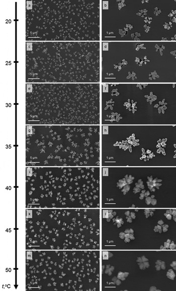
The self-organization of AgNSs in the porous SiO2/p-Si templates occur with a high organization degree of pore filling at all process temperatures that were investigated by SEM as presented in Fig. 1. In addition, the silver deposit in the pores, independently of the process temperature, is characterized by the presence of clearly distinguished priority metal growth fronts along the pore walls. The predominant growth of silver along the edges of the pore is due to the presence of edge defects at the Si–SiO2 boundary at the pore base, which are priority areas for nucleation of silver sediment at the initial stage of AgNSs formation. In this case, the middle of the pore in most cases remains partially unfilled that is clearly visible from SEM studies in Fig. 1.
Scanning electron images of AgNSs morphology evolution in porous SiO2/p-Si templates depending on the silver deposition temperature variation at: (a, b) 20 °C; (c, d) 25 °C; (e, f) 30 °C; (g, h) 35 °C; (i, j) 40 °C; (k, l) 45 °C; (m, n) 50 °C.

The morphology of silver deposited at room temperature is characterized by some variation in the form of obtained AgNSs, which is manifested in the fact that part of the crystallites, which form part of the metallic sediment in the pores have faceted edges and another part have smoothed edges [Figs. 1(a) and 1(b)]. An increasing of silver deposition temperature to 25 °C leads to decreasing of the number of priority growth fronts with a simultaneous increasing of their lateral dimensions, while the structures are smoothed. Differences in the form of AgNSs over the surface are minimal as shown in Figs. 1(c) and 1(d). Further silver deposition temperature increasing to 30 °C does not lead any significant changes in the morphology of the silver sediment in comparison to 25 °C, only insignificant increasing of its size was observed [Figs. 1(e) and 1(f)]. An increasing of the temperature of the solution to 35 °C leads to a radical change in the shape of the NSs: it occurs with an increase of dimensions with filling of the central part of the pore, while on the branches of the AgNSs appear branches of the second order and characteristic for “bud-like” facets of dendrites [Figs. 1(g) and 1(h)]. Further silver growth temperature increasing to 40 °C leads to the destruction of the dendritic structure and formation of AgNSs in different pores with different shapes: part of the metal deposit takes the form of faceted crystallites, and another part takes the form of the elongated smooth interconnected crystallites [Figs. 1(i) and 1(j)]. Further increasing of temperature leads to decreasing of the number of smooth elongated [Figs. 1(k) and 1(l)] crystallites at 45 °C with their complete degeneracy at 50 °C [Figs. 1(m) and 1(n)].
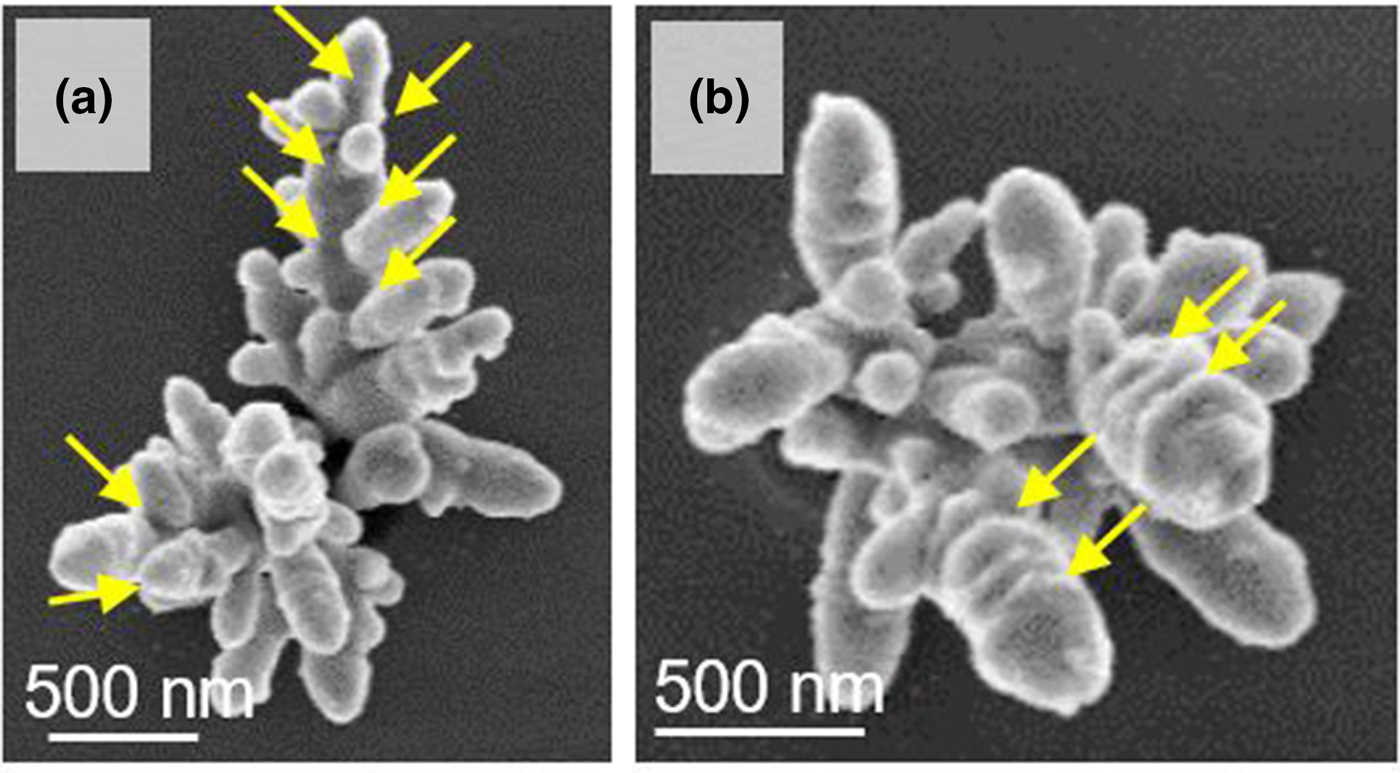
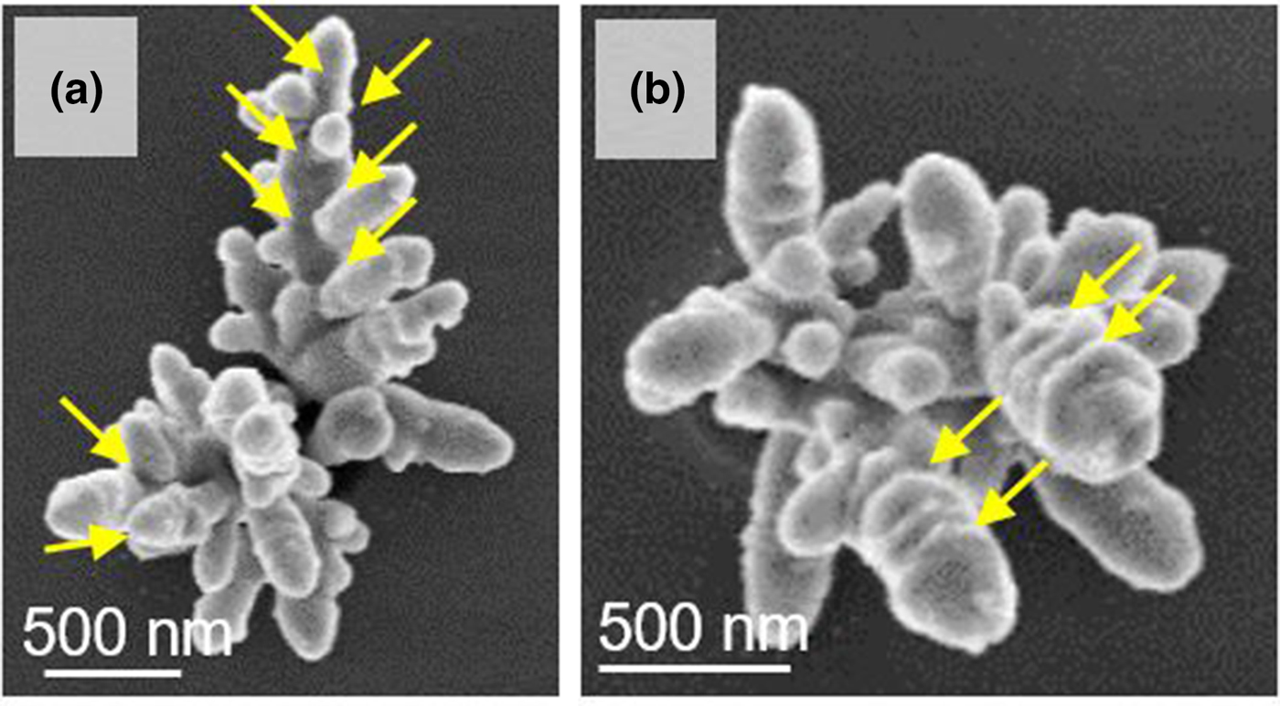
The SERS capabilities of the AgNSs in porous SiO2/p-Si templates can easily be performed by a simple model SERS experiments using the dye molecule (Nile blue) as an agent of interest (analyte). The gaps between the dendrite branches [Fig. 2(a)] and the “bud-like” facets [Fig. 2(b)] are those nanoscale regions for which “hot spot” formation is characteristic.[ Reference Yin, Chen, Zhao, Lv, Shi, Wu, Zhang, Liu, Wang and Xu 27 , Reference Feng, Zhao and Jiang 29 – Reference Qiu, Zhou, Li, Zhang, Lang, Cui and Chu 31 ] Such structures are highly interesting for SERS applications. As it is known, the position of maximums of plasmonic structures extinction depends not only on their shape and size, but also on the polarization of the radiation, which induces plasmon oscillations.[ Reference Yang, Li, Ruan, Li, Ren, Xu and Tian 1 , Reference Feng, Zhao and Jiang 29 ] Accordingly, the same effects will be achieved, when the spatial orientation of the plasmonic structure changes: inclination, turning, etc. SEM images in Fig. 2 show that the resulting structures and their components (individual trunks and branches) are not characterized by strict spatial orientation relative to the template, but have a chaotic inclination and are not identical copies of each other. It is known that dendrites have a large set of “hot spots”.[ Reference Yin, Chen, Zhao, Lv, Shi, Wu, Zhang, Liu, Wang and Xu 27 , Reference Zhao, Lu, Zhang, Fu, Moeendarbari, Shelke, Liu and Hao 30 ] Thus, taking into account the influence of the polarization of the exciting radiation on the distribution of plasmons along the dendrite,[ Reference Feng, Zhao and Jiang 29 ] it can be assumed that even a single silver structure has not one or several distinct bands of plasmon resonance. They are present in large numbers and are distributed throughout the long-wave interval corresponding to plasmon resonance bands of all possible AgNSs.[ Reference Sharma, Frontiera, Henry, Ringe and Van Duyne 32 ]
SEM images of localized regions with “hot spots” on the surface of AgNSs grown at 35 °C in the pores of the SiO2/p-Si template: connecting parts of the branches (a) and the “bud-like” facets (b) of the dendrite.
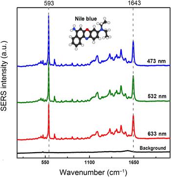
Based on the obtained results, it can be proposed that such structures under consideration are suitable for using in SERS applications. This assumption was verified on SiO2(Ag)/p-Si structures with dendrites in a series of SERS measurements using lasers with different wavelengths: blue (473 nm), green (532 nm), and red (633 nm), as shown in Fig. 3.
Normalized SERS spectra of the Nile blue molecule with a concentration of 10−6 M, obtained on the surface of AgNSs grown at 35 °C in the pores of the SiO2/p-Si template, excited by using different excitation wavelengths (473, 532, and 633 nm), and the Raman spectrum of the substrate's background.

The SERS spectra of Nile blue with a concentration of 10−6 M have brightly expressed characteristic peaks at 592 and 1643 cm−1 (Fig. 3), which correspond to the characteristic lines of Nile blue.[ Reference Li, Zamani, Rivera Gil, Pelaz, Ibanez, Cadavid, Shavel, Alvarez-Puebla, Parak, Arbiol and Cabot 33 , Reference Etchegoin and Le Ru 34 ] The intensity of the signal, when lasers with different wavelengths were used, indicates that the “hot spots” in the spectral region of 473–633 nm are uniformly distributed over the position and intensity of the plasmon resonance. A detailed study of this phenomenon with using of mathematical modeling with the interaction of an external electromagnetic wave with dendrites of different spatial orientations is planned to be carried out in the future work.
Conclusions
AgNSs are formed in the porous SiO2/p-Si template and the interrelation of their morphology with the deposition temperature was determined. It was found that at the temperature of 35 °C the most preferable AgNSs for SERS structures (highly branched dendrites) can be realized. The presence of gaps between the dendrite branches and the “bud-like” facets indicates the presence of “hot spots” in dendritic NSs. Highly reproducible morphology and pore localized synthesis of the silver dendritic NSs in the SiO2/p-Si templates allows one to obtain the SERS-active substrates working in a broad spectral range (473–633 nm). These surfaces are expected to appeal to a wide audience in the nanotechnology related fields, like environmental science, food science, and nanotoxicology, etc., due to the high potential for in-depth studies of substances and track it behavior. From the practical point of view, such silver dendrite NSs in porous SiO2/p-Si matrices are highly perspective for cancer theranostics, food and environment monitoring, e.g., determination of pesticides or harmful additives, water pollution control, and other fields where precise analysis of components in the substances is required.
Acknowledgments
This work was supported by the Scientific-technical ‘program Technology-SG’ (project number 3.1.5.1). VS gratefully acknowledged the German Federal Ministry of Education and Research (BMBF) in the frame of Baltic Sea Network “NanoPhoto” under Grant No. 01DS14017 and National Science Foundation of Germany (DFG) under Grant No. SI-1893/2-1 for the financial support. LAO gratefully acknowledged the Russian Science Foundation (Grant no. 17-12-01386) for the financial support.