Introduction
Haploporidae Nicoll, 1914 is a family of trematode endoparasites of fish distributed worldwide. This family is characterized morphologically by the presence of a hermaphroditic sac and a single testis. In the recent taxonomic revision of the family, Overstreet & Curran (Reference Overstreet, Curran, Jones, Bray and Gibson2005) erected the subfamily Chalcinotrematinae Overstreet & Curran, Reference Overstreet, Curran, Jones, Bray and Gibson2005 to accommodate the genera Chalcinotrema Freitas, 1947, Paralecithobotrys Freitas, 1948, Saccocoelioides Szidat, Reference Szidat1954, Megacoelium Szidat, Reference Szidat1954 and Unicoelium Thatcher & Dossman, 1975.
Species of the genus Saccocoelioides are parasites of freshwater and brackish-water fishes from the Americas (Lumsden, Reference Lumsden1963; Travassos et al., Reference Travassos, Teixera de Freitas and Kohn1969; Szidat, Reference Szidat1970, Reference Szidat1973; Lamothe-Argumedo, Reference Lamothe-Argumedo1974). The taxonomic history, as well as the species composition of this genus, have been controversial (see Yamaguti, Reference Yamaguti1958). In the last review of Haploporidae, Overstreet & Curran (Reference Overstreet, Curran, Jones, Bray and Gibson2005) validated the genus Saccocoelioides, and transferred it to the subfamily Chalcinotrematinae. In North and Middle America three species of Saccocoelioides have been found: S. sogandaresi Lumsden, Reference Lumsden1963 was described from Poecilia latipinna Lesueur from Texas, USA (Lumsden, Reference Lumsden1963). In Mexico, S. sogandaresi has been recorded in poeciliids, characids, cichlids, gobiids, eleotrids and goodeids (Scholz et al., Reference Scholz, Vargas-Vázquez, Moravec, Vivas-Rodríguez and Mendoza-Franco1995; Salgado-Maldonado et al., Reference Salgado-Maldonado, Aguilar-Aguilar, Cabañas-Carranza, Soto-Galera and Mendoza-Palmero2005; Pérez-Ponce de León et al., Reference Pérez Ponce de León, García Prieto and Mendoza Garfías2007; Martínez-Aquino et al., Reference Martínez-Aquino, Mendoza-Palmero, Aguilar Aguilar and Pérez-Ponce de León2014). Saccocoelioides chauhani Lamothe-Argumedo, Reference Lamothe-Argumedo1974 was described from the characid fish Astyanax aeneus Günther and it has also been recorded in poeciliids from Catemaco Lake, Veracruz, Mexico (Jiménez-García, Reference Jiménez-García1996; Pérez-Ponce de León et al., Reference Pérez Ponce de León, García Prieto and Mendoza Garfías2007). Finally, S. lamothei Aguirre-Macedo & Violante-González, 2008 was described from the eleotrid fish Dormitator latifrons Richardson from Tres Palos Lagoon, Guerrero, Mexico (Aguirre-Macedo & Violante-González, Reference Aguirre-Macedo and Violante-González2008), and it has been recorded in other fishes such as gobiids, poeciliids and profundulids (Andrade-Gómez, Reference Andrade-Gómez2015; Pinacho-Pinacho et al., Reference Pinacho-Pinacho, García-Varela, Hernández-Orts, Mendoza-Palmero, Sereno-Uribe, Martínez-Ramírez, Andrade-Gómez, Hernández-Cruz, López-Jiménez and Pérez-Ponce de León2015).
In the present study, we collected haploporids from the fat sleeper Dormitator maculatus (Bloch) (Perciformes: Eleotridae) in six localities along the coast of the Gulf of Mexico. Additionally, adults of S. chauhani and S. lamothei were also collected from their type host and type locality. The morphological examination of the specimens from D. maculatus in combination with information from two nuclear molecular markers suggests that these trematodes represent a new species of Saccocoelioides. The new species is described herein. In addition, new descriptions of S. chauhani and S. lamothei are also provided.
Materials and methods
Sample collection
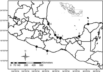
Haploporids were collected from the intestines of four fish species from nine localities in Mexico (see fig. 1): Dormitator latifrons from (1) Tres Palos Lagoon, Guerrero, type host and type locality of S. lamothei (16°48ʹ0ʺN, 99°47ʹ0ʺW), (2) Chacahua Lagoon, Oaxaca (15°58ʹ5ʺN, 97°40ʹ55ʺW); Astyanax aeneus from (3) Catemaco Lake, Veracruz, type host and type locality of S. chauhani (18°25ʹ0ʺN, 95°7ʹ0ʺW); Dormitator maculatus from (4) El Milagro Lagoon, Campeche (18°52ʹ25ʺN, 91°24ʹ39ʺW), (5) Boca del Río, Veracruz (19°6ʹ29ʺN, 96°6ʹ46ʺW), (6) Tamiahua Lagoon, Veracruz (21°15ʹ49ʺN, 97°27ʹ41ʺW), (7) Tlacotalpan, Veracruz (18°36ʹ0ʺN, 95°39ʹ0ʺW), (8) La Palma River, Veracruz (18°33ʹ21ʺN, 95°2ʹ59ʺW) and (9) El Corchito, Progreso, Yucatan (21°16ʹ40ʺN, 89°38ʹ38ʺW) where Gambusia yucatana was also collected. Fish were captured with seine nets or electrofished, transported alive to the laboratory and identified following Miller et al. (Reference Miller, Minckley and Norris2005). Individual fish were euthanized by pithing and immediately examined for helminths. Trematodes were fixed in hot (steaming) 4% formalin or 100% ethanol for DNA analyses.
Sampling sites in Mexico for specimens of Saccocoelioides spp. Legend for localities: 1, Tres Palos Lagoon, Guerrero; 2, Chacahua Lagoon, Oaxaca; 3, Catemaco Lake, Veracruz; 4, El Milagro Lagoon, Campeche; 5, Boca del Río, Veracruz; 6, Tamiahua Lagoon, Veracruz; 7, Tlacotalpan, Veracruz; 8, La Palma River, Veracruz; 9, El Corchito Progreso, Yucatan.

Morphological description
Unflattened digeneans preserved in 4% formalin were stained with Mayer's paracarmine or iron acetocarmine, dehydrated in an ethanol series, cleared with methyl salicylate and mounted in Canada balsam. All the specimens were examined using a bright-field Leica DM 1000 LED microscope (Leica, Wetzlar, Germany). Morphometric measurements were taken using the Leica Application Suite microscope software. Drawings were made with the aid of a drawing tube. All measurements are in micrometres.
Specimens collected in the present study were compared with the original description and with the holotype and paratypes of S. lamothei deposited in the Colección Nacional de Helmintos (CNHE), Instituto de Biología, Universidad Nacional Autónoma de México, Ciudad de México, México (holotype: CNHE 5920; paratypes: CNHE 5921a, 5921b). Type material of S. chauhani could not be examined since they were not available in the CNHE. Voucher specimens from the present study were deposited in the CNHE: S. lamothei from D. latifrons, 4 specimens, CNHE 9373; S. chauhani from A. aeneus, 5 specimens, CNHE 9852.
DNA extraction, sequencing and phylogenetic analyses
For DNA extraction, specimens were placed individually in tubes and digested overnight at 56°C in a solution containing 10 mm Tris–HCl (pH 7.6), 20 mm NaCl, 100 mm disodium EDTA (pH 8.0), 1% Sarkosyl and 0.1 mg/ml proteinase K. Following digestion, DNA was extracted from the supernatant using the DNAzol reagent (Molecular Research Center, Cincinnati, Ohio, USA) following the DNA protocol established by García-Varela & Nadler (Reference García-Varela and Nadler2005). Internal transcribed spacer 2 (ITS2) and the D1–D3 domains of the large subunit (LSU) from the ribosomal DNA (rDNA) were amplified using the forward primer BD3, 5′-GAA CAT CGA CAT CTT GAA CG-3′ (Hernández-Mena et al., Reference Hernández-Mena, García-Prieto and García-Varela2014), and the reverse primer 536, 5′-CAG CTA TCC TGA GGG AAAC-3′ (García-Varela & Nadler, Reference García-Varela and Nadler2005). Polymerase chain reactions (PCR) (25 μl) consisted of 1 μl of 10 μm of each primer, 2.5 μl of 10 × PCR Rxn buffer, 1.5 μl 2 mm MgCl2, 0.5 μl of deoxynucleoside triphosphates (dNTPs) 10 mm, 16.375 μl of water, 2 μl of genomic DNA and 0.125 μl (1 U) of Taq DNA polymerase (Platinum Taq, Invitrogen Corporation, São Paulo, Brazil). PCR cycling parameters for rDNA amplifications included denaturation at 94°C for 1 min; followed by 35 cycles of 94°C for 1 min, annealing at 50°C for 1 min and extension at 72°C for 1 min; followed by a post-amplification incubation at 72°C for 10 min. Sequencing reactions were performed using the forward and reverse primers mentioned above and four internal primers: 504 (5′-CGT CTT GAA ACA CGG ACT AAG G-3′), 502 (5′-CAA GTA CCG TGA GGG AAA GTT GC-3′) (García-Varela & Nadler, Reference García-Varela and Nadler2005), 503 (5′-CCT TGG TCC GTG TTT CAA GAC G-3′) (Stock et al., Reference Stock, Campbell and Nadler2001) and BD2 (5′-TAT GCT TAA ATT CAG CGG GT-3′) (Luton et al., Reference Luton, Walker and Blair1992), with ABI Big Dye (Applied Biosystems, Boston, Massachusetts, USA) terminator sequencing chemistry, and reaction products were separated and detected using an ABI 3730 capillary DNA sequencer. The electropherograms of each sequence were assembled conforming a consensus sequence, and base-calling differences were resolved using Codoncode Aligner version 5.0.2 (Codoncode Corporation, Dedham, Massachusetts, USA).
Sequences obtained in the current research from LSU rDNA and ITS2 were aligned separately with data from another 32 haploporid species downloaded from the GenBank dataset, including a sequence of Saccocoelioides sp. from Nicaragua (see Curran et al., Reference Curran, Tkach and Overstreet2006), plus other species from three families that were used as the outgroup (see supplementary table S1). The alignment consisted of 58 sequences with 1275 nucleotides for the LSU rDNA and 43 sequences with 417 nucleotides for the ITS2. The concatenated alignment contained 43 sequences with 1687 nucleotides. Alignments were constructed using the software Clustal W (Thompson et al., Reference Thompson, Gibson, Plewniak and Jeanmougin1997) with default parameters and adjusted manually with the Mesquite program (Maddison & Maddison, Reference Maddison and Maddison2011). The best model of nucleotide substitution for each dataset was identified with the Akaike Information Criterion (AIC) implemented in jModelTest v0.1.1 (Posada, Reference Posada2008). For ML analyses, the program RAxML v.7.0.4 (Stamatakis, Reference Stamatakis2006) was used with the option GTRGAMMAI and with 10,000 bootstrap replicates. The best model for each dataset was TVM + I + G for ITS2 and GTR + I + G for LSU, which were used with the Bayesian analyses (Huelsenbeck & Ronquist, Reference Huelsenbeck and Ronquist2001). Settings were two simultaneous runs of the Markov chain (MCMC) for 10 million generations, sampling every 1000 generations, a heating parameter value of 0.2 and a ‘burn-in’ of 25%. Trees were drawn using FigTree version 1.3.1 (Rambaut, Reference Rambaut2006). The genetic divergence among taxa was estimated using uncorrected p distances with the program MEGA version 6 (Tamura et al., Reference Tamura, Stecher, Peterson, Filipski and Kumar2013).
Results
Family: Haploporidae Nicoll, 1914.
Subfamily: Chalcinotrematinae Overstreet & Curran, Reference Overstreet, Curran, Jones, Bray and Gibson2005.
Genus: Saccocoelioides Szidat, Reference Szidat1954.
Saccocoelioides lamothei
Description
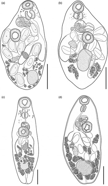
The specimens identified as S. lamothei collected from D. latifrons on Chacahua Lagoon (see fig. 1, locality 2; table 1) are similar to the type material of S. lamothei by having a pyriform to slightly elongated body with numerous eggs (10–11), the tegument with fine spines. Eye-spot remnants dispersed from oral opening to level of anterior margin of hermaphroditic sac. Oral sucker subterminal spherical. Ventral sucker spherical pre-equatorial. An absent prepharynx, pharynx oval, strongly muscular. Caecum sac-like, thick-walled, ending at mid-level of testis in unflattened specimens. Testis single, oval to spherical, terminal (flattened specimens) or subterminal (unflattened). Hermaphroditic sac oval. Internal seminal vesicle oval, filling posterior third of hermaphroditic sac. Ejaculatory duct short, prostatic cells in peripheral region of hermaphroditic sac. Genital pore pre-equatorial. Ovary oval to cylindrical, post-equatorial between ventral sucker and the testis. Uterus extends posterior to the testis; Vitelline follicles irregular in size and shape, can reach terminal or subterminal part of the body (fig. 2a, b).
Saccocoelioides lamothei: (a) holotype (CNHE 5920) from Dormitator latifrons, (b) specimen (CNHE 9373) from D. latifrons. Saccocoelioides chauhani: (c) specimen (CNHE 9852) from Astyanax aeneus, (d) specimen from A. aeneus (from Lamothe-Argumedo, Reference Lamothe-Argumedo1974). Scale bars: (a, b, d) 200 μm; (c) 100 μm.

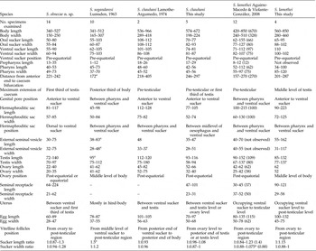
Comparative morphometric data for species of Saccocoelioides from North and Middle America. Data of the holotype of S. lamothei in parenthesis (CNHE 5920).

a Measured from the published figure. *Measured from holotype and paratype.
Remarks
The specimens of S. lamothei show morphological plasticity; for example, metrical data of newly collected material provided lower limits for pharynx length (34–100 this study vs. 52–112 original description) and external seminal vesicle width (31–117 vs. 40–55). Likewise, newly collected material provided higher limits for pharynx width (85–120 vs. 55–97), external seminal vesicle length (55–162 vs. 40–70) and seminal receptacle length (90–121 vs. 30–45) (see table 1).
Saccocoelioides chauhani
Description
The haploporids identified as S. chauhani collected from the intestine of Astyanax aeneus in Catemaco Lake (see fig. 1, locality 3; table 1) are similar to the type material of S. chauhani, by having an elongated body, with spines. Eye-spot remnants dispersed from prepharynx level to hermaphroditic sac level, not represented in original description. Oral sucker subterminal, spherical. Ventral sucker spherical, pre-equatorial. Prepharynx present. Pharynx oval, muscular. Caecum sac-like, ending at level pretesticular or at first third of testis in some specimens. Testis spherical, subterminal, located in the hind body. External seminal vesicle small and spherical. Hermaphroditic sac oval, located at ventral sucker level. Internal seminal vesicle oval, prostatic cells in peripheral region of hermaphroditic sac. Genital pore pre-equatorial. Ovary cylindrical, post-equatorial half between ventral sucker and testis. Uterus between ventral sucker level and testis level or ovary level. Excretory vesicle Y-shaped, bifurcating at anterior level of testis. Miracidium not developed (fig. 2c, d). Vitelline follicles reaching posterior end of body in flattened organisms (original description), and subterminal in unflattened organisms at testis level (fig. 2c, d).
Remarks
The specimens of S. chauhani from type locality and type host show morphological plasticity; for example, metrical data obtained for the newly collected material provided lower limits for body maximum width (198–224 this study vs. 289–418 original description), oral sucker length (70–77 vs. 108–112) and width (82–93 vs. 108–112), ventral sucker length (74–81 vs. 101–105), hermaphroditic sac length (77–105 vs. 112–128), testis length (93–116 vs. 112–120), ovary width (32–40 vs. 52–75) and egg length (70–97 vs. 101–105) (see table 1). Additionally, we also found higher limits on the range for the external seminal vesicle width (28–51 vs. 33–37) (table 1).
Saccocoelioides olmecae n. sp.
Description
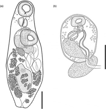
Based on 14 specimens from the type locality (see table 1). Measurements from holotype. Body elongated (fig. 3a), with maximum width at second third of body 527 × 200. Tegument thin, armed with fine spines. Eye-spot remnants dispersed from oral opening to level of anterior margin of hermaphroditic sac. Oral sucker spherical, subterminal 75 × 73. Ventral sucker spherical, larger than oral sucker, pre-equatorial, 90 × 94; sucker length ratio 1:1.2; sucker width ratio 1:1.28. Prepharynx short, 14. Pharynx oval, large, strongly muscular, 42 × 50, shorter than oral sucker length. Oesophagus longer than pharynx 103, extending posterior to level of ventral sucker. Intestinal bifurcation at level of the ovary. Caecum sac-like, thick-walled, ending at level first third of testis. Testis single, oval to spherical, subterminal, located in the posterior third of the body. External seminal vesicle spherical to oval, dorsal to ventral sucker, larger than internal seminal vesicle. Hermaphroditic sac oval to ellipsoidal (fig. 3b), obliquely arched around ventral sucker dorsally, 117 × 82. Internal seminal vesicle elongated–oval, filling posterior third to half of hermaphroditic sac length. Pars prostatic swollen, contiguous to the internal seminal vesicle. Ejaculatory duct and hermaphroditic duct are short and of similar length. Prostatic cells in peripheral region of hermaphroditic sac. Genital pore pre-equatorial, prominent, thick walled. Ovary elongated–oval, equatorial or post-equatorial 40 × 34, contiguous or separated from testis. Uterus occupying from level of ventral sucker opening to hermaphroditic sac, extending to first third of testis, metraterm thick walled. Numerous vitelline follicles (16) usually longitudinally elongate, distributed in two lateral fields, from ovary level, confluent to post-testicular level, reaching posterior end of the body. Eggs 72–89 × 40–41, miracidia not developed; at maximum 3 eggs in uterus from holotype. Excretory vesicle Y-shaped, excretory pore terminal.
Saccocoelioides olmecae n. sp. from Dormitator maculatus: (a) whole worm, holotype, ventral view; (b) hermaphroditic sac, paratype, ventral view. Scale bars: (a) 100 μm; (b) 50 μm.

Taxonomic summary
Type host
Dormitator maculatus (Bloch) (Perciformes: Eleotridae).
Accidental host
Gambusia yucatana Regan (Cyprinodontiformes: Poeciliidae) (see Discussion).
Site
Intestine.
Type locality
Boca del Río, Veracruz, Mexico (19°6′29″N, 96°6′46″W).
Other localities in Mexico
Tamiahua Lagoon, Veracruz (21°15′49″N, 97°27ʹ41ʺW); Tlacotalpan, Veracruz (18°36′0″N, 95°39ʹ0″W); La Palma River, Veracruz (18°33′21″N, 95°2′59″W); El Corchito Progreso, Yucatan (21°16′40″N, 89°38′38″W); El Milagro lagoon, Campeche (18°52′25″N, 91°24′39″W).
Type material
Holotype CNHE: 9846; paratypes CNHE: 9847. Voucher material CNHE: 9848–9851.
Representative sequences
Saccocoelioides olmecae n. sp. D. maculatus [GenBank KU061125–KU061133; KU061135–KU061136 (LSU rDNA gene); GenBank KU061106– KU061113; KU061115–KU061116 (ITS2 rDNA gene)]; S. olmecae n. sp. G. yucatana [GenBank KU061134 (LSU rDNA gene); GenBank KU061114 (ITS2 rDNA gene)].
Etymology
The specific epithet refers to Olmeca, a Mesoamerican civilization that lived along the Gulf of Mexico between 1400 and 1200 bce.
Remarks
The new species possesses the main morphological characters of the genus Saccocoelioides (Overstreet & Curran, Reference Overstreet, Curran, Jones, Bray and Gibson2005). Of the 16 species of genus Saccocoelioides reported from the Americas, 13 species are described from South America (Overstreet & Curran, Reference Overstreet, Curran, Jones, Bray and Gibson2005). Saccocoelioides olmecae n. sp. can be distinguished morphologically from 10 South American species (S. magnus Szidat, Reference Szidat1954; S. elongatus Szidat, Reference Szidat1954; S. szidati Travassos, Freitas & Kohn, Reference Travassos, Teixera de Freitas and Kohn1969; S. octavus Szidat, Reference Szidat1970; S. quintus Thatcher, Reference Thatcher1978; S. saccodontis Thatcher , Reference Thatcher1978; S. magnorchis Thatcher, Reference Thatcher1978; S. antonioi Lunaschi, Reference Lunaschi1984; S. carolae Lunaschi, Reference Lunaschi1984 and S. godoyi Kohn & Miranda-Froes, Reference Kohn and Miranda-Froes1986) by having the smallest body size (340–527 × 150–250) (see Szidat, Reference Szidat1954, Reference Szidat1970; Travassos et al., Reference Travassos, Teixera de Freitas and Kohn1969; Thatcher, Reference Thatcher1978; Lunaschi, Reference Lunaschi1984; Kohn & Miranda-Froes, Reference Kohn and Miranda-Froes1986). Saccocoelioides olmecae n. sp. can be distinguished from S. magniovatus Szidat, Reference Szidat1954 as the latter has a plump body and vitellarium at pre-testicular level vs. body elongated and vitellarium at testicular level in S. olmecae n. sp. Saccocoelioides tarpazensis Díaz & González, Reference Díaz and González1990 differs from the new species by having vitelline follicles that are not confluent at post-testicular level vs. vitelline follicles confluent at post-testicular level in S. olmecae n. sp., a wider oral sucker (80–130 for S. tarpazensis vs. 55–84 for S. olmecae n. sp.) and by the size of eggs (110–130 × 50–125 for S. tarpazensis vs. 60–89 × 28–47 for S. olmecae n. sp.). Finally, S. olmecae n. sp. differs from S. nanii Szidat, Reference Szidat1954 in the distribution of the uterus – in the latter, from ventral sucker level to pre-testicular level vs. from ventral sucker level to testicular level in S. olmecae n. sp. – and by distribution of the vitellarium – in the latter, not confluent at post-testicular level vs. confluent at post-testicular level in S. olmecae n. sp. (Szidat, Reference Szidat1954; Díaz & González, Reference Díaz and González1990).
Three of the 16 species of Saccocoelioides (S. lamothei, S. chauhani and S. sogandaresi) have been recorded in Mexico. Saccocoelioides olmecae n. sp. can be differentiated from S. lamothei by the following characters: a pyriform-elongated body, ovary length and by having vitelline follicles usually longitudinally elongated (see table 1). Saccocoelioides olmecae n. sp. differs from S. sogandaresi by having an elongated body (vs. pyriform, see Fig. 1 in Lumsden, Reference Lumsden1963), a sac-like caecum, vitelline follicles reaching posterior end of body, pre-pharynx length and on the ovary size (see table 1). Finally, S. olmecae n. sp. can be differentiated from S. chauhani on the following characters: body length, ovary size and by having a uterus that extends to the first third of the body (see table 1).
Molecular characterization and phylogenetic analyses
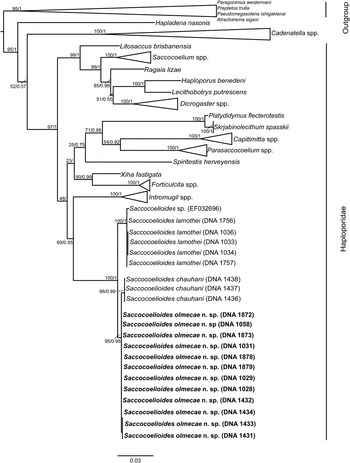
In this study, sequences of the LSU obtained from 12 individuals of S. olmecae n. sp. from six localities along the Gulf of Mexico (see supplementary table S1, fig. 1) were aligned with a LSU dataset containing new sequences of five specimens of S. lamothei and three specimens of S. chauhani from type locality plus Saccocoelioides sp. from Nicaragua (Curran et al., Reference Curran, Tkach and Overstreet2006), in addition to sequences of other haploporids (i.e. Capitimitta Pulis & Overstreet, 1982; Dicrogaster Looss, 1902; Forticulcita, Overstreet, 1982; Platydidymus Overstreet & Curran, 2005; Hapladena Linton, 1910; Haploporus, Looss, 1902; Intromugil, Overstreet & Curran, Reference Overstreet, Curran, Jones, Bray and Gibson2005; Lecithobotrys, Looss, 1902; Litosaccus, Pulis, Cribb & Overstreet, 2014; Parasaccocoelium, Zhukov, 1971; Ragaia Blasco-Costa, Montero, Gibson, Balbuena & Kostadinova, 2009; Saccocoelium, Looss, 1902; Spiritestis, Nagaty, 1948; Skrjabinolecithum, Belous, 1954; Xiha, Andres, Curran, Fayton, Pulis & Overstreet, 2015), and Atractotrema, Goto & Ozaki, 1929, Pseudomegasolena, Machida & Komiya, 1976, Preptetos, Pritchard, 1960, Cadenatella, Dollfus, 1946 and Paragonimus, Braun, 1899 from another four families were used as the outgroup. The LSU dataset was conformed by 1277 characters. The phylogenetic tree inferred with this dataset suggested that Haploporidae and 15 other genera of haploporids are monophyletic. However, this tree suggests that the taxonomy of Hapladena should be examined using morphological and molecular data because this genus is nested among outgroup taxa (fig. 4). The genus Intromugil, which includes trematodes infecting mullet fish, is sister to Saccocoelioides, which is divided into three clades corresponding S. lamothei, S. chauhani and S. olmecae n. sp. (fig. 4). The genetic divergence among the three species of Saccocoelioides was 1%, whereas isolates of S. olmecae n. sp. from D. maculatus and one isolate from G. yucatana were identical among the six localities sampled. The three sequences of S. chauhani were also identical. The five isolates of S. lamothei had a genetic divergence of 1%, including the sequences generated by Curran et al. (Reference Curran, Tkach and Overstreet2006) from an unidentified poeciliid from Nicaragua.
Maximum likelihood tree and consensus Bayesian inference trees inferred from the LSU dataset. Numbers near internal nodes show ML bootstrap clade frequencies and posterior probabilities (BI).

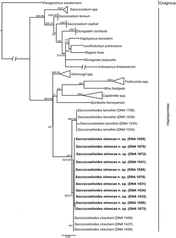
The ITS2 sequences of S. olmecae n. sp. (11 adults) were aligned with three isolates of S. lamothei, four isolates of S. chauhani and other genera of haploporids, forming a dataset of 419 characters. The genetic divergence among S. lamothei, S. chauhani and S. olmecae n. sp. ranged from 1 to 4%. The genetic divergence among the 11 isolates of the new species was very low, ranging from 0 to 1%. The phylogenetic tree inferred from this dataset did not support the monophyly of Haploporidae, and some genera, such as Saccocoelium and Dicrogaster, were paraphyletic with low support of bootstrap and posterior probabilities (fig. 5). This tree also showed that Intromugil is not a sister to Saccocoelioides (fig. 5).
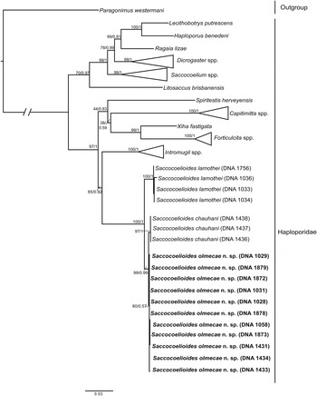
The concatenated alignment (LSU + ITS2) contained 11 specimens of S. olmecae n. sp., four isolates of S. lamothei, three isolates of S. chauhani, plus the same taxa used on the alignments with LSU and ITS2. Maximum likelihood (ML) analysis inferred with LSU, ITS2 and the combined dataset of both (LSU + ITS2) each yielded a single tree that was very similar in topology to the Bayesian inference (BI) consensus tree (figs 4–6). The ML and Bayesian consensus trees inferred with LSU (fig. 4) and that concatenated from both genes (fig. 6), showed that Intromugil is sister to Saccocoelioides, which is formed by S. lamothei, S. chauhani and S. olmecae n. sp. In addition, all the sequences of S. olmecae n. sp. are nested together within a monophyletic clade, with moderate bootstrap support and Bayesian posterior probability values (95/0.95 with LSU, 63/0.80 with ITS2 and 80/0.57 with the concatenated dataset) (figs 4–6).
Maximum likelihood tree and consensus Bayesian inference trees inferred from the ITS2 data set. Numbers near internal nodes show ML bootstrap clade frequencies and posterior probabilities (BI).

Maximum likelihood tree and consensus Bayesian inference trees inferred with the concatenated (LSU + ITS2) dataset. Numbers near internal nodes show ML bootstrap clade frequencies and posterior probabilities (BI).

Discussion
The species analysed in this study belong to Saccocoelioides according to the distribution of vitelline follicles in lateral fields of the body and by parasitizing freshwater fish from the Americas (see Lumsden, Reference Lumsden1963; Lamothe-Argumedo, Reference Lamothe-Argumedo1974; Overstreet & Curran, Reference Overstreet, Curran, Jones, Bray and Gibson2005; Aguirre-Macedo & Violante-González, Reference Aguirre-Macedo and Violante-González2008). However, other morphological characters diagnostic of the genus Saccocoelioides, such as the distribution of the uterus, the presence of a pre-pharynx, caecum sac-like and position of the testis are similar to those of Culuwiya, and therefore a taxonomic debate has emerged regarding Saccocoelioides and Culuwiya. The inclusion of molecular data is necessary to resolve the validity of some species of Saccocoelioides and those of the genus Culuwiya, as some species, such as Culwiya beauforti Hunter & Thomas, 1961; C. overstreeti Bargiela, 1988; C. papernai Bargiela, 1988; C. agonostomus Dyer, Bunkley-Williams & Williams, 1999, originally identified as Saccocoelioides, were transferred to the genus Culuwiya by Overstreet & Curran (Reference Overstreet, Curran, Jones, Bray and Gibson2005).
Our phylogenetic trees, obtained with the concatenated and separate datasets, confirmed that Saccocoelioides is monophyletic, with three sub-clades, each one represented by the three species analysed herein (S. lamothei, S. chauhani and S. olmecae n. sp.), with moderate support of bootstrap and posterior probability values. The phylogenetic analyses inferred with LSU and concatenated datasets showed that Saccocoelioides is sister to Intromugil, which contains four species associated with fish of the genus Mugil distributed along the Atlantic coast (Pulis et al., Reference Pulis, Fayton, Curran and Overstreet2013). The genetic divergence estimated with the LSU dataset among species of Saccocoelioides was 1.0%, and it ranged from 1.0 to 4% with ITS2. These values of genetic divergence among species are similar those of other haploporids. For instance, the genetic divergence found among four species of Saccocoelium (S. cephali Blasco-Costa, Montero, Gibson, Blabuena, Raga & Kostadinova, 2009; S. tensum Looss, 1902; S. obesum Looss, 1902 and S. brayi Blasco-Costa, Balbuena, Raga, Kostadinova & Olson, Reference Blasco-Costa, Balbuena, Raga, Kostadinova and Olson2010) ranged from 0.9 to 4.8% with LSU and from 2.1 to 10.9% with ITS2; between two species of Dicrogaster (D. perpusilla Looss, 1902 and D. contracta Looss, 1902) the genetic divergence was 4.6% and 8.7% with LSU and ITS2, respectively; and among three species of the genus Capitimitta (Capitimitta sp., C. darwinensis Pulis & Overstreet, Reference Pulis and Overstreet2013 and C. costata Pulis & Overstreet, Reference Pulis and Overstreet2013), it ranged from 2.7 to 2.8% with LSU and from 7.3 to 11% with ITS2 (see Blasco-Costa et al., Reference Blasco-Costa, Balbuena, Kostadinova and Olson2009, Reference Blasco-Costa, Balbuena, Raga, Kostadinova and Olson2010; Pulis & Overstreet, Reference Pulis and Overstreet2013).
Currently, Saccocoelioides contains 17 described species distributed in the Americas. In Middle America, S. olmecae n. sp. is the third species of the genus infecting eleotrid fish of the genus Dormitator Gill and it shows some level of host specificity, due to the fact that S. olmecae n. sp. was found in the fat sleeper (D. maculatus) along the Gulf of Mexico. However, a single, poorly developed specimen was collected from the intestine of G. yucatana, suggesting that this fish can act as an accidental host. Saccocoelioides lamothei has been reported from the intestine of D. latifrons from the Pacific coast of Mexico and from another four unrelated families of fish (Eleotridae, Gobiidae, Poeciliidae and Profundulidae) (Aguirre-Macedo & Violante-González, Reference Aguirre-Macedo and Violante-González2008; Andrade-Gómez, Reference Andrade-Gómez2015; Pinacho-Pinacho et al., Reference Pinacho-Pinacho, García-Varela, Hernández-Orts, Mendoza-Palmero, Sereno-Uribe, Martínez-Ramírez, Andrade-Gómez, Hernández-Cruz, López-Jiménez and Pérez-Ponce de León2015). Saccocoelioides sogandaresi has been found in fish of the orders Cyprinodontiformes (Goodeidae, Poeciliidae), Mugiliformes (Mugilidae) and Perciformes (Cichlidae, Gobiidae) from central and south-eastern Mexico (Scholz et al., Reference Scholz, Vargas-Vázquez, Moravec, Vivas-Rodríguez and Mendoza-Franco1995; Pérez-Ponce de León et al., Reference Pérez Ponce de León, García Prieto and Mendoza Garfías2007; Martínez-Aquino et al., Reference Martínez-Aquino, Mendoza-Palmero, Aguilar Aguilar and Pérez-Ponce de León2014). Finally, S. chauhani has only been found mainly in characid and poeciliid fish from Catemaco Lake in Veracruz, Mexico (Pérez-Ponce de León et al., Reference Pérez Ponce de León, García Prieto and Mendoza Garfías2007). In addition, our molecular analyses suggested that the specimen of Saccocoelioides sp. from Nicaragua, found in an unidentified poeciliid, belonged to S. lamothei (see Curran et al., Reference Curran, Tkach and Overstreet2006).
In this study, we combined morphological and molecular data with the aim of describing a new species of Saccocoelioides from along the coast of the Gulf of Mexico, and we also generated new morphological and molecular data of S. chauhani and S. lamothei that could represent the starting point to the study of this group of parasites. The inclusion of other species of Saccocoelioides from South America and North America, such as S. sogandaresi, is fundamental to a better understanding of the phylogenetic history of this complex and fascinating group of parasites.
Supplementary material
To view supplementary material for this article, please visit http://dx.doi.org/10.1017/S0022149X1600047X
Acknowledgements
We are grateful to Luis García Prieto for providing specimens deposited at the CNHE. We also thank Eduardo Hernandez Cruz for his help during the field work.
Financial support
This research was supported by grants from the Programa de Apoyo a Proyectos de Investigación e Innovación Tecnológica (PAPIIT) IN206716 and the Consejo Nacional de Ciencia y Tecnológia (CONACYT) 179048 to M.G.V. L.A.G. is grateful for the support of the Programa de Posgrado en Ciencias Biológicas, Universidad Nacional Autónoma de México, that enabled him to complete his MSc, and to CONACYT for granting a scholarship. C.D.P.P. benefited from a PhD student grant from the Programa de Posgrado en Ciencias Biológicas, UNAM and CONACYT.
Conflict of interest
None.