INTRODUCTION
Microsporidian parasites are obligate intracellular pathogens, currently classified as Fungi (James et al. Reference James, Kauff, Schoch, Matheny, Hofstetter, Cox, Celio, Fraker, Miadlikowska, Lumbsch, Rauhut, Reeb, Arnold, Amtoft, Stajich, Hosaka, Sung, Johnson and O'Rourke2006), that can parasitize most invertebrates and vertebrates (Canning and Vavra, Reference Canning, Vavra, Lee, Leedale and Bradbury2000), often causing severe disease and population collapse (e.g. European Honeybees; Williams et al. Reference Williams, Shafer, Rogers, Shutler and Stewart2008). The culture of parasitic microsporidians has had continued success for many insect- and human-infecting microsporidians (Monaghan et al. Reference Monaghan, Kent, Watral, Kaufman, Lee and Bols2009). Within cell culture, microsporidians are able to complete their life cycle and produce infective spores at production levels for identification, experimentation, preliminary drug screening and biocontrol (e.g. Paranosema locustae; Sokolova et al. Reference Sokolova, Dolgikh, Morzhina, Nassonova, Issi, Terry, Ironside, Smith and Vossbrinck2003; Monaghan et al. Reference Monaghan, Kent, Watral, Kaufman, Lee and Bols2009).
Culturing fish-infecting microsporidians has had some success, but not compared to the level achieved with human- and insect-infecting microsporidians (Monaghan et al. Reference Monaghan, Kent, Watral, Kaufman, Lee and Bols2009). It is possible that the difficulty relates to fish microsporidians displaying higher host specificity than many of the widely infective insect microsporidians. One factor for increased host-specificity in fish could be that the majority of microsporidians develop diffusely within the host body cells, but several species infect cells and transform the host cells into hypertrophic cell–parasite complexes termed xenomas (Lom and Dyková, Reference Lom and Dyková2005). Only a few microsporidian species cause xenoma development – the majority being species that infect fish (Lom and Dyková, Reference Lom and Dyková2005). Xenomas are always single cells that are hypertrophic compared to surrounding cells, and contain all life stages of the parasite. They offer optimal growth conditions for developing microsporidians and their structure varies based on the species and the stage of development (Lom and Dyková, Reference Lom and Dyková2005).
A xenoma-forming fish microsporidian that is of particular interest is Loma salmonae, the causative agent of microsporidial gill disease of salmon. Loma salmonae xenomas develop within gill tissue and elicit chronic inflammatory bronchitis and subsequent respiratory distress (Speare and Lovy, Reference Speare, Lovy, Woo and Buchmann2012). A rainbow trout (Oncorhynchus mykiss)-L. salmonae disease transmission model developed in our laboratory has successfully propagated spores for use in experiments studying the pathogenicity, immunology and life cycle characteristics of this pathogen (Speare and Lovy, Reference Speare, Lovy, Woo and Buchmann2012). Several limitations of the in vivo model include a 6 week waiting period for the infection to mature to propagate spores, reliable drug screening cannot be effectively completed and understanding the early developmental biology of xenomas cannot be easily done.
Of great interest is the development of fish-infecting microsporidians in cell culture, as they often cause high mortality rates in economically important species (Shaw and Kent, Reference Shaw, Kent, Wittner and Weiss1999; Becker and Speare, Reference Becker and Speare2007). Currently, no in vitro model supporting xenoma-forming microsporidia is available to show how xenomas occur and whether microsporidians can develop into a xenoma when the cell is not under the influence of the host organism (Lom and Dyková, Reference Lom and Dyková2005). It would be beneficial to develop L. salmonae at production levels in vitro to reduce fish numbers used in our model, and to propagate spores for vaccine use. The purpose of the present study was to determine whether a rainbow trout gill epithelial cell line (RTG-1) would be permissive to growth of L. salmonae. We outline some preliminary findings that xenoma development can occur within RTG-1 cells and that there is the potential for RTG-1 cells to propagate L. salmonae spores.
MATERIALS AND METHODS
Spore purification
Spores were processed using the methods described in McConnachie et al. (Reference McConnachie, Guselle and Speare2013). Briefly, rainbow trout heavily infected with L. salmonae xenomas in the gills were euthanized by an overdose of benzocaine (120 mg L−1; Sigma-Aldrich). The gills were immediately dissected from the fish and placed in sterile saline containing 2 mg of penicillin/streptomycin and 100 μg gentamicin per mL−1. Gills were homogenized using a tissue grinder, pushed through a cell dissociation sieve, 20 μ m Nytex® mesh and then rinsed with saline. The material was centrifuged (350 g ) and the pellet was resuspended in 20 mL sterile distilled water and vortexed with an equal volume of percoll. The mixture was centrifuged (~1000 g ) and the spores pelleted at the bottom of the tube while the remaining cellular debris was removed in the percoll-water layers. The cells were washed and resuspended in sterile saline containing 0·1 mg units of penicillin/streptomycin and 50 μg gentamicin per mL−1 until further use. Spores were used within 14 days after being purified.
Maintenance of RTG-1 cell line
Rainbow trout gill epithelial cells (RTG-1) were originally obtained from the American Type Culture Collection and grown at 20°C. Cells were maintained in L-15 (Leibovitz) medium with 2 mm l-glutamine, 10% fetal bovine serum and 100 IU mL−1 penicillin/streptomycin (henceforth known as complete medium). Cells were kept in 75 cm2 flasks at ~75% confluence until experimentation. Cells were split and placed into 6-well plates prior to experimentation. Cells were allowed to reach ~80% confluence (~1·0×106 cells) before spore inoculation.
In vitro exposure of L. salmonae spores to RTG-1 cells
Spores were centrifuged and resuspended in complete media. Spores were then counted using a haemocytometer and spore viability was verified using an in-house produced (J. Sheppard; Speare et al. Reference Speare, Daley, Markham, Sheppard, Beaman and Sanchez1998) monoclonal antibody/propidium iodine dye exclusion test. Spore concentrations were adjusted to ensure that a ratio of 10:1 spores to cells was plated onto the RTG-1 cells (Monaghan, Reference Monaghan2011). Approximately 1·0×107 spores were placed into each well, and control wells were maintained. Spores were left on the plates for 3 days and then removed; the monolayer was rinsed with complete media and then re-fed with complete medium. Otherwise, media was replaced weekly.
Detection of spore production on RTG-1 cells
To determine whether spores were developing in RTG-1 cells, a separate experiment was designed. Cells were plated in 12·5 cm2 (Falcon) and 75 cm2 flasks and allowed to reach ~80% confluence. Spores were plated on the 12·5 cm2 flasks at a 10:1 spore to cell ratio. Spores were left on the cell monolayer for 3 days, removed within the supernatant, centrifuged (350 g ) and resuspended in complete medium. The resuspended spores were counted using a haemocytometer and placed back into the 12·5 cm2 flasks on the monolayer. After 7 days, the spores were removed, centrifuged, resuspended in complete medium, counted and placed onto a 75 cm2 flask with RTG-1 cells. Twice a week, spores were removed from the 12·5 cm2 flasks, centrifuged, resuspended, counted and placed back on to the corresponding 75 cm2 flask for up to 4 weeks. Spores on the corresponding 12·5 and 75 cm2 flasks were counted once a week and re-plated. Control flasks were maintained and two replicates were completed. Mean spore numbers produced each week ±s.e. were determined using JMP10 software (SAS Institute, Cary, NC).
Light and transmission electron microscopy
Cells on the six-well plates were observed using bright field and phase contrast light microscopy on an inverted microscope. Previous pilot studies suggested that spore production became noticeable around 10 days post-exposure (PE), and thus samples were removed and processed for transmission electron microscopy (TEM) at 10 days and then weekly for up to 4 weeks. Control cells were sampled at each time point. Cells were removed from the flask by pipetting the media gently and were placed into 2% glutaraldehyde (EM-grade, Canemco & Merivac) in 0·1 m phosphate buffer (pH 7·2). Cells that did not lift by agitation were rinsed with versene (Gibco®), phosphate buffered saline and then exposed to TrypLE (Gibco®) for ~1 min. The TrypLE was deactivated with complete media and the mixture was added to the glutaraldehyde solution. Cells were fixed at room temperature for 1 h, centrifuged and resuspended in fresh 2% glutaraldehyde in 0·1 m phosphate buffer and left for ~12 h. Cells were spun down (~4000 g ) and pellets were washed using phosphate buffer, post-fixed in phosphate buffered 1% osmium tetroxide (Canemco & Merivac) for 1 h and then placed in agar. After several dehydration steps using ethanol and propylene oxide, cell pellets were embedded in Spurr's resin. After resin polymerization, semi-thin (0·5 μ m thick) sections were cut and stained with 1% toluidine blue in 1% sodium tetraborate (Sigma-Aldrich) to observe the cells using light microscopy. When sections containing spores or signs of cellular hypertrophy were observed, thin sections (70–90 nm thick) were cut and stained with uranyl acetate (depleted uranium; Canemco & Merivac), and lead citrate (Canemco & Merivac). Stained thin sections were examined using a Hitachi H7500 TEM (BioTEM, Nissei-Sangyo) operated at 80 kV. Images were captured using the AMTV600 digital image capture engine and an AMT XR 40 digital camera (Danvers, MA, USA).
RESULTS
Light microscopy and detection of spore production on RTG-1 cells
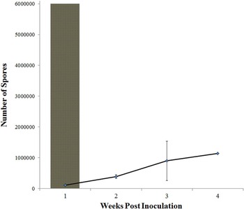
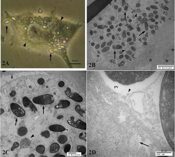
Spores were being produced at 1 week PE, and a steady increase in spore numbers was observed up to 4 weeks (Fig. 1). By 3 days PE, several cells appeared hypertrophic and darker than other cells. Using phase contrast microscopy, spores were evident within the hypertrophic cells by 5 days PE (Fig. 2A). The hypertrophic cells had spores associated with their surface and many spores packed within their cytoplasm surrounding the nucleus (Fig. 2A). Approximately 10 days PE, after spores had been washed from the monolayer, many spores were still observed on the RTG-1 monolayer. The monolayer became ragged and easily detachable by 1 week, but appeared to recover (become adherent) by 4 weeks. After 4 weeks the number of spores being produced decreased in the flasks.
Loma salmonae spore production over a 4 weeks period, within RTG-1 (SYMBOL) as compared to the inoculating dose (6×10⁶ spores). Data are mean (±s.e.) spore numbers produced in each replicate flask (N = 2 replicates).

Phase contrast and TEMs depicting a mature xenoma within RTG-1 containing L. salmonae spores. (A) Phase contrast micrograph of an adhered RTG-1 cell apparently filled with L. salmonae spores, 5 days PE. Spores appear to be on the surface of (arrows), and within (arrowheads) the cytoplasm surrounding the nucleus (N) of the host cell; (B) TEM of a xenoma containing L. salmonae spores, 10 days PE, surrounded by non-infected RTG-1 cells (*). Many mature spores (arrows), and various degraded parasite developmental stages (arrowheads) are present throughout the cytoplasm of the xenoma; (C) higher magnification of the same xenoma showing empty spores (*) along with mature spores (large arrows) and degraded developmental stages (arrowheads) within the cytoplasm and the distinct cell membrane (small arrow) adjacent to surrounding non infected cells; (D) TEM showing blebbing (arrow) of portions of the peripheral cytoplasm and surrounding plasma membrane of the xenoma. Tubular elements (arrowhead) are evident within the parasitophorous vacuole (PV) containing mature L. salmonae spores.

Transmission electron microscopy
Control and uninfected RTG-1 cells were epithelial-like with attenuated cytoplasmic processes and typical spherical nuclei. Secondary lysosomes containing material phagocytized from the media (e.g. dead cells and membranous components) were commonly observed within control and spore-exposed cells. Within spore-exposed samples, intact spores were found surrounding the cells with no cellular associations. Several cells did contain single, intact spores within their cytoplasm that were often associated with secondary lysosomes and some of these spores appeared degraded with a loss in electron density.
Serial, ultrastructural examination of a hypertrophic, spore filled cell, first observed in a toluidine stained semi-thin section revealed the structure was a xenoma containing mostly intact mature spores that demonstrated the characteristic 15–17 polar tube coils (Fig. 2B and C). The xenoma host cell was much larger than the surrounding, non-infected cells. Many mature spores, empty spores and various, degraded parasite developmental stages were present within large irregular-shaped parasitic vacuoles throughout the cytoplasm of the xenoma (Fig. 2C and D). The cytoplasm of the xenoma appeared to be less electron dense compared to surrounding cells, and portions of the peripheral cytoplasm and surrounding plasma membrane of the xenoma exhibited blebbing (Fig. 2C and D). Tubular elements were evident within parasitic vacuoles containing mature and empty spores (Fig. 2D).
DISCUSSION
This study presents the first example of microsporidian xenoma development within cell culture, verified by ultrastructural examination. Our observations of a complete life cycle of L. salmonae within cell culture suggest that there is a potential to produce this parasite in vitro, using RTG-1 cells. It was also determined that we are able to propagate spores, albeit at low numbers. Further manipulations of this model are underway to determine if this type of spore development can be optimized.
It is interesting to note that the xenoma observed at 10 days PE was mature and approaching the degradation stage. Within fish, L. salmonae xenomas do not mature until around 6 weeks PE (Speare and Lovy, Reference Speare, Lovy, Woo and Buchmann2012). Our observations of hypertrophic, spore-filled cells by 3 days PE, and the mature state of the xenoma at 10 days PE suggest that the development of the xenoma occurs in a short period of time compared to development in vivo. Even so, the length of the developmental period in vivo may reflect the selective xenoma development within fish gill tissue. Previous work has shown that xenoma production in the gills is biphasic, and although the majority of xenomas form around weeks 5–6 PE, there are others that form as early as 3 weeks PE (Speare et al. Reference Speare, Daley, Markham, Sheppard, Beaman and Sanchez1998). Loma salmonae circulates within the body of fish in vivo for 2 weeks prior to being detected in the gill by in situ hybridization, and thus mature xenomas by 3 weeks would suggest a 1 week maturation period, consistent with our observations (Speare et al. Reference Speare, Daley, Markham, Sheppard, Beaman and Sanchez1998). Additionally, non-ideal culture conditions may have contributed to early degradation (e.g. nutrient availability). Further in vivo and TEM studies are underway to capture development of the parasite within 10 days of being exposed to RTG-1 cells.
Microsporidian xenomas have not previously been described to develop within cultured cells. One study of a human microsporidian did elicit some xenoma-like growth within E6 cells (Leitch et al. Reference Leitch, Shaw, Colden-Stanfield, Scanlon and Visvesvara2005). The growths were determined not to be true xenomas, but rather aggregates of multiple E6 cells fusing together to form large cells with many developing spores within them (Leitch et al. Reference Leitch, Shaw, Colden-Stanfield, Scanlon and Visvesvara2005). Lores et al. (Reference Lores, Rosales, Mascaró and Osuna2003) elicited development of fish-infecting Glugea sp. spores within the mosquito Aedes albopictus cell line without true xenoma formation. More recently, development of Loma morhua within a larval cod-derived cell line (GML-5) was shown to occur slowly (~36 days PE) and at extremely low infection levels although xenoma development was not confirmed or discussed (McLeod, Reference McLeod2012). Loma morhua specifically infects Atlantic cod (Gadus morhua) and failed to develop within haddock embryo and RTG-1 cells, further suggesting that host specificity is extremely important in xenoma-forming microsporidians (McLeod, Reference McLeod2012). RTG-1 cells appear to be permissive for xenoma development by L. salmonae, which is intuitive since the major site of infection in salmonids is within gill tissue in endothelial, and other epithelial-type cells (Speare and Lovy, Reference Speare, Lovy, Woo and Buchmann2012).
Our results indicate that xenoma formation can occur in cell culture without influence from the host organism. With improvements, our L. salmonae-RTG-1 cell model has the potential to be useful for further study and propagation of L. salmonae and other xenoma-forming microsporidians. Further studies probing how, when, and why xenoma development occurs may be possible using our model. It may even be possible to determine the specific inducing factor(s) that elicit hypertrophic growth and early development in microsporidian xenomas (Lom and Dyková, Reference Lom and Dyková2005). Further development and optimization of an in vitro technique supporting the entire life cycle of this microsporidian will also provide, when compared to the current in vivo model, a more functional platform for broad and in depth assessment of host–pathogen relationships at the cellular level.
AKNOWLEDGEMENTS
We would like to thank Dr Dorota Wadowska for her technical assistance in the electron microscopy laboratory. We greatly appreciate the support this work received from the Natural Sciences and Engineering Research Council of Canada (NSERC) through the Canadian Integrated Multi-trophic Aquaculture Network (CIMTAN) in collaboration with its partner Fisheries and Oceans Canada, the University of New Brunswick, Cooke Aquaculture Inc., Kyoquot SEAfoods Ltd and Marine Harvest Canada Ltd.
FINANCIAL SUPPORT
This work was supported by the Natural Sciences and Engineering Research Council of Canada (NSERC) through the Canadian Integrated Multi-trophic Aquaculture Network (CIMTAN): NSERC Strategic Network Grant #NETGP 372815-08.