

Introduction
Didymoceras is a heteromorphic ammonoid that is common to abundant in late Campanian rocks of the Western Interior Seaway (Kennedy et al. Reference Kennedy, Landman, Cobban and Scott2000). The abundance of this genus has led to the use of several species as biostratigraphic index fossils in the Western Interior of the United States (Larson et al. Reference Larson, Jorgensen, Farrar and Larson1997; Kennedy et al. Reference Kennedy, Landman, Cobban and Scott2000; Cobban et al. Reference Cobban, Walaszczyk, Obradovich and McKinney2006; Landman et al. Reference Landman, Kennedy, Cobban and Larson2010). While much attention has been paid to biostratigraphy, little is known about the constraints on hydrostatics, locomotion, and mode of life for these aberrantly coiled ammonoids. Didymoceras, along with other genera within Nostoceratidae, can be characterized by irregular helical whorls that terminate in a retroversal U-shaped hook at maturity. Embryonic shells (ammonitellae) of D. stevensoni and D. cheyennense are characterized by 1.5 to 2.5 loosely coiled whorls, while the ammonitella of D. nebrascense is unknown (Larson et al. Reference Larson, Jorgensen, Farrar and Larson1997; Kennedy et al. Reference Kennedy, Landman, Cobban and Scott2000). The juvenile phase of Didymoceras (beyond the ammonitella) is relatively orthoconic (straight shelled) to slightly curved. This general morphology can vary considerably, and the apical-most portions are poorly known (Kennedy et al. Reference Kennedy, Landman, Cobban and Scott2000). The profound changes in coiling schemes during ontogeny are expected to result in changes in the hydrostatic properties acting on these cephalopods during life. This variation contrasts with that in most planispiral ammonoids, which do not undergo such drastic changes in shell morphology or hydrostatics throughout ontogeny (see Klug [2001] for description of some notable exceptions). Changing hydrostatic properties imply differences in the constraints influencing orientation, locomotion, and mode of life. These are first-order questions about ammonoid paleobiology that, once answered, can shed light on the paleoecology of this enigmatic group of cephalopods. Here, syn vivo hydrostatic properties for multiple phases of ontogeny are investigated for three species of Didymoceras (D. stevensoni, D. nebrascense, and D. cheyennense). While these species are mostly known from the Western Interior Seaway, their physical properties are more broadly relevant for the cosmopolitan nostoconic morphotype.
Ectocochleate Cephalopod Hydrostatics
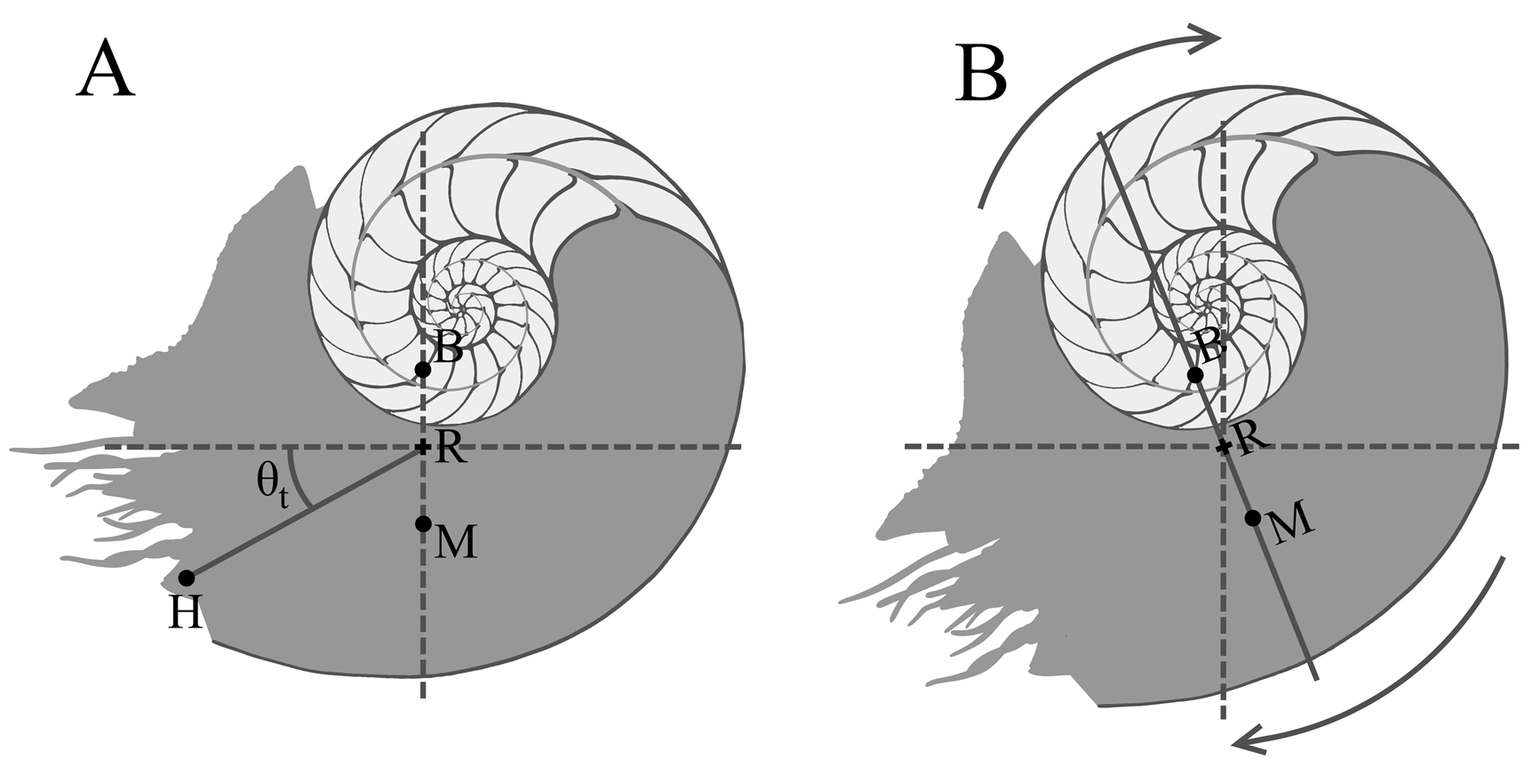
Like other ectocochleate (externally shelled) cephalopods, Didymoceras has a shell consisting of a body chamber and a phragmocone. The phragmocone acts as a buoyancy apparatus to reduce organismal weight relative to the weight of the water displaced by the entire animal. A neutrally buoyant condition occurs when these two quantities are equal. The orientation of an ectocochleate during life and its hydrostatic stability can be determined by understanding the relative positions of the centers of buoyancy and mass (Fig. 1). The center of buoyancy is equal to the center of volume of the displaced seawater, while the center of mass depends upon the total 3D distribution of organismal mass (which is influenced by each material of unique density in the living ectocochleate). The equilibrium orientation occurs when the centers of buoyancy and mass are vertically aligned (Fig. 1). Hydrostatic stability is proportionate to the separation between these two centers (Trueman Reference Trueman1941; Raup and Chamberlain Reference Raup and Chamberlain1967; Saunders and Shapiro Reference Saunders and Shapiro1986; Okamoto Reference Okamoto, Landman, Tanabe and Davis1996; Tajika et al. Reference Tajika, Naglik, Morimoto, Pascual-Cebrian, Hennhöfer and Klug2015b; Naglik et al. Reference Naglik, Rikhtegar and Klug2016; Peterman et al. Reference Peterman, Barton and Yacobucci2019a,Reference Peterman, Ciampaglio, Shell and Yacobuccib). A stability of zero would result in a condition similar to zero gravity, while higher stabilities would impart larger restoring moments acting to return the living cephalopod to its equilibrium orientation (Okamoto Reference Okamoto, Landman, Tanabe and Davis1996; Peterman et al. Reference Peterman, Ciampaglio, Shell and Yacobucci2019b). The thrust angle (Fig. 1) describes the efficiency of locomotion in the direction of movement and the rocking behavior that occurs in response to normal locomotion via jet propulsion. This property is defined as the angle between the center of rotation (midpoint between centers of buoyancy and mass) and the location of the animal's hyponome (source of thrust via expelled jet of water). Idealized horizontal movement without rocking occurs when the location of the hyponome and center of rotation are aligned horizontally (thrust angle approaches zero). Lower thrust angles where the aperture is facing downward would result in idealized upward vertical movement. However, this alignment depends on differential drag and lift, which could skew movement direction, especially at higher velocities (Klug and Korn Reference Klug and Korn2004). All of these hydrostatic properties act as constraints on locomotion and therefore have implications for understanding how the living cephalopod could have swum, captured prey, escaped predators, migrated, and mated.
Hypothetical diagram of the hydrostatic metrics used in this study: center of buoyancy (B), center of mass (M), center of rotation (R), location of hyponome (H), thrust angle (θt). A, Static setting in equilibrium (i.e., B and M aligned vertically). B, Cephalopod displaced from equilibrium, imparting a restoring moment (arrows). Light gray, phragmocone; dark gray, soft body, based on living Nautilus.

Previous Modeling of Ectocochleate Cephalopods
It is difficult to model heteromorphic ammonoids due to their complex coiling schemes and constantly changing occupation of 3D space throughout ontogeny. The earliest models of ectocochleate hydrostatics were accomplished by finding the approximate centers of buoyancy and mass from 2D medial sections (Moseley Reference Moseley1838; Trueman Reference Trueman1941; Westermann Reference Westermann1977, Reference Westermann2013; Saunders and Shapiro Reference Saunders and Shapiro1986). A 2D approach may approximate the hydrostatics of simple, bilaterally symmetric shapes, but is not possible for heteromorphs that coil about multiple axes. Morphogenetic models have been created in three dimensions to investigate the growth of ammonoids and other mollusks (Okamoto Reference Okamoto1988b, Reference Okamoto, Landman, Tanabe and Davis1996; Checa et al. Reference Checa, Okamoto and Keupp2002; Urdy et al. Reference Urdy, Goudemand, Bucher and Chirat2010). Heteromorphic ammonoids in particular were generated in three dimensions with models that simulate a growing tube (Okamoto Reference Okamoto1988b,Reference Okamoto, Landman, Tanabe and Davis1996) and that accommodate changes in coiling through ontogeny. An alternate method is proposed in the current study, which builds upon these biological growth models, but also accounts for the total 3D distribution of mass and internal morphology to compute various hydrostatic properties. Tomographic techniques have been increasingly used to generate very accurate 3D models of ectocochleate conches. Computed tomography (CT) scanning can be used to image 3D volumes based on X-ray attenuation factors within a matrix containing fossil ectocochleates (Hoffmann et al. Reference Hoffmann, Schultz, Schellhorn, Rybacki, Keupp, Gerden, Lemanis and Zachow2014, Reference Hoffman, Lemanis, Naglik, Klug, Klug, Korn, De Baets, Kruta and Mapes2015, Reference Hoffmann, Lemanis, Falkenberg, Schneider, Wesendonk and Zachow2018; Lemanis et al. Reference Lemanis, Zachow, Fusseis and Hoffmann2015, Reference Lemanis, Korn, Zachow, Rybacki and Hoffmann2016; Inoue and Kondo Reference Inoue and Kondo2016). Similarly, serial grinding tomography can create 3D models by successively grinding through the fossil and partitioning the objects of interest (Tajika et al. Reference Tajika, Morimoto, Wani, Naglik and Klug2015a,Reference Tajika, Naglik, Morimoto, Pascual-Cebrian, Hennhöfer and Klugb; Naglik et al. Reference Naglik, Monnet, Goetz, Kolb, De Baets, Tajika and Klug2015a, Reference Naglik, Rikhtegar and Klug2016). However, these methods require significant contrasts in physical properties to distinguish the fossil conch from its surroundings. Tomographic techniques also require relatively complete and undeformed specimens, which occur very rarely for most nostoceratid heteromorphs, especially Didymoceras (Kennedy et al. Reference Kennedy, Landman, Cobban and Scott2000). Such difficult morphotypes can be theoretically modeled with array generation algorithms, similar to the morphospace parameters of Raup (Reference Raup1967), and photogrammetric models of reconstructed specimens (see methods of Peterman et al. Reference Peterman, Barton and Yacobucci2019a,Reference Peterman, Ciampaglio, Shell and Yacobuccib; Peterman and Barton Reference Peterman and Barton2018). These methods allow the 3D analysis of complicated shapes to determine hydrostatic properties.
Paleobiology of Didymoceras
Didymoceras is traditionally thought of as a poor swimmer due to its aberrant shape (Westermann Reference Westermann, Landman, Tanabe and Davis1996). This shape would have caused considerable hydrodynamic drag, and the cumbersome orientation of the aperture may have further hindered rapid movement and migration over long distances (Landman et al. Reference Landman, Kennedy, Cobban and Larson2010). Fossils of this genus from the Western Interior Seaway of the United States yield isotopic compositions similar to those of benthic inoceramids, suggesting they lived very close to the seafloor (He et al. Reference He, Kyser and Caldwell2005; Slattery et al. Reference Slattery, Clementz and Johnson2007). Didymoceras is known from methane seeps in the Western Interior Seaway, which may have served as oases in terms of food supply and oxygenation on a seafloor that was otherwise rather poor in prey (Kauffman et al. Reference Kauffman, Arthur, Howe and Scholle1996; Landman et al. Reference Landman, Cochran, Larson, Brezina, Garb and Harries2012, Reference Landman, Cochran, Slovacek, Larson, Garb, Brezina and Witts2018). However, ammonoids occurring at these seeps are usually common elsewhere in the basin as well. Ammonoid jaws can shed light on their diet and paleoecology within their corresponding habitats (Tanabe and Landman Reference Tanabe, Landman, Summesberger, Histon and Daurer2002; Kruta et al. Reference Kruta, Rouget, Landman, Tanabe and Cecca2009, Reference Kruta, Landman, Rouget, Cecca and Larson2010). Kruta et al. (Reference Kruta, Landman, Rouget, Cecca and Larson2010) described the jaws of Didymoceras nebrascense, reporting a beak-like morphology that may have served a grasping and cutting function. They suggest this morphology corresponds to a macrophagous diet and reject an opercular function due to the very thin calcitic covering of the lower jaw. Other closely related nostoceratids likely had similar jaw morphologies (Tanabe and Landman Reference Tanabe, Landman, Summesberger, Histon and Daurer2002; Kruta et al. Reference Kruta, Landman, Rouget, Cecca and Larson2010). These lines of evidence support the interpretation of Didymoceras as a slow-moving predator that fed on benthic prey, at least in later life stages. However, the exact nature of this ammonoid's orientation and locomotory ability—and how those may have changed through ontogeny—is still unclear. Hence, the investigation of nostoconic hydrostatics presented here provides important insights into the paleobiology of this extraordinary cephalopod.
Methods
The virtual reconstruction of three species of Didymoceras (D. stevensoni, D. nebrascense, and D. cheyennense) largely follows the methods of Peterman et al. (Reference Peterman, Barton and Yacobucci2019a) and Peterman and Barton (Reference Peterman and Barton2018) with some improvements described below. Specimens used to create the virtual 3D models of these species are housed in the National Museum of Natural History (USNM) and Wright State University Lake Campus collections (WSU). Final digital models of the three species of Didymoceras are stored online at the data-sharing website morphosource.org (Project: Didymoceras Theoretical Models; Peterman Reference Peterman2019).
Creating Hydrostatic Model Components
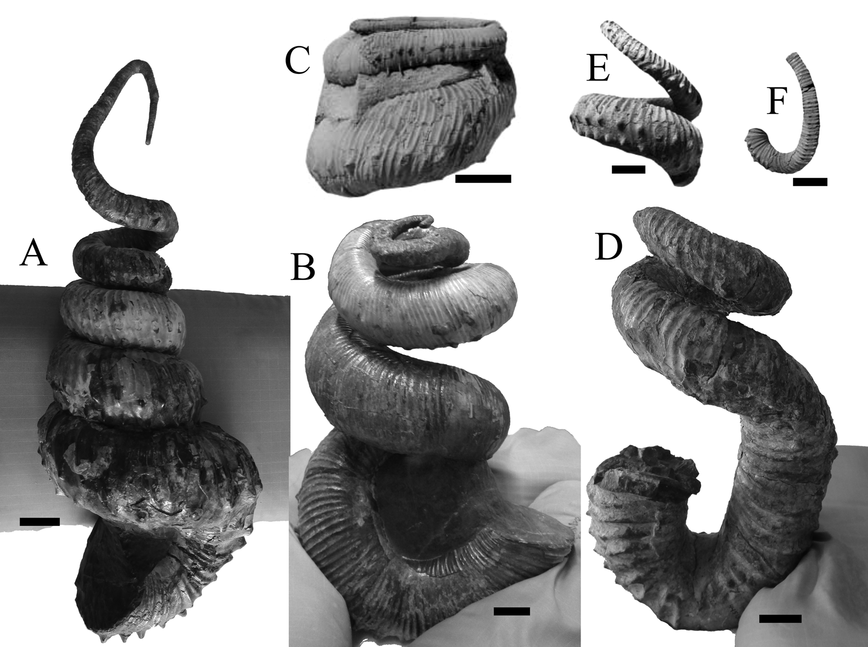
Exterior 3D models of Didymoceras were created via photogrammetry to serve as stencils for virtual reconstruction. The software 3DF Zephyr (3DFlow 2018) was used to create the initial models, which involved taking ~50 photographs each around the object of interest and using geomatic algorithms to create 3D models of the object's exterior. Well-preserved, remarkably complete, and properly reconstructed specimens of D. stevensoni (USNM 430976), D. nebrascense (USNM 430977), and D. cheyennense (USNM 445155) served as the initial models to serve as stencils (Fig. 2). Missing apical portions of the models of D. nebrascense and D. cheyennense were filled in with the specimens USNM 450333 and USNM 445099/482458, respectively. However, the juvenile-most pieces of Didymoceras just after the ammonitella are poorly known. Therefore, each apical model extrapolates the juvenile phase to the approximate location of the ammonitella, then terminates. In the current study, we do not investigate the hydrostatic properties at earlier ontogenetic states than the initial uncoiled juvenile shell.
USNM specimens used for model construction. Didymoceras stevensoni (A, USNM 430976). Majority of D. nebrascense (B, USNM 430977), apical portion of D. nebrascense (C, USNM 450333). Majority of D. cheyennense (D, USNM 445155), apical portions of D. cheyennense (E, USNM 445099; F, 482458). C, E, and F are modified from Kennedy et al. (Reference Kennedy, Landman, Cobban and Scott2000). Scale bars, 2 cm.

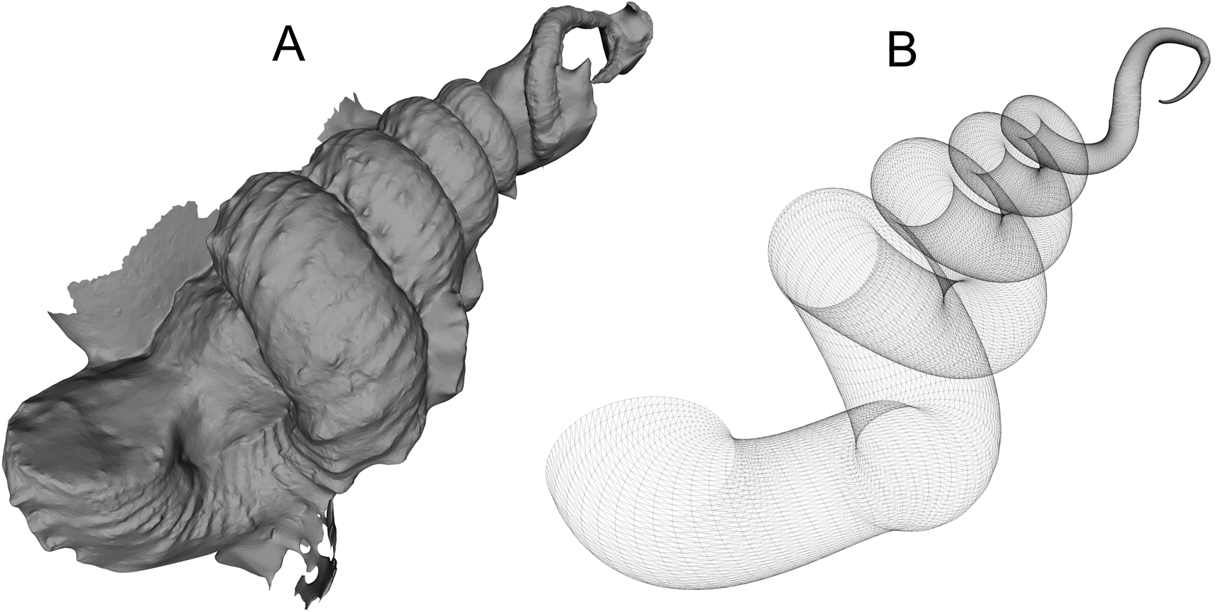
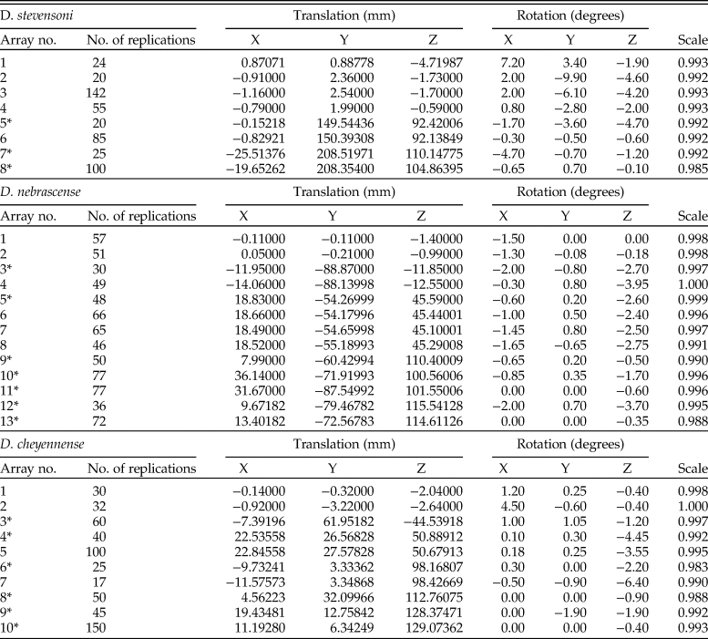
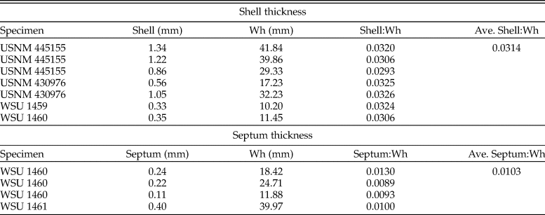
The 3D computer graphics software Blender v. 2.76 (Blender Online Community 2017) was used to replicate the whorl section fitted to the terminal aperture on each model with array instructions (Table 1) similar to the parameters of Raup (Reference Raup1967). This process translated, rotated, and scaled the whorl section to match the coiling of the photogrammetry model (Fig. 3). Due to the complex coiling scheme of Didymoceras, array instructions were used in a piecewise manner to change shell coiling throughout ontogeny. Toward the apical direction for each model, the origin of the arrays had to be periodically reset in order to make tighter coils (marked with asterisks in Table 1). Because the models are built from aperture to apex, smaller scaling values represent larger whorl expansion rates throughout ontogeny. The array algorithms create an infinitely thin 3D mesh of the exterior of the shell. Shell thickness can be defined by using the same array instructions on a smaller whorl section, representing the internal interface of the shell. The difference in radii between these two whorl sections is equal to the average ratio of shell thickness to whorl height (0.0314 measured from USNM 445155, USNM 430976, WSU 1459, and WSU 1460; Table 2).
A, Exterior 3D model of Didymoceras stevensoni (USNM 430976) created with photogrammetry in 3DF Zephyr. B, Wireframe view of exterior shell created with array instructions in Blender v. 2.76 after using the photogrammetry model as a stencil (Table 1).

Array instructions used to build each model of Didymoceras. Each array is used in a piecewise manner to replicate the initial whorl section, while simultaneously translating, rotating, and scaling. Asterisks mark the arrays where the origin must be reset to accommodate tighter whorls. Adult apertures were replicated with the following diameters measured or extrapolated from the photogrammetry models: D. stevensoni (X 48.600 mm, Y 48.600 mm), D. nebrascense (X 43.150 mm, Y 46.660 mm), and D. cheyennense (X 40.591 mm, Y 46.619 mm).

Shell thicknesses (Shell) and septum thicknesses (Septum) at respective whorl heights (Wh) measured from specimens of Didymoceras. These measurements were used to compute the shell thickness to whorl height (Shell:Wh) and septum thickness to whorl height (Septum:Wh) ratios. The average (ave.) of these two ratios were used to extrude the shell and septum thicknesses of the Didymoceras models of the current study.

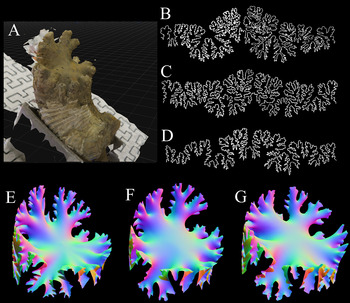
The septa were constructed by modeling with photogrammetry (Fig. 4A) an exposed septum of D. nebrascense (WSU 1459) that preserved the first- and second-order folds (see Peterman and Barton Reference Peterman and Barton2019). The finer-scale corrugations were constructed for each species by using their respective suture patterns (Fig. 3B, C, and D; WSU 1461, WSU 1462, and WSU 1463, respectively). Each suture pattern was recorded with a digital camera lucida, then wrapped around the whorl section of the phragmocone in the program Meshmixer (Autodesk Inc. 2017a), and extruded inward. The extruded suture and septal photogram were merged together and smoothed to create a reconstructed septum (Fig. 3E–G). Thickness was defined for the septum by using the solidify modifier in Blender v. 2.76. The average septal thickness to whorl height ratio was measured to be 0.0103 from specimens WSU 1460 and WSU 1461 (Table 2). The extruded septa were replicated with the same array instructions used to build the shell of each corresponding species. Extra septa were deleted until the distal elements of each septum were barely tangential to each other. The external and internal interfaces of the shell and the replicated septa were repaired and unified together with Netfabb v. 2017.3 (Autodesk Inc. 2017b) to create a single, manifold 3D mesh capable of volumetry.
Creation of Didymoceras septa. A, Model septum of D. nebrascense (WSU 1459) preserving first- and second-order folds. B, Suture of D. stevensoni (WSU 1461). C, Suture of D. nebrascense (WSU 1462). D, Suture of D. cheyennense (WSU 1463). E, Model septum of D. stevensoni. F, Model septum of D. nebrascense. G, Model septum of D. cheyennense.

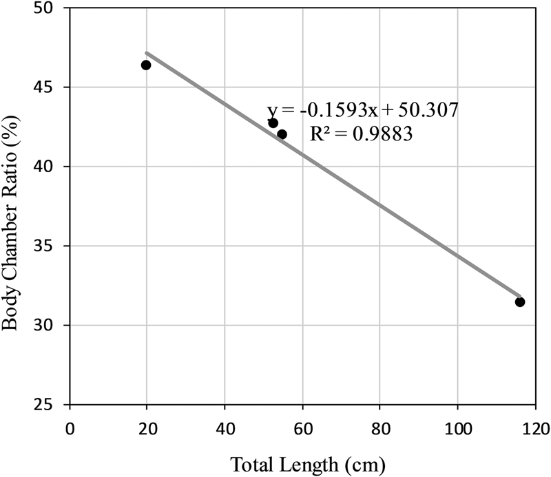
The ratio of the body chamber length to total length was measured from an adult Didymoceras (USNM 430976) and three progressively more juvenile specimens (USNM 482449, USNM 482445, and USNM 482441). The apical portions of the juvenile specimens were incomplete, so the positions of their apertures and terminal septa were marked on the virtual 3D model of a complete Didymoceras. The curvilinear distance along the venter between the aperture to terminal septum and between the aperture to apex were recorded from Blender (by summing the spaces between the adjacent replicated whorl sections). The body chamber ratio (length of body chamber to total length) seems to vary from approximately 1:3 in adults to 1:2 in the earliest juvenile specimen (Fig. 5). It should be noted that the body chamber ratio in nostoceratids has been reported to vary widely during adulthood (Cobban Reference Cobban1974). Body chamber ratios were extrapolated from the recorded relationship to examine several ontogenetic snapshots of each species of the modeled Didymoceras. Three-dimensional models of the body chambers were created by isolating the internal interface of the shell from the aperture to terminal septum. The faces of the mesh were inverted so that the exterior was pointing outward, then the aperture was filled in Meshmixer. A uniform-density soft body flush with the aperture was applied similar to Peterman et al. (Reference Peterman, Barton and Yacobucci2019a). Having a similar density to seawater, this has minimal influence in a static setting.
Body chamber ratios as a function of total length recorded from USNM 482411, USNM 482445, USNM 482449, and USNM 430976 (listed from earliest juvenile to adult). These values were used to extrapolate body chamber ratio for the modeled specimens for each ontogenetic stage.

The siphuncle diameter to whorl height ratio was measured at 0.041 from WSU 1461 and WSU 1463. This value allowed a cross section of the siphuncle to be replicated with the corresponding array instructions for each species. The siphuncle 3D model was then unified to the model of the soft body and subtracted from the model of the shell to create siphuncular foramina in each septum.
A model of the water displaced by each species is required to determine buoyancy and other hydrostatic calculations. This model was created by isolating the external interface of the shell and soft body that come into contact with the surrounding water.
Hydrostatic Computations
A neutrally buoyant condition occurs when the sum of the organismal mass is equal to the mass of the water displaced (Archimedes’ principle):
 $$m_{{\rm sb}} + m_{{\rm sh}} + m_{{\rm cl}} + m_{{\rm cg}} = m_{{\rm wd}}$$
Where m sb is the mass of the soft body, m sh is the mass of the shell, m cl is the mass of cameral (chamber) liquid, m cg is the mass of cameral gas, and m wd is the mass of water displaced by the cephalopod.
The proportion of the phragmocone volume to be emptied of cameral liquid for neutral buoyancy (Φ) is found by setting equation (1) in terms of volume and density:
 $${\rm \Phi } = {\rm \;}\displaystyle{{\left({\displaystyle{{V_{{\rm wd}}{\rm \rho }_{{\rm wd}}-V_{{\rm sb}}{\rm \rho }_{{\rm sb}}-V_{{\rm sh}}{\rm \rho }_{{\rm sh}}} \over {V_{{\rm ct}}}}} \right)-\lpar {{\rm \rho }_{{\rm cl}}} \rpar } \over {\lpar {{\rm \rho }_{{\rm cg}}-{\rm \rho }_{{\rm cl}}} \rpar }}{\rm \;\;}$$
Where V wd and ρwd are the volume and density of the water displaced, V sb and ρsb are the volume and density of the soft body, V sh and ρsh are the volume and density of the shell, ρcl is the density of cameral liquid, ρcg is the density of cameral gas, and V ct is the total volume of all camerae. A soft-body density of 1.065 g/cm3 is reported in Westermann (Reference Westermann2013) and is assumed to uniformly fill the body chamber of the current models. A shell density of 2.62 g/cm3 is commonly accepted (Reyment Reference Reyment1958; Okamoto Reference Okamoto, Landman, Tanabe and Davis1996; Klug et al. Reference Klug, Zatoń, Parent, Hostettler, Tajika, Klug, Korn, De Baets, Kruta and Mapes2015). The density of cameral liquid is assumed to be equal to that of seawater (1.025 g/cm3; Greenwald and Ward Reference Greenwald, Ward, Saunders and Landman1987). The density of cameral gas is negligible, but fixed at 0.001 g/cm3 to complete the hydrostatic computations. All volumes were computed in MeshLab v. 1.3.3 (Cignoni and Ranzuglia Reference Cignoni and Ranzuglia2014).
The volume of cameral gas can be found by multiplying the volume of all camerae by Φ, and conversely, the volume of cameral liquid is found by multiplying the cameral volumes by 1- Φ. After these volumes are determined, their spatial distribution in three dimensions, along with the distribution of the shell and soft body, is required to find the total center of mass:
 $$M = \displaystyle{{\sum \lpar {L\ast m_{\rm o}} \rpar } \over {\sum m_{\rm o}}}$$
Where M is the x, y, or z component of the total center of mass, L is the x, y, or z component of the center of mass of each material with unique density (soft body, shell, cameral liquid, and cameral gas) measured with respect to an arbitrary datum, and m o is the mass of each of these objects of unique density.
The center of buoyancy (B) is equal to the center of volume of the water displaced. The hydrostatic stability of an object is proportional to the separation between the center of buoyancy and the total center of mass (M). Okamoto redefined the stability index of Raup (Reference Raup1967) to apply to heteromorphic ammonoids by dividing the distance between the centers of buoyancy and gravity by the organismal volume (equal to the volume of water displaced):
 $$S_{\rm t} = \;\displaystyle{{\overline {BM} } \over {\root 3 \of V }}$$
It should be noted that the hydrostatic stability in the current study differs from previous studies, which assumed the center of mass was located in the center of the body chamber (Truman Reference Trueman1941; Raup Reference Raup1967; Raup and Chamberlain Reference Raup and Chamberlain1967; Okamoto Reference Okamoto, Landman, Tanabe and Davis1996).
The syn vivo orientation of each model occurs when the centers of buoyancy and mass are vertically aligned. The apertural angle θa was found by appropriately orienting each model, then using the protractor function in Blender to measure the angle between the aperture and the vertical plane. Therefore, θa equal to −90° corresponds to downward-facing orientations, +90° corresponds to upward-facing orientations, and 0° indicates a horizontally facing aperture.
The thrust angle (θt) was computed by using the protractor function in Blender to measure the angle between the center of rotation and the location of the hyponome (Fig. 1). The location of the hyponome (along the venter of the aperture) serves as the center of thrust. Therefore, θt values of zero would result in optimal horizontal translation with minimal rotation. Note that θa herein differs from that of Ebel (Reference Ebel1990) and Okamoto (Reference Okamoto, Landman, Tanabe and Davis1996). These two studies use the center of mass instead of the center of rotation to measure the thrust angle. Both the organismal weight and buoyancy impact the total restoring moment; therefore, the midpoint between the two is the proper pivot point in a hydrostatic setting, as modeled here. However, it should be noted that in a dynamic setting, it is possible to change the pivot point based on rotational drag and differential lift acting on the object.
Results
Hydrostatic Models of Didymoceras
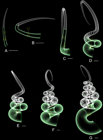
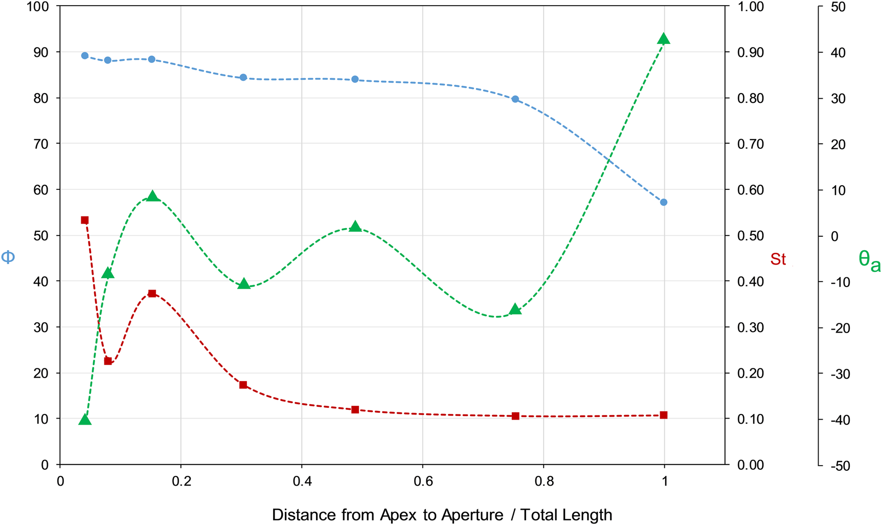
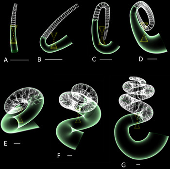
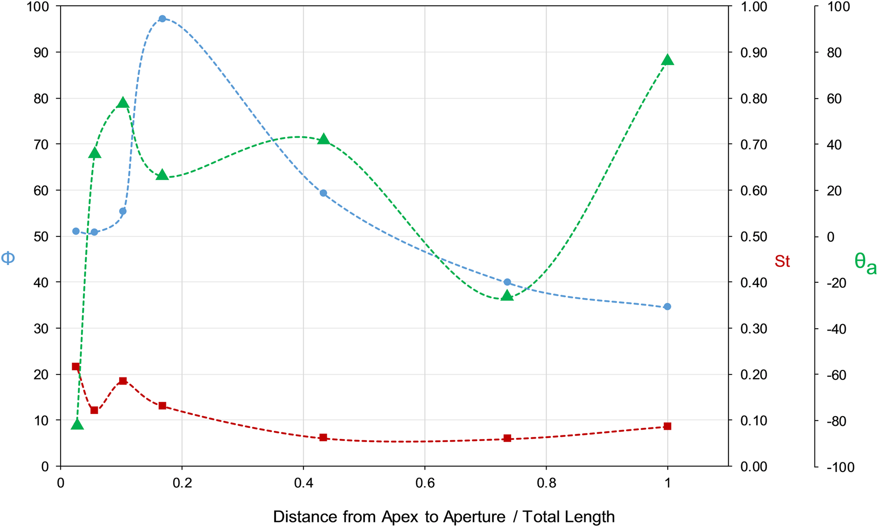
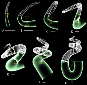
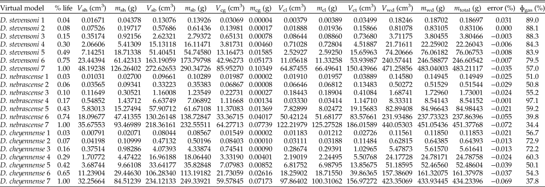
Hydrostatic models of D. stevensoni (Figs. 6 and 7), D. nebrascense (Figs. 8 and 9), and D. cheyennense (Figs. 10 and 11) were examined at several key ontogenetic stages to investigate how their hydrostatic properties changed throughout growth. The progress from an early-juvenile stage to the terminal (gerontic) age is proxied by the curvilinear distance from the apex to aperture relative to the total curvilinear length at adulthood for each species (i.e., percentage of their way through life). All of the models at each examined ontogenetic stage have the capacity for neutral buoyancy. The ontogenetic changes of this property, however, are not straightforward. Neutral buoyancy largely depends upon the body chamber ratio and the rate at which the aperture expands during growth. An expansion of the aperture requires a decrease in cameral liquid within the phragmocone for the animal to remain neutrally buoyant. As the shell continues to grow, this region eventually becomes septate and empty, promoting greater buoyancy and allowing Φ to stabilize. Such whorl expansions and changes in Φ are most prominent in juvenile to early intermediate stages of D. nebrascense and D. cheyennense (Figs. 9 and 11, respectively). It should be noted that the Φ values reported here are dependent upon the arbitrarily chosen life stages, which do not perfectly correlate among the chosen species.
A–G, Hydrostatic models of Didymoceras stevensoni throughout ontogeny showing static syn vivo orientations and the locations of the centers of buoyancy (tip of upper cone, circle) and centers of mass (tip of lower cone, x). Scale bars, 1 cm.

Hydrostatic metrics computed for Didymoceras stevensoni for each of the seven ontogenetic stages represented in Fig. 6. The percentage of the cameral volumes emptied for neutral buoyancy (Φ; circles), hydrostatic stability index (S t; squares), and the angle of the aperture measured from vertical (θa; triangles) are reported for each stage. Age is proxied by the curvilinear distance from the apex to aperture at each stage divided by total curvilinear length at adulthood.

A–G, Hydrostatic models of Didymoceras nebrascense throughout ontogeny showing static syn vivo orientations and the locations of the centers of buoyancy (tip of upper cone, circle) and centers of mass (tip of lower cone, x). Scale bars, 1 cm.

Hydrostatic metrics computed for Didymoceras nebrascense for each of the seven ontogenetic stages represented in Fig. 8. The percentage of the cameral volumes emptied for neutral buoyancy (Φ; circles), hydrostatic stability index (S t; squares), and the angle of the aperture measured from vertical (θa; triangles) are reported for each stage. Age is proxied by the curvilinear distance from the apex to aperture at each stage divided by total curvilinear length at adulthood.

A–G, Hydrostatic models of Didymoceras cheyennense throughout ontogeny showing static syn vivo orientations and the locations of the centers of buoyancy (tip of upper cone, circles) and centers of mass (tip of lower cone, x). Scale bars, 1 cm.

Hydrostatic metrics computed for Didymoceras cheyennense for each of the seven ontogenetic stages represented in Fig. 10. The percentage of the cameral volumes emptied for neutral buoyancy (Φ; circles), hydrostatic stability index (S t; squares), and the angle of the aperture measured from vertical (θa; triangles) are reported for each stage. Age is proxied by the curvilinear distance from the apex to aperture at each stage divided by total curvilinear length at adulthood.

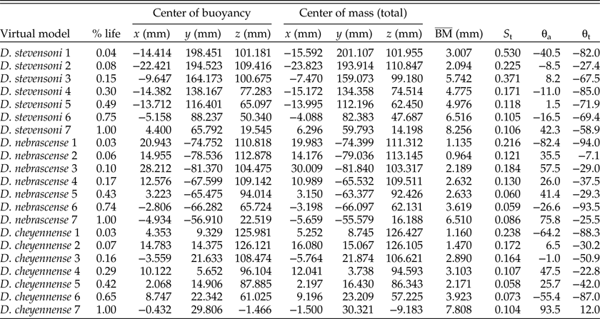
Throughout the ontogenetic trajectory of each species, hydrostatic stability (S t) follows the same general pattern. Each species starts with an orthoconic phase with relatively high stability, then stability gradually decreases throughout ontogeny as the organismal mass is concentrated toward the center of the helical whorls. The helical portion of D. nebrascense and D. cheyennense has a shorter duration than D. stevensoni, resulting in a slight increase in hydrostatic stability after the formation of the retroversal U-shaped hook (compare Figs. 6–11).
A remarkably similar pattern in syn vivo orientation, based on the computed apertural angle θa, occurs throughout ontogeny for all three species of Didymoceras. This pattern can be divided into three distinct phases: (1) a juvenile orthoconic phase resulting in a downward-facing aperture, (2) a helical phase in which the apertural angle oscillates around the horizontal plane, and (3) the formation of the retroversal U-shaped hook yielding an upturned aperture at adulthood.
The thrust angles for each of the three examined species of Didymoceras somewhat correspond to the pattern of syn vivo orientation throughout ontogeny (Fig. 12). Each species starts with low thrust angles, best suited for upward propulsion. After the first deviation from the orthoconic phase (i.e., first elbow bend in the shell), each of these species is better suited for slow horizontal movement for a relatively short period of time. Afterward, each of the models has thrust angles suited for diagonal upward thrust. Finally, at adulthood, the thrust angle increases to a more horizontal direction of motion for each species, though the shift is modest in D. stevensoni compared with the other two species.
Thrust angle (θt) measured from the horizontal vs. age for each of the seven examined ontogenetic stages of D. stevensoni (Fig. 6), D. nebrascense (Fig. 8), and D. cheyennense (Fig. 10). Low thrust angles (–90°) correspond to idealized upward movement, while thrust angles of zero correspond to idealized horizontal-backward movement.

The Φ values, masses, and volumes of each model component for hydrostatic calculations are organized in Table 3. The centers of mass and buoyancy, their separation, hydrostatic stability, apertural angles, and thrust angles for each model are organized in Table 4. Ideally, the sum of the masses of each model component should be equal to the mass computed from the water displaced. Discrepancies between these values result in a mass surplus or absence that influences all of the hydrostatic calculations. The mass discrepancies for the virtual models are all less than 0.1% (Table 3), suggesting that hydrostatic calculations are not significantly impacted. The absence of ornamentation in the models could cause shell mass to be slightly lower for smooth models compared with actual specimens (potentially raising Φ values). The assumption of isometric shell and septal thicknesses also may contribute some error. However, Peterman et al. (Reference Peterman, Barton and Yacobucci2019a) compared an isometric Nautilus shell with a CT-scanned specimen from Hoffmann et al. (Reference Hoffmann, Schultz, Schellhorn, Rybacki, Keupp, Gerden, Lemanis and Zachow2014) and found only a 4% difference in mass. The isometric shell thicknesses in the current study likely have a similarly low error. Additionally, the mass of the soft body is much more influential on the hydrostatics and mass distribution than the shell (see Table 3). Other potential sources of error are the discrepancies in septal mass resulting from differences between each of the specimens used to record septa and changes of the models during processing. Sutures were recorded from different positions on each whorl and from differently sized specimens (ranging from whorl heights of 24 mm to 31mm). Suture patterns were also assumed to remain constant through ontogeny. Additionally, some of the finest lobules and folioles were smoothed during the extrusion process (Fig. 4). However, the normalized surface areas of each septum are less than 5% different from each other and probably do not contribute much to the total error in mass, especially because the shell masses are consistently lower than those of the soft body (Table 4).
The volumes (V) and masses (m) for each component of the Didymoceras models (sh, shell; sb, soft body; cg, cameral gas; cl, cameral liquid; ct, camerae total; wd, water displaced) for each ontogenetic stage (denoted by a number after the species name: 1 = earliest juvenile to 7 = adult). Age (% life) is proxied by the curvilinear distance between the apex and aperture divided by total curvilinear length at adulthood. The percent error (mass discrepancies between the total model mass and the mass of the water displaced) and the percentage of the cameral volume that must be emptied of liquid to achieve a neutrally buoyant condition (Φ) are listed for each model.

The x, y, and z components of the centers of buoyancy and mass are reported with respect to an arbitrary datum (the center of the adult whorl section). Hydrostatic metrics ( $\overline{\rm BM}$, the distance between the centers of buoyancy and mass; S t, hydrostatic stability index; θa, apertural angle; and θt, thrust angle) are reported as a function of age (% life = curvilinear distance from apex to aperture/total curvilinear length).

Discussion
The three species of Didymoceras investigated in the current study can each be divided into three ontogenetic stages based on shell coiling: (1) an early, generally straight phase; (2) a helical intermediate phase; and (3) an adult phase with a retroversal U-shaped hook. This hook probably represents the attainment of sexual maturity and may have been formed relatively quickly (Bucher et al. Reference Bucher, Landman, Klofak, Guex, Landman, Tanabe and Davis1996). The relative duration of the intermediate helical phase for the examined species increases from D. cheyennense to D. nebrascense and reaches a maximum in D. stevensoni. It is unknown, however, whether the extra helical portion of D. stevensoni corresponds to a larger amount of time spent building this part of shell. The hydrostatic calculations presented here reveal differences between each of the examined species of Didymoceras and also have implications for the nostoconic morphotype as a whole.
All virtual models of Didymoceras have the capacity for neutral buoyancy throughout each ontogenetic stage and must leave some amount of cameral liquid in the phragmocone to maintain this condition. Therefore, while isotopic studies suggest this genus was demersal and lived very close to the benthos (He et al. Reference He, Kyser and Caldwell2005, Reference He, Kyser and Caldwell2016), it still retained the ability to hover around, close to the seafloor. This life mode would enable the ammonoid to push up off the seafloor with minimal effort to evade predators or pounce on prey items. The degree of chamber emptying required for neutral buoyancy would have varied as the ammonoid grew. The largest constraint on neutral buoyancy is the mass of the soft body (Table 3). The soft-body mass is directly influenced by the whorl expansion and body chamber ratio, both of which change throughout ontogeny in a very complicated fashion in Didymoceras. Interestingly, the whorl expansion of D. stevensoni changes more gradually than that of D. nebrascense or D. cheyennense. This lower whorl expansion rate results in gradual changes in Φ throughout ontogeny (Fig. 7). Conversely, the amount of cameral liquid in the phragmocone rapidly decreases for D. nebrascense (Fig. 9) and D. cheyennense (Fig. 11) as they build the “elbow” that starts the intermediate helical phase. It should be noted that the conditions for neutral buoyancy (Φ) must fluctuate during actual growth, because each septum is precipitated in water, then emptied of cameral liquid (Ward Reference Ward1979). Therefore, Φ probably does not change as regularly as the interpolated dashed lines on Figures 7, 9, and 11 depict. Furthermore, decreases in Φ can be compensated by reducing the body chamber ratio (Klug et al. Reference Klug, Korn, Richter and Urlichs2004).
While the soft-body density is very close to that of water, it is still three orders of magnitude larger than that of the gas filling the camerae. This difference fundamentally weights the distribution of mass toward the body chamber for all ectocochleates and influences the stability index (Peterman et al. Reference Peterman, Barton and Yacobucci2019a,b). For each model, hydrostatic stability (S t) is at a maximum during the earliest juvenile, orthoconic phase. Orthocones have very high hydrostatic stability values because they distribute the mass away from the phragmocone and toward the soft body in a linear fashion (see Peterman et al. Reference Peterman, Ciampaglio, Shell and Yacobucci2019b). Any deviation from the straight line (coiling of any kind) would consequently reduce stability. Throughout growth, total organismal mass becomes concentrated in response to coiling. The helical whorls in the intermediate growth stages also influence the volume of water displaced and cause the centers of buoyancy and mass to be placed closer together, reducing stability. The formation of the retroversal, U-shaped hook causes hydrostatic stability to slightly increase for all three examined species of Didymoceras, though the increase is quite small in D. stevensoni. The adult body chamber deviates from the earlier whorls, distributing mass farther from the center of buoyancy. This stability increase is more prominent for D. nebrascense and D. cheyennense, because they devote a smaller portion of their life to coiling during the intermediate stage. Although hydrostatic stability decreases throughout ontogeny for each of the examined species, they all have adult values around twice the stability of the modern Nautilus (0.050; Peterman et al. Reference Peterman, Barton and Yacobucci2019a). While the stability index of Nautilus is a seemingly low number, it still imparts a restoring moment large enough so that the living animal does not pinwheel during locomotion (Saunders and Shapiro Reference Saunders and Shapiro1986; Peterman et al. Reference Peterman, Ciampaglio, Shell and Yacobucci2019b). Selective pressures on hydrostatic stability represent a trade-off in locomotory efficiency: a high stability prevents excessive rocking or pinwheeling during jet propulsion but also makes it more difficult for the animal to move into an orientation other than its hydrostatically defined one. The hydrostatic stability of each of the Didymoceras virtual models is large enough that syn vivo orientation is probably unable to be significantly modified with active locomotion, or at least very costly in terms of energy use. This difficulty in movement is especially true in earlier ontogenetic stages. In a dynamic setting, the larger hydrostatic stability values at juvenile stages may be negligible depending on the Reynolds number of the system (the ratio of inertial forces to viscous forces). When moving horizontally at one body width per second, the adult model has a Reynolds number on the order of 101 to 102, while the early juvenile has a Reynolds number less than 100. This lower Reynolds number means that the surrounding seawater would seem more viscous for the early-juvenile ammonoids, hindering movement and possibly the effects of the restoring moment. From the time of an early hatchling, most ammonoids were probably passive and planktic (Jacobs and Chamberlain Reference Jacobs, Chamberlain, Landman, Tanabe and Davis1996; Landman Reference Landman, Wiedmann and Kullmann1988; Mapes and Nützel Reference Mapes and Nützel2008; Ritterbush et al. Reference Ritterbush, De Baets, Hoffmann and Lukeneder2014; Naglik et al. Reference Naglik, Tajika, Chamberlain, Klug, Klug, Korn, De Baets, Kruta and Mapes2015b; Tajika et al. Reference Tajika, Nützel and Klug2018). If ocean currents within the habitat of Didymoceras were not strong enough to facilitate the dispersal of their hatchlings (Tajika and Wani Reference Tajika and Wani2010), the hatchlings may have remained close to their hatching sites, as it would be relatively difficult for the early hatchlings to move any distance via active locomotion.
The life orientation of ectocochleates is vital to understanding how the living cephalopods would have swum and interacted with their surroundings (Saunders and Shapiro Reference Saunders and Shapiro1986; Okamoto Reference Okamoto1988a; Klug Reference Klug2001; Naglik et al. Reference Naglik, Tajika, Chamberlain, Klug, Klug, Korn, De Baets, Kruta and Mapes2015b). While the orientation of the aperture (θa) may not directly indicate a particular mode of life, it nonetheless imposes some important limitations, especially when the orientation of the aperture cannot be significantly modified with active locomotion due to a high stability index (S t). Each of the Didymoceras models have downward-facing apertures during the earliest juvenile orthoconic phase (consistent with the baculite models of Peterman et al. [2019b]). A downward orientation in Didymoceras hatchlings is consistent with feeding either within the water column or off the bottom. Afterward, the apertures oscillate around the horizontal, forward-facing plane as the helical intermediate stage progresses. This orientation could have allowed the juvenile ammonoid to feed or scavenge along the seafloor and to catch slow demersal prey items. Each model terminates with an upward apertural orientation after the U-shaped hook is developed. This adult orientation away from the seafloor suggests either a different mode of feeding or possibly the absence of feeding in reproductive adults, which is well-documented in various modern coleoid cephalopod groups (Rocha et al. Reference Rocha, Guerra and González2001; Hanlon and Messenger Reference Hanlon and Messenger2018). The duration of the intermediate helical phase with a generally horizontal apertural orientation is larger for D. stevensoni and progressively lower for D. nebrascense and D. cheyennense. Changes in the duration of each phase indicate that there may have been differences among these species in how long an individual maintained the life habit consistent with that orientation. Alternatively, it may be a consequence of larger whorl expansion rates in D. nebrascense and D. cheyennense relative to D. stevensoni. This is evidenced by the differences in total length along the venter at adulthood (116 cm, 96 cm, and 78 cm for D. stevensoni, D. nebrascense, and D. cheyennense, respectively). It is unknown whether or not this difference in shell length corresponds to differences in terminal ages or in metabolic growth rates.
The upturned aperture at adulthood is enigmatic for ammonoids that lived close to the seafloor. This orientation may be incompatible with scavenging or searching for prey items in this setting (Klinger Reference Klinger, House and Senior1981; Landman et al. Reference Landman, Cochran, Larson, Brezina, Garb and Harries2012). However, a U-shaped hook is a common adult modification for many taxa of heteromorphic ammonoids within Nostoceratidae, Scaphitidae, Diplomoceratidae, Ancyloceratidae, and Macroscaphitidae, among others, spanning multiple higher taxonomic levels (Klug et al. Reference Klug, Zatoń, Parent, Hostettler, Tajika, Klug, Korn, De Baets, Kruta and Mapes2015). One adaptive explanation for the prevalence of the adult hook is that, by shifting the aperture to face up, brooding adults could hold their egg masses (assuming they had them) up and away from benthic predators (Walton et al. Reference Walton, Korn and Klug2010). Further complicating the interpretation of an upturned aperture, it is unknown how mutable the direction of thrust was for such ammonoids. Were they able to stretch the hyponome over the ventral aperture and direct their jet thrust downward, enabling them to move in the direction they were facing (like the extant Nautilus; Packard et al. Reference Packard, Bone and Hignette1980)? It is easier for extant ectocochleates (nautilids) to manage their buoyancy by being very slightly negatively buoyant (Ward Reference Ward1979). Was this the case for ammonoids with U-shaped body chambers at maturity? If so, were they able to counteract this difference in buoyancy? The largest hurdle in answering these questions is that unequivocal preservation of soft tissues has yet to be discovered (Klug and Lehmann Reference Klug, Lehmann, Klug, Korn, De Baets, Kruta and Mapes2015). The functional significance of such an adult modification should be further explored.
The thrust angle (θt; Figs. 1, 12) is a useful tool to understand the efficiency and constraints on the direction of locomotion for animals that move via jet propulsion (Okamoto Reference Okamoto, Landman, Tanabe and Davis1996; Klug and Korn Reference Klug and Korn2004; Naglik et al. Reference Naglik, Tajika, Chamberlain, Klug, Klug, Korn, De Baets, Kruta and Mapes2015b). Ideally, the optimal horizontal movement would occur when the thrust angle is zero. This would transmit the energy produced from thrust into more translational movement and less rotational movement. Conceptually, a downward-facing orthocone would have trouble moving horizontally due to its very low thrust angle (around −90°) and large distance between the center of rotation and location of thrust (in addition to higher drag in this configuration; Peterman et al. Reference Peterman, Ciampaglio, Shell and Yacobucci2019b). Similar to the other hydrostatic properties computed in this study, the thrust angle has similar patterns throughout ontogeny for each of the examined species of Didymoceras, at least in coarse terms (Fig. 12). The early-juvenile orthoconic phase seems to be well adapted for upward vertical movement; however, at smaller sizes, the low Reynolds number may have made locomotion in any direction more difficult. The thrust angle then changes rapidly toward zero (horizontal alignment) just before the helical phase develops for each species. The horizontally oriented aperture and low thrust angles would permit efficient horizontal movement. These properties could have strongly enhanced the dispersal ability of juvenile Didymoceras, especially in the absence of a current near the seafloor. As the helical phase develops, the thrust angles seem more variable; however, they generally do not favor horizontal movement, especially in D. stevensoni, even though the apertures are more or less horizontally oriented. This intermediate helical phase resembles the torticonic ammonoids belonging to Turrilitidae. Klinger (Reference Klinger, House and Senior1981) suggests that this morphology could have been adept at rotating along the z-axis with an improved turning ability compared with monomorphs. For Didymoceras the center of mass is displaced laterally outside the body chamber, in the center of the helical whorls (similar to the probable mass distribution of turrilitids; Figs. 6, 8, 10). A pirouetting mode of life assumed during the helical phase of Didymoceras could have allowed it to scan larger portions of the water column for plankton by rotating, while expending less energy compared with lateral movement. At adulthood, thrust angles increase once again, with the increase being much more pronounced in D. nebrascense and D. cheyennense than in D. stevensoni. This increase is due to the U-shaped body chamber raising the aperture with respect to the center of rotation. Therefore, the U-shaped adult modification functions to improve horizontal locomotion by raising the thrust angle relative to the earlier helical stage (as mentioned previously by Klug et al. [2015]), even though the aperture faces up in these adult forms. This modification could have been significant during the time of mating, enabling horizontal movements to approach potential mates. The aforementioned differences in the duration of the helical phase between each of the examined species also influences the thrust angle. Didymoceras stevensoni has more negative thrust angles at adulthood, while D. nebrascense and D. cheyennense are closer to zero. This is due to D. stevensoni's more helical whorls concentrating the centers of mass and buoyancy in the apical direction. Conversely, fewer helical whorls result in the centers of buoyancy and mass being pulled closer to the U-shaped body chamber, causing the thrust angle to approach zero. These differences in adult Didymoceras suggest that D. cheyennense was best suited for horizontal-backward movement, while D. nebrascense was slightly less optimized for this style of locomotion, and D. stevensoni remained better suited for more vertical-upward movement during the last stage of life. Subtle differences can result depending upon the position of the terminal aperture, which may be somewhat variable for these species.
Didymoceras stevensoni differed somewhat from the other two Didymoceras species investigated here in its growth rate and hydrostatic properties through ontogeny, implying differences in the mode of life of this species relative to its congeners, including a longer intermediate juvenile stage and a more vertical component of locomotion through later ontogenetic stages. Didymoceras stevensoni is intermediate in age between the other two species (Kennedy et al. Reference Kennedy, Landman, Cobban and Scott2000), so it does not represent an early or late species in a phylogenetic sequence. Rather, the differences seen in D. stevensoni's hydrostatic parameters may indicate an enhanced degree of interspecific variability in modes of life within the genus. More generally, the results presented here indicate that selective pressures varied through ontogeny in these ammonoids, resulting in different shell forms well adapted to different life habits. Hence, the “aberrant” nostoconic morphotype reflects evolutionary solutions to functional challenges at different stages in the life course of these extraordinary ammonoids.
Conclusions
Virtual 3D models of D. stevensoni, D. nebrascense, and D. cheyennense reveal hydrostatic properties throughout ontogeny that are characteristic of the nostoconic morphotype, as well as several differences among each of the examined species. All of the models suggest that Didymoceras had the capacity for neutral buoyancy and left some amount of cameral liquid in the phragmocone to maintain this condition. Hydrostatic stability was probably too large for the living animal to significantly modify its orientation. The orientation of the aperture for each species starts downward-facing, oscillates around the horizontal plane as the helical whorls develop, and terminates with an upward-facing aperture after formation of the U-shaped adult body chamber. Thrust angles during the helical intermediate phase suggest that thrust energy would most efficiently be transmitted into pirouetting (rotation about the vertical axis). The thrust angles throughout ontogeny do not favor efficient horizontal movement, except for two distinct phases: (1) the formation of the first “elbow” before the helical phase develops and (2) after completion of the U-shaped adult body chamber. These hydrostatic properties suggest that the seemingly aberrant coiling of Didymoceras and similar heteromorphic ammonoids may reflect a method to their madness. These ammonoids probably exploited a low-energy lifestyle near the seafloor, either scavenging or capturing slow demersal prey items. At two life stages, the coiling scheme of Didymoceras would enhance lateral movement: for the potential dispersal of juveniles and the location of a mate at adulthood.
Acknowledgments
We thank L. Chang, K. Hollis, H. Little, and M. Florence (Smithsonian National Museum of Natural History) for their help with the specimens of Didymoceras used for photogrammetry. We also appreciate the help of D. Clements (North Carolina Museum of Natural Sciences) for donating WSU 1461, which aided in construction of the virtual models. We would like to thank M. Hopkins, C. Klug, and an anonymous reviewer for their constructive comments on our article.