Turner’s syndrome is a condition caused by complete or partial loss of the second X chromosome (45, XO) with typical clinical features. Associated congenital cardiovascular malformations are well-known in this population, with a prevalence ranging from 17 to 45%.Reference Gotzsche, Krag-Olsen, Nielsen, Sorensen and Kristensen 1 – Reference Hou, Hwu, Tsai, Lee, Wang and Lue 4 Among these cardiac anomalies, the bicuspid aortic valve is often associated with dilatation of the ascending aorta. Nevertheless, dilatation of the ascending aorta may well occur in patients with Turner’s syndrome in the absence of aortic valve anomalies.Reference Matura, Ho, Rosing and Bondy 5 , Reference Lin, Lippe and Rosenfeld 6 Clinical monitoring of the ascending aorta in girls and women with Turner’s syndrome, therefore, becomes essential for early detection and prevention of secondary dramatic incidents such as the dissection of the ascending aorta.Reference Carlson and Silberbach 7 , Reference Gravholt, Landing-Wilhelmsem and Stochholm 8
The aortic dissection in these patients occurs at a younger age, and with a higher prevalence compared with the general population.Reference Carlson and Silberbach 7 , Reference Gravholt, Landing-Wilhelmsem and Stochholm 8 Age, systemic hypertension, karyotype, and pregnancy have been identified as risk factors for aortic dissection in patients with Turner’s syndrome. In addition, underlying CHDs and structural aortic anomalies, in particular, such as bicuspid aortic valve with or without stenosis and coarctation of the aorta, have been identified as independent risk factors for aortic dilation in general and in patients with Turner’s syndrome in particular.Reference Matura, Ho, Rosing and Bondy 5 , Reference Lin, Lippe and Rosenfeld 6 Nevertheless, little is known about the risk of aortic dilation in patients with Turner’s syndrome in the absence of cardiac anomalies; therefore, the aim of this study was to assess the prevalence and the progress rate of dilatation of the ascending aorta of Turner’s syndrome patients free of structural cardiac anomalies.
Methods
In this retrospective study, we included all patients with a genetic diagnosis of Tuner’s syndrome followed-up at the outpatient clinics of the division of paediatric cardiology, CHU Sainte-Justine, University of Montreal, between 1992 and 2010. Patients who had at least two echocardiograms performed during the study period were eligible. Patients with significant associated cardiac anomalies – for example, bicuspid aortic valve, coarctation of the aorta, and aortic stenosis – previous cardiac surgery, and those with missing important data were excluded from the analysis. Patients who were under vasoactive medication for systemic hypertension were also excluded. This study was granted ethics board approval.
Basic anthropometric data were collected from medical charts, which included age, weight, and height at the time of echocardiography. Other pertinent data including karyotype and concurrent medication – anti-hypertensive drugs and growth hormones – were similarly collected.
Echocardiography measurements were performed on commercially available sonographs, the Vivid 7 (GE Healthcare, Louisville, Kentucky, United States of America) and the iE33 (Philips, Einthoven, The Netherlands). Ascending aorta diameters were retrieved from the local echocardiography database. In our centre, the first echocardiography studies are completed according to the same local protocol, and subsequent studies are focussed towards target lesions. Missing diameters were measured from recorded studies by the same experienced echocardiography technician. Ascending aorta was measured in the long parasternal-axis view at the level of projection of the right pulmonary artery from inner edge to inner edge.Reference Warren, Boyd, O’Connell and Dodds 9 Aortic valve and root measurements were similarly recorded.
Ascending aorta diameter was adjusted for body surface area and expressed as a Z-score, calculated using an equation derived from over 1200 healthy patients (0 to 20 years old) from our institution.Reference Altit, Sarquella-Brugada and Dahdah 10 The ascending aorta was considered dilated whenever the actual measurement was superior to two standards deviations. We compared the ascending aorta Z-scores of the first and last echocardiographies for each patient to assess the progression of ascending aorta diameters during follow-up. Z-scores along the follow-up period were taken into consideration for new-onset dilatation. Z-score at the last follow-up was considered the final outcome. Patients with dilatation of the ascending aorta at the first echocardiography were defined as early aortic dilatation. Patients with dilatation at the last echocardiography were defined as late aortic dilatation. We evaluated the freedom from aortic dilatation with the event being the earliest occurrence of aortic dilatation during the follow-up, including patients who had resolution of their dilatation. We also compared the survival with freedom of dilatation of the ascending aorta among patients with Turner’s syndrome according to growth hormone intake and chromosomal status (45 XO versus mosaicism).
Finally, to account for weight gain in patients, we also used a second multivariate model equation to calculate ascending aorta Z-scores in comparison with a more traditional bi-variate model that we used for our study.Reference Dallaire, Bigras, Prsa and Dahdah 11 Repeated analyses were then performed, similar to those implemented with the previous body surface area-based regression equation.
Statistical analysis
Continuous data are expressed as mean±standard deviation. The Student’s t-test was used to compare continuous data with normal distribution, but the Mann–Whitney U test was used otherwise. Categorical data were analysed using the χ2 or the Fisher exact test. The Kaplan–Meier survival curve was used to evaluate the freedom from aortic dilatation. Log rank test was used to compare Kaplan–Meier survival curves between sub-groups. Statistical significance was defined with p<0.05.
Results
There were a total of 120 patients diagnosed with Turner’s syndrome who were followed-up at our clinics during the study period, aged 9.8±8.0 years at the first cardiology evaluation. Of those, 87 (73%) had no significant cardiac malformations, among whom 28 (31%) were excluded due to missing data, and another 15 (25.4%) were excluded from the main analysis due to anti-hypertensive therapy (Fig 1). Of the retained patients, 4.9±2.6 (median 4; with a range from 2 to 11) reliable echocardiography studies were available per patient.
Study population.

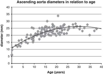
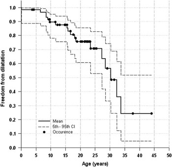
The final study population consisted of 44 patients, aged 11.9±7.4 years at the first echocardiogram and 17.9±7.3 years at the last follow-up. The mean follow-up duration was 6.0±3.7 years. Ascending aorta dimensions varied from 6.43 to 34 mm, as depicted in absolute values against age (Fig 2). Of the 44 study patients, 13 (29.5%) patients exhibited aortic dilatation at either the first, last, both, or intermediate echocardiography studies during the follow-up period, suggesting an actuarial estimate of the freedom from aortic dilatation dropping from 86 to 70% and then to 37% at 10, 20, and 30 years of age, respectively (Fig 3). Nevertheless, eight (18.1%) patients had early aortic dilatation, among whom only two (4.5%) maintained aortic dilatation at the last evaluation. In this group, the maximum ascending aorta Z-score varied between 2.11 and 2.98 with a statistically significant regression from 2.52±0.30 to 1.43±1.00 at last follow-up (p=0.02). As expected, absolute ascending aorta dimensions did not regress; they either remained relatively stable or increased with age. The other five (11.3%) patients had normal initial aortic Z-score of 0.83±1.05 and progressed to aortic dilatation status with a Z-score of 2.40±0.40 at the last follow-up (p=0.03) at the age of 8.9±4.8 years.
Absolute ascending aorta measurements in relation to age in patients with Turner’s syndrome.

Freedom from aortic dilatation (mean and 95th percentile) in patients with Turner’s syndrome free of associated cardiac structural anomalies.

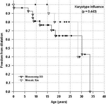
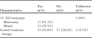
Genetic testing results were available for all the patients but for four; 27/40 (67.5%) had monosomy (45, XO), and 13 (32.5%) had mosaicism (Table 1). Aortic dilatation occurred in eight (29.6%) patients with monosomy versus three (23%) with mosaicism (p=1.0). Actuarial survival analysis did not reveal any significant difference from this perspective (p=0.381) (Fig 4).
Freedom from aortic dilatation according to karyotype.

Clinical features among patients with no associated cardiac anomalies (total 44).

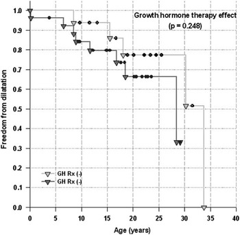
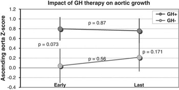
Growth hormone therapy was being administered to 25 patients either during or before the study (0.3 mg/kg per week) versus none in 17 patients (Table 1). Growth hormone treatment was started at a mean age of 10.7±3.4 years and ended at a mean age of 15.6±2.9 years. The mean duration of growth hormone treatment was 4.8±2.2 years. Aortic dilatation was noted in 8/25 patients (32%) treated with growth hormones compared with 3/17 (17.6%) untreated patients (p=0.477). Patients under growth hormone treatment had a borderline higher initial Z-score (p=0.07), which persisted over follow-up but decreased in importance becoming a non-significant difference at the last evaluation (p=0.171) (Fig 5). The progression of the ascending aorta Z-score during follow-up was not statistically significant between the two groups either (p=0.575); from 0.80±1.22 at the initial study to 0.76±1.19 at the last echocardiography in patients under growth hormone treatment (p=0.686) and from 0.04±1.43 to 0.22±1.19 (p=0.563) in the other patients. This was further confirmed by survival analysis with a freedom from aortic dilatation of 94 versus 88% at 8 years, 75 versus 86% at 15 years, and 66 versus 77% at 18 years of age, respectively (p=0.248) (Fig 6).
Impact of GH therapy on the progression of ascending aorta Z-score at early and last echocardiograms. GH=growth hormone.

Freedom from aortic dilatation according to growth hormone treatment.

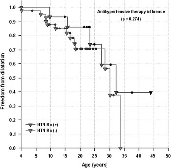
The effect of anti-hypertensive therapy was assessed separately, as drugs such as β-blockers and renin-angiotensin modulators are thought to stabilise and/or reduce aortic dilatation. This included a total of 15 (25.4%) patients. The indication for treatment initiation was the combination of systemic hypertension and aortic dilatation in five patients and isolated systemic hypertension in 10. Therapy included β-blockers, angiotensin-converting enzyme inhibitors, and angiotensin receptor antagonists, including a combination of three medications in one patient. Among those, aortic dilatation was present in 5/15 (33%) compared with 13/44 (31%) free of anti-hypertensive medication (p=1.0), with no significant difference in terms of freedom from aortic dilatation (p=0.274) (Fig 7).
Freedom from aortic dilatation according to anti-hypertensive medication.

As a potentially excessive weight gain during follow-up could have impacted significantly body surface area in some patients and, therefore, explain in part the regression in ascending aorta Z-score, we calculated patients’ body mass index at their first and last echocardiography studies. Classification according to the World Health Organisation (2006)Reference Onis, Onyango, Borghi, Siyam, Nishida and Siekmann 31 has been used to categorise patients as normal weight (body mass index under 25), overweight (body mass index between 25 and 30), and obesity (body mass index above 30). In total, 10 patients were overweight, and three patients were obese at their initial echocardiography. At the last echocardiography, the body mass index category increased in nine patients: five patients with previously normal weight became overweight and four overweight patients reached obesity; three patients previously categorised as overweight retrieved to normal weight category. We specifically compared the average change of ascending aorta Z-score using the multivariate equation,Reference Lacro, Guey and Dietz 29 which compensates for the extremes of body mass index. Accordingly, mean Z-scores were significantly lower at the first echocardiography (0.31±0.46 versus 0.59±1.40; p=0.02) and comparable at the last echocardiography (0.49±0.49 versus 0.50±1.44) than the bi-variate formula. Interestingly, no patient exhibited aortic dilatation (Z-score >2) at any time with the multivariate formula. The percentage change of Z-score between the last and first echocardiography was comparable between the two Z-score formulae (p=0.69).
Finally, as different aortic sites of dilatation have been reported,Reference Dore, Brochu and Baril 14 – Reference Lopez, Arheart and Colan 16 we calculated Z-scores for aortic valve and aortic root diameters and compared early and last echocardiography results. The mean aortic valve dimension was (15.6±3.6 mm) at early echo and (17.0±2.4 mm) at last echo (p=0.03). Although none had aortic valve dilatation at the first evaluation, two patients developed aortic valve dilatation on last echo – one of whom had associated aortic root dilatation – but none had concomitant ascending aortic dilatation. The aortic root dimensions instead increased at the last echocardiogram in three patients from normal initial values, early dilatation in two remained so during the last echo, and one early dilatation normalised at the last evaluation. For three patients, early dilatation of the aortic root coincided with early dilatation of the ascending aorta. These numbers were too small to contrast with confounding variables, however.
Discussion
In this retrospective study, we evaluated a longitudinal series of patients with Turner’s syndrome for the risk and prevalence of aortic dilation. Our findings are within the reported range of aortic dilatation prevalence (6.8 to 32%) based on cross-sectional reports on all age.Reference Matura, Ho, Rosing and Bondy 5 , Reference Hjerrild, Mortensen and Sorensen 12 , Reference Cleemann, Mortensen and Holm 13 Aortic dilatation has been particularly associated with coarctation of the aorta, bicuspid aortic valve, or systemic hypertension.Reference Warren, Boyd, O’Connell and Dodds 9 , Reference Dore, Brochu and Baril 14 , Reference Kim, Roman and Cavallini 15 Different sites of aortic dilatation have been reported, such as dilatation of the aortic valve annulus (12%), the sinus of Valsalva (20%), and the ascending aorta (30%).Reference Lopez, Arheart and Colan 16 A report from the National Institute of Health, United States of America, describes similar results in a series of 250 patients,Reference Sachdev, Matura and Sidenko 17 but the reported sites may vary among various studies. For example, in a study of 107 patients with no associated cardiac lesions, dilatation of the sino-tubular junction was the main finding but not that of the ascending aorta compared with controls.Reference Prandstraller, Mazzanti and Giardini 18 Although a case–control study may be an acceptable methodology for adults, normalisation to body habitus, to body surface area in particular, is the current standard methodology for the paediatric population. The normalisation bi-variate equation we applied is based on the best mathematical fit models and were duplicated with two different formulae reported in the literatureReference Warren, Boyd, O’Connell and Dodds 9 , Reference Gautier, Detaint and Fermanian 19 with similar trends (data not shown). Above all, Z-score normalisation in children allows longitudinal assessment, which takes into account expected structural growth rate with age. In this study, we took the analysis further to account for potential excessive weight variation during follow-up using our recently published Z-score formula that uses a multivariate model. This formula reduces the bias of the body mass index on proximal aorta diameters Z-scores in comparison with bi-variate models.Reference Dallaire, Bigras, Prsa and Dahdah 11 This model shows interestingly significant lower Z-scores when compared with the first formula. We could not demonstrate a significant difference of Z-scores variation in time between the two methods; the multivariate model also calculates regression of Z-scores in some patients. Other possibilities may include variation of proximal aortic growth patterns with different age stages or due to error in measurements and precision by echocardiography.
Both, the age of appearance of aortic dilatation in children with Turner’s syndrome and the evolution of the dilatation, are still unknown. Our study, therefore, is the first to represent the freedom from aortic dilatation in the paediatric population using the longitudinal observational methodology we adopted. Indeed, the observed aortic dilatation in our cohort seemed to be rather mild (Z-score between 2.11 and 2.98) on one hand, readily regressing with time without medical intervention. Patient follow-up is essential to disease understanding in general. Nevertheless, there is only one echocardiography-based study where patients were prospectively assessed for the progression of aortic dimensions over a period of 37 months.Reference Lanzarini, Larizza, Prete, Calcaterra and Klersy 20 Accordingly, aortic diameters were seen to enlarge, but no factor known to represent a risk for aortic dissection could be associated with the observed enlargement. A more recent study assessed the progression of aortic diameters by MRI prospectively in adults with Turner’s syndrome over a mean period of 2.4 years. In this study, diameters increased at the aortic sinus level, at the sino-tubular junction, and in the mid-ascending aorta with a growth rate of 0.1–0.4 mm/year, which was more likely to occur in patients with bicuspid aortic valve.Reference Mortensen, Hjerrild and Stochholm 21 Yet, the paucity of published data regarding the progression of aortic dilatation in Turner’s syndrome patients renders the clinical follow-up planning somewhat challenging. Although one might argue that associated structural cardiac anomalies may be simply extrapolated to patients without such anomalies, our findings in this particular setting – that is, freedom of structural aortic anomalies – are rather re-assuring regarding severity and progress of dilatation in this cohort.
Other findings deserve particular attention as well, such as growth hormone therapy. In this respect, our data do not show a specific risk of aortic dilatation in girls under growth hormone therapy. In previous studies, growth hormone deficiency was associated with wall thickening of the large arteries and decreased wall compliance.Reference Irving, Carson, Webb and Walker 22 , Reference Smith, Evans and Wilkinson 23 Growth hormone treatment in patients with growth hormone deficiency, in contrast, decreased arterial wall thickness and increased compliance.Reference Irving, Carson, Webb and Walker 22 , Reference Bruel and Oxlund 24 In an animal study, growth hormone therapy was found to increase aortic distensibility in rats by influencing collagen metabolism and the mixture of fibrous elements in the aortic wall.Reference Bruel and Oxlund 24 In a retrospective study, growth hormone intake in Turner’s syndrome patients was associated with increased aortic annulus diameter.Reference Lopez, Arheart and Colan 16 Other reports, however, suggested that growth hormones have no impact on aortic and ventricular size when adjusted to height and body surface area.Reference Bondy, Van, Bakalov and Ho 25 , Reference Matura, Sachdev, Bakalov, Rosing and Bondy 26 The two latter studies are cross-sectional, however. One study has found a positive effect of growth hormone therapy on aortic distensibility and was dosage-related, with increased distensibility with higher growth hormone dosage.Reference Van den Berg, Bannick, Wielopolski, Pattynama, Muinck Keizer-Schrama and Helbing 27 Although sometimes conflicting, these reports warrant further assessment in prospective, randomised trials.
Other factors specific to Turner’s syndrome patients, such as chromosomal arrangement, should be taken into account. In this perspective, the monosomy versus mosaicism status did not seem to affect aortic Z-scores in our series. The possible difference in expression between these genotype variants has been already reported with regard to CHDs, with a higher prevalence attributed to monosomy karyotype.Reference Gotzsche, Krag-Olsen, Nielsen, Sorensen and Kristensen 1 , Reference Ostberg, Brookes, McCarthy, Halcox and Conway 28 In adults, larger diameters at the sino-tubular junction and ascending aorta levels were also associated with monosomy karyotype.Reference Prandstraller, Mazzanti and Giardini 18 Once again, the concurrent structural lesions may have been the leading reason for such observation.
Above all concerns, chasing aortic dilatation is particularly important for the prediction of the eventual dramatic event of aortic dissection. Such events are real concerns in Turner’s syndrome patients owing to their increased prevalence and their occurrence at an earlier ageReference Carlson and Silberbach 7 , Reference Gravholt, Landing-Wilhelmsem and Stochholm 8 compared with the general population. Although these events are more likely to occur in patients with associated structural anomalies, about 10% of adult Turner’s syndrome patients who died of aortic dissection were free of such malformations.Reference Carlson and Silberbach 7 , Reference Gravholt, Landing-Wilhelmsem and Stochholm 8 Post-mortem reports from patients below 20 years of age have also been reported, all of whom had aortic valve disease or coarctation, however.Reference Gravholt, Landing-Wilhelmsem and Stochholm 8 In our series, including patients who were not eligible for this analysis, aortic dissection was never observed. It is possible that implementing anti-hypertensive therapy early during childhood is protective against significant aortic dilatation. β-blockers and angiotensin receptor blockers, in particular, represent the mainstream therapy for the prevention of aortic dissection in Marfan syndrome patients for instance.Reference Lacro, Guey and Dietz 29 In our series, patients who were on anti-hypertensive medications did not exhibit an increased risk for aortic dilatation; however, our data are underpowered by the small number to draw significant conclusions.
Study limitations
There are several limitations to this study besides its retrospective nature, which is compensated for, in part, by the longitudinal observation and the actuarial analysis. As all echocardiography studies were performed with a specific attention to aortic diameters, any concern regarding the appropriateness of the site of measurement should be downplayed. Similarly, the spatial ultrasound resolution for relatively large-sized vessels, such as the ascending aorta, is favourable for appropriate reproducibility.Reference Tierney, Levine and Chen 30 Compared with MRI, the precision of echocardiographic measurements could be raised. Although this may be true for the adult population with limited acoustic windows, this is typically not an issue in children. In addition, the high cost associated with MRI and the need of deep sedation in very young children should be kept in the balance of imaging modality.
In this series, we preferred to exclude patients under anti-hypertensive medication from the core analysis to eliminate potential confounders of the effect of such medication on the expansion of the aorta. In aortopathies, angiotensin-converting enzyme inhibitors and angiotensin receptors antagonists may play beneficial supplemental roles in preventing aortic dilation rather than by lowering systemic pressure. Impact of treatment on the progression of the disease should warrant attention and should be assessed.
Conclusion
Although the prevalence of aortic dilatation in patients with Turner’s syndrome free of structural aortic anomalies is comparable with those with associated lesions, the extent of the dilatation does not reach an alarming perspective. This is the first paediatric series to study specifically Turner’s syndrome patients in the absence of other associated cardiac lesions. In this anatomical patient profile, growth hormone therapy and karyotype were not associated with increased prevalence of aortic dilatation. Longitudinal follow-up in this population is still warranted to better delineate the progression of aortic dilatation and the impact of risks factors in a prospective manner.
Acknowledgements
None.
Financial Support
This research received no specific grant from any funding agency, commercial, or not-for-profit sectors.
Conflicts of Interests
None.
Ethical Standards
This project was approved by the CHU Ste-Justine Ethics and Scientific review boards.