The intestines are the primary site for digestion, nutrient absorption and defence against pathogens. However, young animals, particularly piglets, are highly susceptible to intestinal damage due to underdeveloped immune system and compromised gut barrier integrity, which significantly increases their vulnerability to enteric pathogens(Reference de Groot, Fariñas and Cabrera-Gómez1). During the weaning process, piglets face numerous physiological and environmental stressors – such as abrupt dietary changes, maternal separation and microbial challenges – that collectively disrupt intestinal homeostasis(Reference Tang, Xiong and Fang2). This cascade of disturbances often results in intestinal inflammation, barrier dysfunction and pathogenic diarrhoea, ultimately leading to reduced growth performance and higher mortality rates(Reference Wijtten, van der Meulen and Verstegen3). As a result, post-weaning gastrointestinal disorders cause substantial economic losses to the global swine industry each year, underscoring the urgent need for effective nutritional interventions to enhance gut resilience during this critical developmental phase.
Lipopolysaccharide (LPS) derived from pathogenic bacteria serves as a critical contributor to intestinal injury in piglets(Reference Wang, Kuang and Lv4). Emerging evidence indicates that LPS-induced intestinal injury is closely linked to necroptosis, a form of programmed cell necrosis(Reference Zhu, Wang and Wang5). To date, TNF-α-triggered necroptosis has been most extensively investigated(Reference Gao, Wang and Zhou6). During necroptotic signalling, receptor-interacting protein kinase 1 (RIPK1) interacts with RIP3 through their RIP homotypic interaction motif, leading to RIPK3 activation and autophosphorylation. This interaction facilitates the formation of a RIPK1/RIPK3 signalling complex known as the necrosome(Reference Cho, Challa and Moquin7). Subsequently, RIPK3 phosphorylates its substrate, mixed lineage kinase domain-like protein (MLKL), which oligomerizes and translocates to cellular membranes. This translocation induces membrane depolarisation, plasma membrane rupture and necrotic cell death(Reference Yan, Wan and Choksi8). Notably, our research group has provided the first evidence that LPS administration induces intestinal necroptosis through activation of the canonical necroptosis signalling pathway, accompanied by significant histological and functional impairments to intestinal integrity(Reference Liu, Xu and Wang9). These findings collectively suggest that the regulation of necroptosis may offer a promising therapeutic strategy to mitigate LPS-induced intestinal damage in piglets.
Some studies have found that substituting fat sources with purified medium-chain fatty acid (MCFA) or alternative natural sources rich in MCFA can improve the performance and intestinal health of weaned piglets(Reference Weng10). Coconut oil, extracted from coconut kernels, is characterised by its high content of saturated fats (> 80 %), predominantly comprising medium-chain triglycerides (∼54 %) like lauric acid, capric acid and caprylic acid. Unlike purified MCFA blends or other rich sources like palm kernel oil, coconut oil is particularly rich in lauric acid (45–50 %), which has been reported to exhibit potent antimicrobial and anti-inflammatory properties. From an application perspective, coconut oil is a readily available, cost-effective and practical feed ingredient. Comparing its efficacy with purified MCFA, which is more expensive and less commonly used in commercial pig feed, adds practical value to our research. Unlike long-chain fatty acids, medium-chain triglycerides are easily digestible and rapidly metabolised via the portal vein for immediate energy. Additionally, lauric acid and its derivative monolaurin exhibit potent antimicrobial effects, disrupting pathogens (e.g., Staphylococcus aureus) while sparing beneficial bacteria(Reference Borrelli, Varriale and Dipineto11,Reference López-Colom, Castillejos and Rodríguez-Sorrento12) . Coconut oil, often revered for its potential benefits on overall well-being, has recently gained attention for its potential influence on gut health(Reference López-Colom, Castillejos and Rodríguez-Sorrento12,Reference Pinheiro, Watanabe and Araújo13) . For example, Braundmeier-Fleming et al. confirmed that coconut oil could enhance the absorption of nutrients and the production of digestive enzymes, promoting better digestion and assimilation of nutrients(Reference Braundmeier-Fleming, Skenandore and Gil14). However, the potential of coconut oil to modulate intestinal inflammation and injury induced by LPS in piglets remains largely unclear.
In this study, we proposed that coconut oil might reduce intestinal damage and inflammation in pigs exposed to LPS by influencing necroptosis signalling pathways. We used Escherichia coli LPS to induce intestinal injury in weaning piglets for the purpose of studying gut health impacts. The piglet model is valuable in studying intestinal development and nutrition due to its anatomical and physiological similarities to humans. This work reveals the role and potential mechanism of coconut oil in alleviating small intestinal damage and provides an effective nutritional intervention for the prevention of intestinal diseases.
Materials and methods
Animal study
All animal experimental procedures complied with the Animal Care and Use Committee of Wuhan Polytechnic University and were approved by the Animal Ethics Committee of Wuhan Polytechnic University (Approval No. WPU2023080902). A total of twenty-four healthy castrated barrows (Duroc × Landrace × Large White), 35 (sem 1) d of age, with an initial body weight (BW) of 8·17 (sem 0·75) kg, were allocated to four treatment groups in a 2 × 2 factorial design. The main factors included dietary treatment (piglets fed a diet containing 3 % soybean oil or 3 % coconut oil) and LPS challenge (piglets challenged with LPS or saline). The dietary inclusion level of 3 % for both soybean oil and coconut oil was selected based on our previous study(Reference Guo, Chen and Zhou15). Following the 28-d feeding period, piglets from each dietary group were randomly allocated to receive an intraperitoneal injection of LPS (E. coli serotype 055: B5, Sigma) at 100 μg/kg BW or an equivalent volume of saline. The dosage and treatment time of LPS are consistent with our earlier research(Reference Wang, Kuang and Lv4). Each treatment consisted of six piglets. Piglets were housed individually in metabolic cages (one cage per piglet) in a controlled environment at 22–25°C with free access to water and feed. Fatty acid profiles of soybean oil and coconut oil are shown in online Supplementary Table 1. The nutrient composition of the diet met National Research Council (NRC, 2012) nutrient requirements (online Supplementary Table 2). Coconut oil and soybean oil were obtained from Yihai Oils & Grains Industries Co., Ltd (Shanghai, China).
Sample collection
After 4 h of saline or LPS injection, 8-ml blood samples were collected from the jugular vein of each piglet. The sampling time point at 4 h post-injection was selected based on the established kinetics of the systemic inflammatory response to LPS, aiming to capture the peak of acute cytokine production and the initial phase of intestinal injury(Reference Liu, Xu and Wang9). The samples were centrifuged at 3000 g for 20 min at 4°C following a 30-min cooling period on ice to collect the serum. Following blood collection, piglets were euthanised via intramuscular sodium pentobarbital injection (80 mg/kg BW). The abdomen was immediately opened, and the jejunum was isolated and washed with ice-cold PBS. A 3-cm segment from the mid-jejunum was fixed in 4 % paraformaldehyde for morphological analysis, while a 10-cm section of the jejunum was longitudinally opened, cleaned with PBS, and mucosal tissue was collected by scraping with a glass slide. The mucosal samples were snap-frozen in liquid nitrogen and stored at −80°C for subsequent analysis.
Intestinal histology
The intestinal samples were subjected to standard paraffin embedding protocols for fixation. Three 5-μm cross-sections from each segment were then stained with haematoxylin and eosin. From each section, measurements of villus height and crypt depth were taken from ten well-preserved villi and their associated crypts. The villus-to-crypt ratio was determined by dividing the villus height by the crypt depth.
Oxidative stress parameters
The total antioxidant capacity (no. A015-1), superoxide dismutase (no. A001-1), GSH peroxidase (no. A005-1) activity and malondialdehyde (MDA, no. A003-1) concentration in plasma were measured using commercial assay kits from Nanjing Jiancheng Bioengineering Institute (Nanjing, China), following the manufacturer’s instructions. Total antioxidant capacity and superoxide dismutase activity were quantified as U/ml, while MDA concentration was reported as nmol/ml.
TUNEL histology assay
The TUNEL assay was performed using TUNEL Apoptosis Assay Kits (Vazyme, no. A112-01) according to the manufacturer’s protocol. After fixation, the intestinal tissues were embedded in paraffin, and 4-μm-thick sections were prepared. After washing with PBS, the sections were incubated with the TUNEL reaction mixture (including terminal deoxynucleotidyl transferase and labelled 2’-Deoxyuridine-5’-triphosphate [dUTP]). The reaction was carried out at 37°C for 1 h, followed by washing with PBS. For visualisation, the sections were incubated with a substrate solution containing diaminobenzidine, which stains apoptotic cells green. Finally, sections were counterstained with haematoxylin to visualise the nuclei, and the slides were mounted for examination under a light microscope. Apoptotic cells were identified by the presence of green staining in the nuclei.
Western blot analysis
The intestinal mucosal samples were homogenised in a lysis buffer (KeyGEN, no. KGP250/KGP2100) containing 1 mmol/l Phenylmethylsulfonyl fluoride (PMSF) to inhibit protease activity. Following homogenisation, the samples were centrifuged at 12 000 g for 15 min, and the supernatant was collected. Protein concentration was determined using the bicinchoninic acid assay (Beyotime, Beijing, China). Equal amounts of protein were loaded onto 10 % SDS-PAGE gels and transferred to Polyvinylidene difluoride (PVDF) membranes. The proteins were probed with primary antibodies: rabbit anti-ZO-1 (no. 10315, Santa Cruz), rabbit anti-Occludin (no. Ab31721, Abcam), rabbit anti-phospho-RIPK1 (no.38662, Cell Signaling), rabbit anti-RIPK1 (no. ab72139, Abcam), rabbit anti-phospho-RIPK3 (no. 91702, Cell Signaling), rabbit anti-RIPK3 (no. 13526, Cell Signaling), rabbit anti-phospho- MLKL (no. 37333, Cell Signaling), rabbit anti- MLKL (no. 37705, Cell Signaling) and rabbit anti-β-actin (no. 3700, Cell Signaling). Protein bands were visualised using the Clarity Max Western ECL Substrate (Bio-Rad, no. 1705062S), and protein quantification was done using the Image Lab system (Bio-Rad). The relative levels of the target proteins were expressed as a ratio of target protein to β-actin.
RNA extraction and quantitative RT-PCR
RNA extraction from jejunal mucosa was performed using TRIzol reagent (Invitrogen), following the instructions provided by the manufacturer. RT of 1 μg of total RNA was carried out using the PrimeScript RT reagent kit with gDNA eraser (TaKaRa, no. RR047A). For the quantitative RT-PCR, the SYBR Premix Ex Taq (Tli RNase H Plus) qPCR kit (TaKaRa, no. RR420A) was employed, and amplification was performed on the 7500 Real-Time PCR system (Applied Biosystems). The relative mRNA expression was normalised against β-actin as an invariant control. Primer sequences are detailed in online Supplementary Table 3.
Statistical analysis
Statistical analysis was performed using ANOVA with a 2 × 2 factorial design, applying the general linear model procedures in SAS (version 9.2). The model incorporated the effects of the challenge (saline or LPS), diet (soybean oil or coconut oil) and their interactions. Post hoc testing was conducted using Duncan’s multiple comparison test. Results are shown as the mean (sem), and GraphPad Prism 10 was used for processing. P ≤ 0·05 was considered statistically significant, while 0·05 < P < 0·10 were considered indicative of trends.
Result
Growth performance
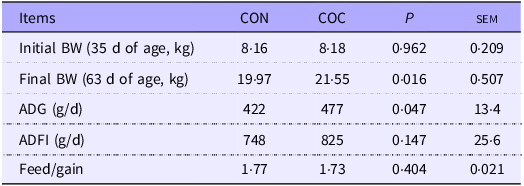
As shown in Table 1, piglets in the coconut oil group had significantly higher BW than those in the control group after 28 d (P < 0·05). Additionally, their average daily weight gain was significantly greater than that of the control group during the (P < 0·05). No difference was observed in average daily feed intake and feed/gain between two groups (P > 0·05).
Effect of coconut oil on the growth performance of piglets

BW, body weight; ADG, average daily gain. ADFI, average daily feed intake, n 6 pigs/group.
Intestinal morphology
As shown in Figure 1(a), piglets injected with saline exhibited normal intestinal villus morphology and intact structure in the jejunum. After LPS stimulation, the jejunum of the piglets showed partial villus shedding, fragmentation and multiple areas of blood cell infiltration. The inclusion of coconut oil in the diet slightly alleviated these changes caused by LPS. Pigs challenged with LPS had lower villus height and villus height/crypt depth (P < 0·001) in the jejunum (Figure 1(b) and (d)). Pigs fed coconut oil had higher villus height (P = 0·020) and tended to have higher villus height/crypt depth (P = 0·072) compared with those fed soybean oil (Figure 1(b) and (d)). An LPS × diet interaction was observed for villus height (P = 0·026) in the jejunum (Figure 1(b)). Coconut oil supplementation increased villus height and villus height/crypt depth in LPS-treated piglets, whereas no effect was observed in saline-injected pigs (Figure 1(b) and (d)). However, neither LPS nor diet affected crypt depth in the jejunum (Figure 1(c)).
Effects of coconut oil on intestinal morphology in piglets challenged with LPS. (a) Representative images and histomorphometry analysis of jejunum tissue section of weaned piglets stained with haematoxylin and eosin (H&E) (magnification: 200×). *indicates partial villus shedding and fragmentation; #indicates blood cell infiltration. (b)–(d) Villus height (b), crypt depth (c), villus height to crypt depth ratio (d) in the jejunum. Values are means (sem), n 6. abcMeans without a common letter differ significantly (P < 0·05).

Tight junction protein expression
As shown in Figure 2, pigs challenged with LPS exhibited significantly lower ZO-1 and Occludin (P < 0·01) protein expression in the jejunum compared with those treated with saline (Figure 2(a)–(c)). Pigs fed coconut oil had higher Occludin expression (P = 0·005) and tended to have higher ZO-1 protein expression (P = 0·089) compared with those fed soybean oil (Figure 2(a)–(c)). An interaction between LPS and diet was observed for ZO-1 protein expression (Figure 2(c)). Coconut oil supplementation alleviated the LPS-induced reduction of jejunal ZO-1 and Occludin protein expression (Figure 2(a)–(c)).
Effects of coconut oil on intestinal barrier function in piglets challenged with LPS. (a) Representative bands of tight junction protein. (b)–(c) Protein expression of ZO-1 (b) and Occludin (c) in the jejunum. (d)–(k) The mRNA expression of VILLIN (d), ALPI (e), mucin 2 (MUC2) (F), lysozyme (LYZ) (g), olfactomedin 4 (OLFM4) (h), chromogranin A (CHGA) (i), leucine-rich-repeat-containing G-protein-coupled receptor 5 (LGR5) (j) and achaete-scute family BHLH transcription factor 2 (ASCL2) (k) in the jejunum. Values are means (sem), n 6. abcMeans without a common letter differ significantly (P < 0·05).

Pigs challenged with LPS showed lower mRNA expression of VILLIN and ALPI (absorptive cell markers), lysozyme (Paneth cell marker), chromogranin A (enteroendocrine cell marker) and leucine-rich-repeat-containing G-protein-coupled receptor 5 and achaete-scute family BHLH transcription factor 2 (intestinal stem cell markers) in the jejunum (P < 0·05) compared with those injected with saline (Figure 2(d)–(k)). Pigs fed coconut oil exhibited higher mRNA expression of jejunal Villin (P = 0·005) compared with those fed soybean oil (Figure 2(d)). An LPS × diet interaction was observed for mucin 2 and chromogranin A expression (P < 0·05) in the jejunum (Figure 2(f) and (i)). Neither LPS nor diet affected olfactomedin 4 (intestinal stem cell marker) mRNA expression in the jejunum (Figure 2(h)).
Antioxidant function
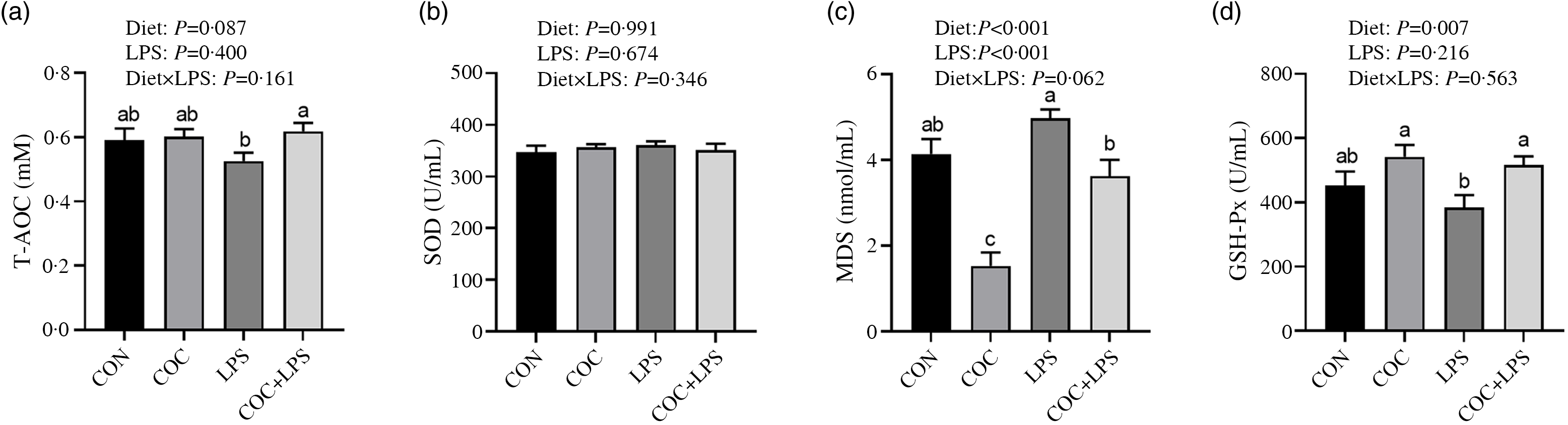
Compared with pigs injected with saline, pigs challenged with LPS exhibited a significantly higher MDA level (P < 0·001) in plasma (Figure 3(c)). Pigs fed coconut oil showed a trend towards higher plasma total antioxidant capacity (P = 0·087), significantly increased plasma GSH peroxidase activity (P = 0·007) and significantly lower MDA level (P < 0·001) compared with those fed the control diet (Figure 3(a)–(d)). A trend for interaction between LPS and diet was observed for plasma MDA level (Figure 3(c)). Coconut oil supplementation significantly increased plasma total antioxidant capacity level and GSH peroxidase activity in LPS-treated piglets, whereas no effect was observed in saline-injected pigs (Figure 3(a) and (d)). Neither LPS nor diet affected superoxide dismutase level in plasma (Figure 3(b)).
Effects of coconut oil on plasma antioxidant parameters in piglets challenged with lipopolysaccharide (LPS). (a) Total antioxidant capacity (T-AOC) level; (b) Superoxide dismutase (SOD) activity; (c) malondialdehyde (MDA) concentration; (d) GSH peroxidase (GSH-Px) activity. Values are means (sem), n 6. abcMeans without a common letter differ significantly (P < 0·05).

Pro-Inflammatory cytokines
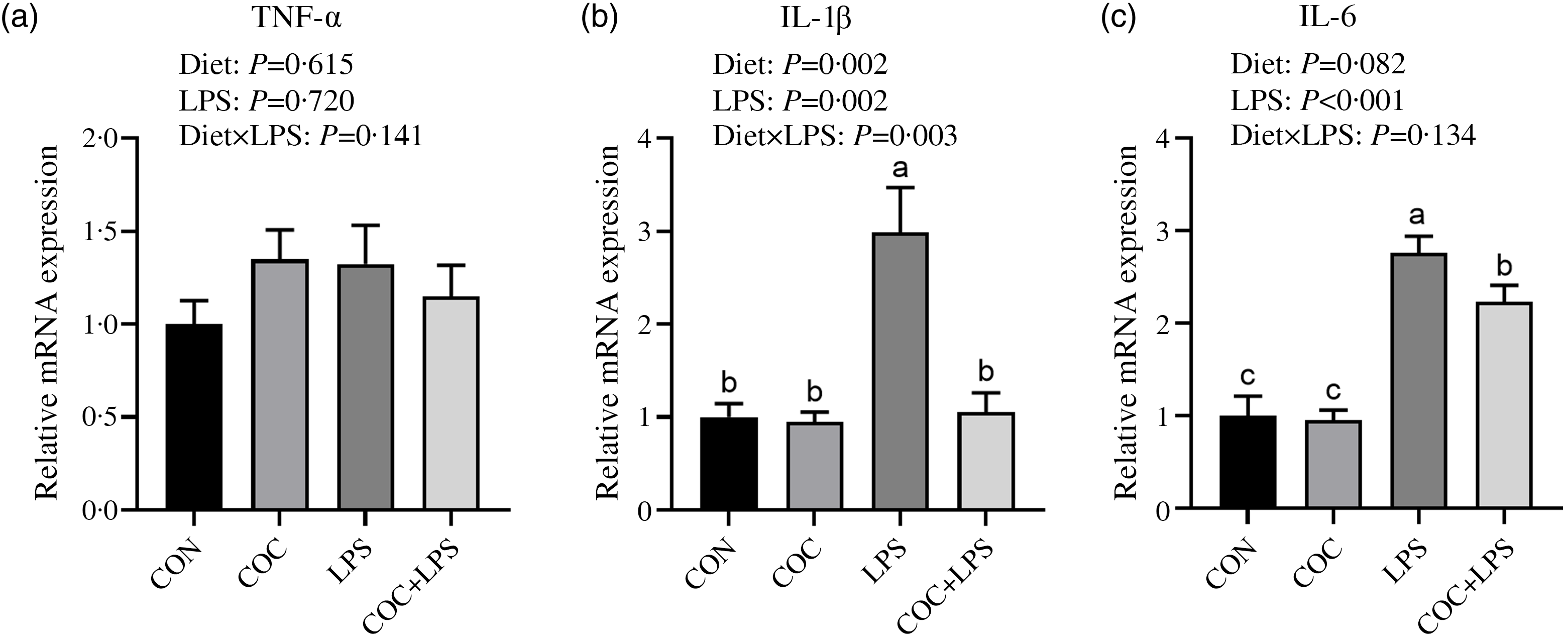
Pigs challenged with LPS exhibited higher mRNA expression of interleukin-1β (IL-1β) and IL-6 in the jejunum (P < 0·05) compared with those treated with saline (Figure 4(b)–(c)). Pigs fed coconut oil showed lower jejunal IL-1β mRNA expression (P = 0·002) and tended to have lower IL-6 mRNA expression (P = 0·082) compared with those fed soybean oil (Figure 4(b)–(c)). No interaction between LPS and diet was observed for TNF-α and IL-6 expression (Figure 4(a) and (c)), while an interaction between LPS and diet was found for IL-1β expression (Figure 4(b)). Coconut oil supplementation reduced jejunal IL-1β and IL-6 mRNA expression in LPS-treated pigs, while no differences were observed in saline-treated pigs (Figure 4(b)–(c)). Neither LPS nor diet affected TNF-α mRNA expression in the jejunum (Figure 4(a)).
Effects of coconut oil on intestinal inflammatory cytokines in piglets challenged with lipopolysaccharide (LPS). (a)–(c) Relative mRNA expression of TNF-α, IL-1β and IL-6 in the jejunum. Values are means (sem), n 6. abMeans without a common letter differ significantly (P < 0·05).

Apoptosis-related gene expression
Pigs challenged with LPS exhibited a higher number of TUNEL-positive cells (P < 0·001) and lower mRNA expression of proliferating cell nuclear antigen (proliferation marker, P < 0·001) compared with those treated with saline (Figure 5(a) and (e)). Pigs fed coconut oil showed a tendency for a reduced number of TUNEL-positive cells (P = 0·065), as well as lower mRNA expression of CASPASE 8 and CASPASE 9 (P < 0·05) in the jejunum compared with those fed soybean oil (Figure 5(a)–(c)). No interaction was found between LPS and diet for CASPASE 8 and KI67 (Figure 5(b) and (e)), while a significant interaction between LPS and diet was observed for the number of TUNEL-positive cells and CASPASE 9 mRNA expression (Figure 5(a) and (c)). Coconut oil supplementation decreased the number of TUNEL-positive cells and the mRNA expression of CASPASE 8 and CASPASE 9 in LPS-treated pigs, while no significant differences were observed in saline-treated pigs (Figure 5(a)–(c)). Neither LPS nor diet influenced KI67 mRNA expression in the jejunum (Figure 5(d)).
Effects of coconut oil on intestinal cell death and proliferation in piglets challenged with lipopolysaccharide (LPS). (a) Representative images and quantification of cell death number by TUNEL staining in the jejunum. (b)–(e) The mRNA expression of CASPASE 8 (b), CASPASE 9 (c), KI67 (d) and proliferating cell nuclear antigen (PCNA) (e) in the jejunum. Values are means (sem), n 6. abcMeans without a common letter differ significantly (P < 0·05).

Necroptosis-related gene expression
Compared with the saline-injected pigs, LPS-treated pigs showed higher mRNA levels of RIPK3 and MLKL (P < 0·05) in the jejunum (Figure 6(b)–(c)). Pigs supplemented with coconut oil exhibited lower mRNA expression of MLKL (P = 0·027) and protein expression of p-RIPK3 and p-MLKL (P < 0·05) compared with those fed the basal diet (Figure 6(c), (f)–(g)). A significant LPS × diet interaction was observed for RIPK3 and MLKL mRNA expression (Figure 6(b)–(c)), as well as for p-MLKL protein expression (Figure 6(g)). Coconut oil supplementation in the diet mitigated the LPS-induced increases in RIPK3 and MLKL mRNA expression (Figure 6(b)–(c)), as well as p-RIPK3 and p-MLKL protein expressions in the jejunum (Figure 6(f)–(g)). Neither LPS nor the diet had an effect on RIPK1 mRNA expression or p-RIPK1 protein expression in the jejunum (Figure 6(a) and (e)).
Effects of coconut oil on intestinal necroptosis signalling pathway in piglets challenged with lipopolysaccharide (LPS). (a)–(c) Relative mRNA expression of receptor-interacting protein kinase 1 (RIPK1), RIPK3 and mixed lineage kinase domain-like protein (MLKL) in the jejunum. (d) Representative protein bands of RIPK1, RIPK3 and MLKL. (e)–(g) Protein expression of RIPK1, RIPK3 and MLKL in the jejunum. Values are means (sem), n 6. abcMeans without a common letter differ significantly (P < 0·05).

Discussion
Because of its structural complexity, the intestinal epithelium is highly susceptible to oxidative stress and infections(Reference Guo, Li and Duan16,Reference Ma, Lyu and Song17) . In this experiment, LPS was used to develop an intestinal injury model in piglets. Intraperitoneally injected LPS is absorbed into the systemic circulation and induces intestinal inflammation through a cascade of systemic immune events. In the blood, LPS binds to its pattern recognition receptor, toll-like receptor 4, on immune cells and endothelial cells. This binding triggers potent intracellular signalling cascades, leading to a massive release of pro-inflammatory cytokines (e.g., TNF-α, IL-1β, IL-6) into the bloodstream. These circulating inflammatory mediators then orchestrate damage to the intestinal mucosa by increasing intestinal permeability. This study investigated the effects of coconut oil supplementation on LPS-induced intestinal injury and inflammation in weaned piglets. Beyond its relevance for swine production, our study may hold implications for human nutrition and health. The piglet is a well-established translational model for human infant gastrointestinal physiology and immunology, sharing remarkable similarities in gut development, digestive processes and immune function(Reference Lunney, Van Goor and Walker18). Therefore, our findings that dietary coconut oil can ameliorate LPS-induced intestinal barrier dysfunction and necroptosis provide a compelling rationale for exploring similar nutritional strategies in human contexts. Given the widespread consumption of coconut oil and the established safety of MCFA in the human diet, our results suggest that coconut oil supplementation could be a nutritional intervention to prevent intestinal diseases.
One of the primary outcomes of this study was the improvement in growth performance observed in piglets supplemented with coconut oil. Piglets in the coconut oil group exhibited significantly higher BW and average daily weight gain compared with those in the control group. These findings align with previous research that demonstrated the growth-promoting effects of MCFA, including lauric acid, a major component of coconut oil(Reference Thomas, Woodworth and Tokach19). The observed increase in feed intake is likely attributed to the superior palatability of the coconut oil-supplemented diet. Rich in MCFA, coconut oil has a characteristic aroma profile that is often more attractive to young pigs than the less distinct aroma of long-chain fatty acid-rich soybean oil. This enhanced palatability appears to be a primary contributor to the stimulated voluntary feed consumption, resulting in greater overall energy and nutrient intake that directly supported the improved growth rates and final BW. In addition, MCFA are known to be easily absorbed and rapidly metabolised, providing an immediate source of energy, which may contribute to enhanced growth performance in piglets(Reference Gebhardt, Thomson and Woodworth20). Additionally, the fast metabolism of MCFA helps maintain an optimal energy balance, counteracting the detrimental effects of inflammation and oxidative stress, which often disrupt growth performance. Due to the acute LPS injection model we used, we did not observe the effect of coconut oil on growth performance under the LPS-induced inflammatory condition. LPS is a potent inflammatory factor that can trigger a systemic inflammatory response, leading to impaired growth, increased energy expenditure and reduced nutrient absorption(Reference Yang, Zhong and Zhang21). If coconut oil alleviates LPS-induced growth retardation, its anti-inflammatory and antioxidant properties may support piglet health and development under stress.
The intestinal integrity of piglets is highly susceptible to damage during weaning or bacterial infection(Reference Tang, Laarveld and Van Kessel22,Reference Xiao, Zou and Li23) . Disruption of the intestinal integrity is often accompanied by a decrease in digestion and absorption. The results from histological analysis revealed that LPS significantly compromised the intestinal morphology, as evidenced by villus shedding, fragmentation and blood cell infiltration. These histological changes are indicative of severe intestinal injury and dysfunction, consistent with previous studies showing that LPS-induced inflammation leads to structural damage in the small intestine(Reference Liu, Chen and Odle24). However, coconut oil-fed piglets showed higher villus height and villus height/crypt depth ratios, suggesting that coconut oil may have a protective effect on intestinal morphology, particularly in the condition of inflammation. The restoration of intestinal morphology may be due to the beneficial effect of coconut oil on the intestinal barrier. The intestine’s barrier function is maintained by tight junction proteins like Occludin and ZO-1, which prevent harmful pathogens and toxins from entering(Reference Suzuki25). The significant reduction in tight junction protein expression after LPS exposure highlights its detrimental effect on intestinal barrier function. Conversely, coconut oil supplementation preserved and even boosted ZO-1 and Occludin expression, suggesting it may help maintain intestinal barrier integrity under inflammatory stress. These findings align with studies suggesting that MCFA improve gut health by modulating tight junction proteins and enhancing barrier function(Reference Jiao, Zheng and Zhuang26).
Oxidative stress plays a pivotal role in the pathophysiology of intestinal inflammation and injury. LPS exposure is known to increase the production of reactive oxygen species, which in turn promotes inflammation and tissue damage(Reference Cho, Zhang and Lazrak27). In this study, piglets challenged with LPS exhibited a marked increase in plasma MDA, a biomarker of oxidative stress, while coconut oil supplementation attenuated this oxidative damage by significantly lowering MDA levels and increasing plasma total antioxidant capacity and GSH levels. The ability of coconut oil to mitigate oxidative stress aligns with previous research indicating that MCFA possess antioxidant properties(Reference Mett and Müller28). Coconut oil’s antioxidant effects may reduce the systemic oxidative burden caused by LPS, thereby protecting intestinal cells from damage and supporting overall health. In addition to clearing lipid peroxidation, coconut oil’s ability to enhance antioxidant enzyme activities may contribute to the maintenance of gut health during inflammation. Higher GSH levels indicate better redox balance and reduced oxidative damage, supporting intestinal tissue repair and regeneration after injury(Reference Reid, Berardinelli and Ray29). This further supports the hypothesis that coconut oil’s beneficial effects on intestinal health may be mediated by its capacity to modulate oxidative stress pathways.
Inflammation is a central component of LPS-induced intestinal injury, and the modulation of pro-inflammatory cytokines is critical in reducing tissue damage(Reference Wang, Xiao and Yu30). In the present study, LPS challenge led to increased expression of IL-6 and IL-1β in the jejunum, which are key cytokines involved in the inflammatory response. The administration of coconut oil reduced the expression of these pro-inflammatory cytokines, suggesting that coconut oil has potent anti-inflammatory properties. This finding is consistent with the known anti-inflammatory effects of coconut oil, which have been attributed to its ability to modulate the expression of cytokines and other inflammatory mediators(Reference Pardiñas López, García-Caro and Vallejo31). The ability of coconut oil to attenuate LPS-induced inflammation is likely due to its bioactive components, including lauric acid, which has been shown to exhibit anti-inflammatory effects by inhibiting the activation of toll-like receptor 4/NF-κB signalling pathway(Reference Khan, Aamir and Jusuf32). These results suggest that the modulation of inflammatory responses by coconut oil may reduce intestinal injury severity, promoting faster recovery and improving overall intestinal function.
A novel and critical aspect of this study is the investigation of necroptosis as a potential mechanism underlying coconut oil’s protective effects against LPS-induced intestinal injury. Necroptosis, a form of programmed cell death, is characterised by the activation of RIPK3 and MLKL, which together form the necrosome and induce cell death(Reference Yan, Wan and Choksi8). Increasing evidence suggests that necroptosis is crucial in the development of LPS-induced pathological conditions(Reference Wang, Wang and Li33,Reference Shan, Pan and Najafov34) . For example, our previous study showed that inhibition of necroptosis by Nec-1 decreased LPS-induced intestinal inflammation in piglets(Reference Liu, Xu and Wang9). The present study provides compelling evidence that coconut oil supplementation significantly reduced the expression of RIPK3 and MLKL, suggesting that coconut oil inhibits necroptosis in the jejunum of LPS-challenged piglets. The findings are in line with previous research suggesting that medium-chain TAG supplementation alleviated LPS-induced intestinal injury and inflammation by inhibiting the activation of necroptosis signalling in piglets(Reference Xu, Chen and Wang35). Interestingly, a notable finding in our study was the lack of significant change in the expression of RIPK1, a canonical upstream regulator of necroptosis. This suggests that LPS-induced systemic inflammatory response may have led to direct activation of RIPK3 and subsequent MLKL phosphorylation, bypassing RIPK1. This phenomenon has been reported in several studies where excessive cytokine production triggers necroptosis without changes in RIPK1 expression or phosphorylation(Reference Moujalled, Cook and Okamoto36). In addition, this could be due to alternative activation pathways or post-translational modifications that bypass RIPK1 involvement. Therefore, our data definitively identify RIPK3 and MLKL as the critical mediators of necroptosis in this model, while also highlighting the context-dependent regulation of this cell death pathway. By modulating necroptosis signalling, coconut oil may prevent excessive cell death, thereby promoting the recovery and regeneration of the intestinal epithelium. This represents a novel therapeutic approach to mitigating LPS-induced intestinal injury, highlighting the potential of coconut oil as a dietary supplement for enhancing gut health.
In conclusion, this study provides strong evidence that coconut oil supplementation offers significant protection against LPS-induced intestinal injury and inflammation in weaned piglets. The beneficial effects of coconut oil on growth performance, barrier function, antioxidant capacity, inflammatory responses and necroptosis signalling pathways suggest that coconut oil may serve as an effective dietary intervention to support gut health in piglets under stressful conditions. Further research is warranted to explore the long-term effects of coconut oil supplementation and its potential as a therapeutic strategy for improving intestinal health and resilience in weaned animals.
Supplementary material
For supplementary material/s referred to in this article, please visit https://doi.org/10.1017/S000711452510620X
Acknowledgements
This research was financially funded by the Project of National Key R&D Program of China (2022YFD1300403).
D. W.: conceptualisation, data curation, writing – original draft, writing – review and editing. D. W.: conceptualisation, data curation, visualisation. W. H.: investigation. X. Z.: data curation. J. C.: methodology. A. L.: investigation. H. Z.: methodology. Y. L.: conceptualisation, writing – review and editing.
The authors declare no conflict of interest.