Introduction
Helicoverpa armigera (Hübner) (Lepidoptera: Noctuidae) is a fast adapting pest, and different field populations that are resistant to various classes of pesticides have emerged (McCaffery, Reference McCaffery1998; Yang et al., Reference Yang, Chen, Wu, Yang and Wu2007). As a viable alternative control strategy, protease inhibitors are promising agents (Tamhane et al., Reference Tamhane, Giri, Sainani and Gupta2007). The serine class consists of main proteolytic enzymes, and the use of serine protease inhibitors in the control of herbivory has been well reported in the literature (Tamhane et al., Reference Tamhane, Chougule, Giri, Dixit, Sainani and Gupta2005, Reference Tamhane, Giri, Sainani and Gupta2007). However, more effective results have been established when cysteine protease inhibitors were coupled with serine protease inhibitors (Senthilkumar et al., Reference Senthilkumar, Cheng and Yeh2010). In addition to serine proteases, cysteine proteases, carboxypeptidase-A, aminopeptidase-N and aspartic proteases have also been reported from the gut extract (Bown et al., Reference Bown, Wilkinson and Gatehouse1998; Patankar et al., Reference Patankar, Giri, Harsulkar, Sainani, Deshpande, Ranjekar and Gupta2001; Chougule et al., Reference Chougule, Giri, Sainani and Gupta2005; Angelucci et al., Reference Angelucci, Barrett-Wilt, Hunt, Akhurst, East, Gordon and Campbell2008). Serine proteases are by far the most abundant, with one study reporting 18 genes for trypsin, 14 for chymotrypsin and two for elastase from larvae fed on an inhibitor-free diet (Bown et al., Reference Bown, Wilkinson and Gatehouse1997). In a separate study, Bown et al. (Reference Bown, Wilkinson and Gatehouse1998) reported 27 genes for trypsin alone. Furthermore, Angelucci et al. (Reference Angelucci, Barrett-Wilt, Hunt, Akhurst, East, Gordon and Campbell2008) reported seven genes for aminopeptidase-N in the larval midgut.
The protease profile of the larval gut is not constant and changes with the developmental stage of the larva. In addition, the presence of inhibitors in the diet affects the expression of proteases as the insect adjusts its protease profile to compensate for enzyme-specific inhibition (Chougule et al., Reference Chougule, Giri, Sainani and Gupta2005). Inhibition of trypsin by the soyabean Kunitz trypsin inhibitor increases the activity of carboxypeptidase-A (Bown et al., Reference Bown, Wilkinson and Gatehouse1998). Moreover, in nature, H. armigera feeds on different parts of its host at different larval stages (Damle et al., Reference Damle, Giri, Sainani and Gupta2005). Experiments using specific inhibitors have revealed the presence of serine, cysteine, aspartyl and metalloproteases in second-instar larvae, but serine proteases only in the fifth instar (Patankar et al., Reference Patankar, Giri, Harsulkar, Sainani, Deshpande, Ranjekar and Gupta2001).
Thus, an estimation of various protease inhibitors at different stages of larval development would be helpful in identifying important targets for inhibition, and would improve our knowledge on the complex profiles of gut proteases. In this study, specific proteolytic substrates were used to detect and estimate the levels of protease activities in the gut extract of the third-, fourth-, fifth- and late fifth-instar larvae. The profiles of gut proteases in different larval instars were further elucidated using specific chemical inhibitors.
Materials and methods
Insects
Larvae were raised on a chickpea-based diet (Nagarkatti and Prakash, Reference Nagarkatti and Prakash1974). The composition for 625 ml of the diet was as follows: 84 g chickpea; 11 g yeast extract; 5 g casein; 2 g p-hydroxy benzoate; 3 g l-ascorbic acid; 1 g sorbic acid; 0.2 g cholesterol; 0.2 g streptomycin; 11 g agar. The larvae were raised in separate sample containers at a constant temperature of 27 °C. Progression through the larval stages was identified by the moulting of skin and head capsules. Each larval instar lasted for 2 to 4 days.
Chemicals
Substrates and enzymes were obtained from Sigma-Aldrich (St Louis, Missouri, USA), which are as follows: substrates – N α-p-tosyl-l-arginine methyl ester (TAME); hippuryl-l-phenylalanine (HLP); N α-benzoyl-l-arginine ethyl ester (BAEE); N-benzoyl-l-tyrosine ethyl ester hydrochloride (BTEE); l-alanine 4-nitroanilide hydrochloride (l-Ala-PNA); l-leucine 4-nitroanilide (l-Leu-PNA); l-pyroglutamyl-l-phenylalanyl-l-leucine-p-nitroanilide (PFLNA); azocasein; enzymes – carboxypeptidase-A (bovine pancreas); aminopeptidase-N (Aeromonas proteolytica); papain (Carica papaya); trypsin (bovine pancreas); chymotrypsin (bovine pancreas). Protease inhibitors were also obtained from Sigma-Aldrich, which are as follows: phenylmethylsulphonyl fluoride (PMSF); sodium ethylenediaminetetraacetic acid (EDTA-Na); N-(N-(l-3 trans-carboxirane-2-carbonyl)-l-leucyl)-agmatine (E-64); aprotinin; 4-(2-aminoethyl)-benzenesulphonyl-fluoride (AEBSF); N-acetyl-leu-leu-Nle-CHO (ALLN); antipain; bestatin; chymostatin; leupeptin; pepstatin; phosphoramidon.
Preparation of gut protease extracts
To obtain a sufficient amount of gut protease extracts, guts from 20 larvae of the third instar (as the size of this larvae was considerably smaller than that of the fourth and fifth instars) and from 10 larvae of the fourth, fifth and late fifth instars were dissected in ice-cold buffer (Tris–Cl 50 mM, pH 7.5; cysteine HCl 5 mM) and homogenized separately with a pre-chilled Dounce homogenizer. The homogenized guts were centrifuged at 13,000 g for 10 min at 4 °C to remove debris and fat deposits. The supernatant was collected and stored at − 20 °C (gut protease extract) until analysis. Protein concentrations of the gut extracts were estimated following the Bradford method using bovine serum albumin as the standard (Bradford, Reference Bradford1976). Gut protease activities were estimated using equal amounts of proteins from each instar. For the third-, fourth- and fifth-instar larvae, gut protease extracts were obtained within 24 h of the onset of each larval stage; for the late fifth-instar larvae, the gut extract was obtained after 96 h of onset when the larvae almost stopped feeding and were about to metamorphose into pupae.
Optimum pH
Optimum pH for proteolytic activity of the gut extract was determined by the azocasein assay (Secades and Guijarro, Reference Secades and Guijarro1999) using the following buffering agents: 50 mM sodium citrate (pH 3–6); 50 mM Tris–Cl (pH 7–10); glycine-NaOH (pH 11–13). The gut extracts (10 μg) were added to 490 μl azocasein (1%, w/v) substrate solutions and incubated at 28 °C for 30 min. After incubation, 500 μl of 10% trichloroacetate were added to each solution, placed on ice for 30 min and then centrifuged at 20,400 g for 10 min at 4 °C. To 800 μl of each supernatant, 200 μl of 1.8 N NaOH were added for colour development; absorbance was read at 420 nm. The pH of the buffer preparation was adjusted with HCl or NaOH using a pH meter.
Protease activity assays
Gut extracts from the third-, fourth-, fifth- and late fifth-instar larvae were assayed for proteolytic activity using various substrates. Serine protease activities were estimated using the substrates TAME, BAEE and BTEE. TAME (1 mM) was prepared in 100 mM Tris–Cl buffer (pH 8); absorbance was read at 247 nm (Wirnt, Reference Wirnt and Bergmeyer1974b ). BAEE (56 mM) was prepared in 200 mM Tris–Cl buffer (pH 6.2) containing 5 mM l-cysteine HCl, 2 mM EDTA-Na and 300 mM sodium chloride; absorbance was read at 253 nm (Arnon, Reference Arnon1970). BTEE (0.55 mM) was prepared in 38 mM Tris–Cl buffer (pH 7.8) containing 50 mM calcium chloride; absorbance was read at 256 nm (Wirnt, Reference Wirnt and Bergmeyer1974a ). Carboxypeptidase-A activity was estimated using the substrate hippuryl l-phenylalanine (0.5 mM) dissolved in 25 mM Tris–Cl buffer (pH 7.5) containing 500 mM sodium chloride; absorbance was read at 254 nm (Bergmeyer et al., Reference Bergmeyer, Gawehn, Grassl and Bergmeyer1974). Aminopeptidase-N activities were estimated using the substrates l-Ala-PNA (1.66 mM) and l-Leu-PNA (1.6 mM) dissolved in 10 mM Tris–Cl buffer (pH 7.2) containing 50 mM magnesium chloride; absorbance was read at 405 nm (Peleiderer, Reference Peleiderer, Perlmann and Lorand1970). Thiol proteases were estimated using the substrate PFLNA (2 mM) dissolved in 100 mM Tris–Cl buffer (pH 6.5); absorbance was read at 405 nm (Filippova et al., Reference Filippova, Lysogorskaya, Oksenoit, Rudenskaya and Stepanov1984). In all assays, 10 μg of gut extract proteins were used. Changes in absorbance were recorded in a continuous spectrophotometric mode using a Hitachi U-2800 spectrophotometer (Hitachi High-Technologies Corporation; Tokyo, Japan). General proteolytic activities were estimated by the azocasein assay. Proteolytic enzyme assays were also performed at pH 10.2 using 200 mM Tris–Cl buffer.
Protease inhibition assays
Gut protease inhibition assays were performed by the incubation of the gut extracts with inhibitors at 30 °C for 10 min followed by a normal azocasein assay (Secades and Guijarro, Reference Secades and Guijarro1999).
Insect bioassay
To assay the influence of protease inhibitors on the growth and development of larvae, four sets of an artificial diet were prepared. In the first set, the larvae were fed on a diet containing 0.1% PMSF; in the second set, the larvae were reared on a diet containing 0.1% (w/w) EDTA-Na; in the third set, the larvae were reared on a diet containing both 0.05% (w/w) PMSF and 0.05% (w/w) EDTA-Na. The total inhibitor concentration was kept constant. The fourth set was the control in which the larvae were reared on an inhibitor-free diet based on chickpea. For each set of the diet, 10 larvae were used. The wet weight taken at days 2, 5, 9 and 12 of larval culture was considered to be a measure of growth in terms of weight gain compared with that of the control group.
Statistical analysis
A one-way analysis of variance was used for the statistical analysis of the activities of gut proteases across the different instars using specific substrates or inhibitors. It was also used to analyse the larval weights of the different treatment groups in the insect bioassay. Each pairwise multiple comparison was made by the Holm–Sidak method. All statistical analyses were performed using SigmaPlot 11.0 software (SigmaPlot Software; San Jose, California, USA).
Results
Gut protease activities of larval instars
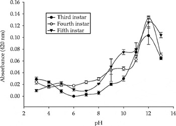
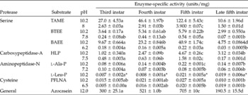
Proteolytic activity in the lysate from the gut extracts of all the three larval stages (third to fifth) was optimal at pH 12, with the activity being observed between pH 3 and pH 13 (Fig. 1). The activities of trypsin, chymotrypsin, cysteine, carboxypeptidase-A and aminopeptidase-N were detected in the gut extract using specific substrates (Table 1). The differences in the specific activities of proteases were compared across the different instars and found to be statistically significant. Their P values were less than 0.05 except for the activity of aminopeptidase-N with l-Leu-pNA, indicating that the differences in specific activities were greater than would be expected by chance.
Protease activity levels in the gut extracts of Helicoverpa armigera at different pH levels.

Protease activities in the gut extracts from the different larval instars of Helicoverpa armigera at different pH levels

Values are mean ± SE of three replicates. Mean values within a row with unlike letters were significantly different (P< 0.05). *P= 0.083.
Inhibition of gut proteases of larval instars
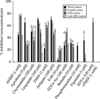
Various specific and broad-range protease inhibitors were used to assay the activities of proteases in the gut extracts (Fig. 2). In the third-instar larvae, aprotinin, leupeptin and antipain were effective inhibitors of protease activity, but E-64 was not effective. In the fourth- and fifth-instar larvae, AEBSF was the optimal inhibitor, whereas in the fourth-, fifth- and late fifth-instar larvae, pepstatin and phosphoramidon exhibited minimum or no inhibition. In the late fifth-instar larvae, PMSF showed the maximum inhibition of gut protease activities. ALLN, an inhibitor specific to calpain and other neutral cysteine proteases, did not inhibit gut protease activities in any of the instars. The metalloendopeptidase inhibitor phosphoramidon (10 mM) inhibited gut protease activities only in the third instar. The aspartic protease inhibitor pepstatin (1 μM) inhibited gut protease activities only in the third and fourth instars. Bestatin (10 μM), a specific inhibitor of aminopeptidase-N, did not inhibit the activities of proteases in the gut extracts of the late fifth instar.
Inhibition of protease activity in different instars of H. armigera with different inhibitors. Each value is the mean of three replicates ± sd. Similar letters within a group are used to denote statistically insignificant difference. All tested groups have P value < 0.05. The experiment (*) using EDTA (100 mM)+PMSF (1 mM) was performed against 5th instar larvae only.

Insect bioassay
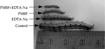
Larval growth was retarded in all the three treatment groups compared with the control group (Table 2 and Fig. 3). PMSF (0.1%, w/w) caused 80% mortality and a combination of PMSF (0.05%, w/w) and EDTA-Na (0.05%, w/w) in the larval diet led to 40% mortality on day 9 of the larval culture. However, EDTA-Na (0.1%, w/w) did not cause mortality, although it inhibited larval growth. The differences in larval wet weight among the different treatment groups on days 2, 5 and 9 were statistically significant (P< 0.001). Pupation occurred on day 12 in the control group and the lone survivor of the PMSF-treated group; in the EDTA-Na-treated group, pupation was delayed and occurred on day 17. There were no survivors in the combined EDTA-Na and PMSF-treated group.
Effects of EDTA-Na, PMSF and EDTA-Na+PMSF on the growth of the larvae

n, number of larvae.
Mean values within a column were significantly different (P< 0.001).
1 Pupa.
2 Larvae in the EDTA-Na-treated group pupated at day 17.
3 No survivor.
Differences in the growth and development of Helicoverpa armigera larvae reared on different diets containing PMSF (0.1%), EDTA-Na (0.1%) and a combination of PMSF (0.05%) and EDTA-Na (0.05%) at day 9 of larval culture.

Discussion
The gut protease complement of H. armigera is complex. Serine proteases, such as trypsin and chymotrypsin, are the main constituents along with the subordinate presence of carboxypeptidase-A, aminopeptidase-N, cysteine and aspartic proteases. Several genes encode digestive proteases. The expression of these genes is not constant throughout the stages of larval growth; proteases are either up- or down-regulated depending on the diet and developmental stage (Patankar et al., Reference Patankar, Giri, Harsulkar, Sainani, Deshpande, Ranjekar and Gupta2001; Chougule et al., Reference Chougule, Giri, Sainani and Gupta2005). Pauchet et al. (Reference Pauchet, Muck, Svatos, Heckel and Preiss2008) characterized the proteome of the gut lumen of fifth-instar larvae, and found nine serine proteases and two carboxypeptidase-A enzymes.
The optimum pH for total proteolytic activity was 12. The activity was also detected at low pH and was least around pH 6. Özgür et al. (Reference Özgür, Yücel and Öktem2009) also reported a highly alkaline optimum pH for gut protease activity.
In experiments using specific substrates, the levels of proteolytic activity did not remain constant. There was a general increase in protease activities as the larvae progressed to the fifth instar stage and a decrease thereafter. This was true for all proteases evaluated in this study, which is consistent with the observation of Patankar et al. (Reference Patankar, Giri, Harsulkar, Sainani, Deshpande, Ranjekar and Gupta2001) showing the same trend for trypsin and total gut proteases. The rate of food ingestion increased by a factor of 3 and the volume of food by 3.5 from the fourth to the fifth instar; the length of the feeding time remained equal (Browne and Raubenheimer, Reference Browne and Raubenheimer2003). In our study, the specific activity of trypsin (TAME, pH 10.2) increased by a factor of 2.6 between the fourth and fifth instars. The activities of carboxypeptidase-A (HLP, pH 10.2) increased by a factor of 1.9, aminopeptidase-N (l-Ala-pNA, pH 10.2) by 1.5 and cysteine (pGlu-Phe-Leu-pNA, pH 10.2) by 1.3. The increase in protease activities seemed to be a direct response to the increased consumption of food because when the larvae stopped feeding at the onset of pupation in the late fifth instar, the specific activity of all proteases decreased. In addition, all classes of gut proteases seem to have become adapted to the alkaline conditions of the gut as highly significant differences in specific activity were recorded at pH 6–8 and pH 10–12. The serine protease substrates, TAME, BAEE and BTEE, were most hydrolysed by gut proteases followed by the carboxypeptidase-A substrate HLP in all larval instars studied. The substrates for aminopeptidase-N and cysteine proteases were the least hydrolysed.
Gut protease activities were detected directly using specific substrates and indirectly with chemical inhibitors. All major classes of proteases were detected, although serine proteases were the most abundant. The levels of inhibition of every inhibitor varied across the larval instars and reflected the complexity of the gut protease profile. The levels of protease inhibition by AEBSF specific for serine, E-64 for cysteine and EDTA-Na for metalloproteases increased as the larvae matured from the third to the fifth instar and decreased in the late fifth instar (Fig. 2). The inhibitor bestatin for aminopeptidase-N expressed maximum activity in the third instar, which then declined towards zero in the late fifth instar. The levels of protease inhibition by PMSF, a dual inhibitor of serine and cysteine proteases, increased as the larvae matured. The absence of calpain and other neutral cysteine proteases was indicated by the absence of inhibition by ALLN. Pepstatin inhibited the activities of gut proteases in the third and fourth instars, indicating the presence of aspartic proteases in younger larvae (Fig. 2). Patankar et al. (Reference Patankar, Giri, Harsulkar, Sainani, Deshpande, Ranjekar and Gupta2001) using the inhibitor pepstatin also inferred the presence of aspartic proteases in the second instar and their absence in the fifth instar.
The strategy of using serine protease inhibitors is successful to an extent since serine proteases are the major proteolytic enzymes; however, a limiting factor is the presence of other classes of gut proteases. When a combination of trypsin and cysteine protease inhibitors was used (Senthilkumar et al., Reference Senthilkumar, Cheng and Yeh2010), it very effectively reduced the survivability of larvae. Similarly, Oppert et al. (Reference Oppert, Morgan and Kramer2004) also achieved a better control of the coleopteran pest Tribolium castaneum by using a combination of serine and cysteine protease inhibitors. The use of both types of inhibitors countered the insect's adaptive response of switching digestive enzymes when only one type of inhibitor was used. In this study, the effect of EDTA-Na, a metalloprotease inhibitor, was examined. EDTA-Na (0.1%, w/w) in the larval diet inhibited larval growth, but did not cause mortality. PMSF at the same concentration induced greater inhibition of growth as well as 90% mortality on day 12 of larval culture. The combination of PMSF (0.05%, w/w) and EDTA-Na (0.05%, w/w) resulted in total mortality on day 12 of larval culture, and this inhibition of larval growth was comparable to that induced by PMSF (0.1%) alone. A combination of the two inhibitors at lower doses seemed as, if not more, effective as a higher dose of PMSF alone. Since exopeptidases digest the proteolytic product of endopeptidases, its inhibition should work in synergy with the inhibition of serine proteases. Thus, exopeptidases may be inhibited along with the dominant serine endoproteases for the effective control of herbivory by H. armigera.
Acknowledgements
The authors are grateful to Dr G.T. Gujar, Indian Agricultural Research Institute (IARI), New Delhi, India for providing the initial eggs of H. armigera. Lunminlal Kipgen acknowledges the Council for Scientific and Industrial Research (CSIR), India for research fellowship (CSIR F.No.09/806 (0012)/2008-EMR-I).