




No CrossRef data available.
Ultrasound diagnosis of cystic echinococcosis: updates and implications for clinical management
Published online by Cambridge University Press: 17 April 2024
Abstract
The diagnosis of cystic echinococcosis (CE) is based on imaging. Detection of a focal lesion with morphological characteristics of Echinococcus granulosus sensu lato metacestode is the starting point for the diagnostic workup. In organs explorable with ultrasound (US), this is the method of choice for both aetiological diagnosis of CE and staging of the CE cyst. Staging in terms of lesion morphology is also needed when serology is added to the diagnostic workflow when imaging alone is inconclusive. Finally, staging guides the clinical management of uncomplicated CE, especially in the liver. This commentary provides an overview of the most up-to-date evidence backing the above-mentioned role of US in the diagnosis and clinical management of CE. Finally, we outline future perspectives for the improvement of CE diagnosis.
Information
- Type
- Commentary
- Information
- Copyright
- © The Author(s), 2024. Published by Cambridge University Press
References
Brunetti, E, Kern, P, Vuitton, DA; Writing Panel for the WHO-IWGE (2010). Expert consensus for the diagnosis and treatment of cystic and alveolar echinococcosis in humans. Acta Tropica 114, 1–16. https://doi.org/10.1016/j.actatropica.2009.11.001.CrossRefGoogle ScholarPubMed
Brunetti, E, Tamarozzi, F (2023). Watch-and-wait approach for inactive echinococcal cysts: Scoping review update since the issue of the WHO-IWGE Expert Consensus and current perspectives. Current Opinion in Infectious Diseases 36, 326–32. https://doi.org/10.1097/QCO.0000000000000943.CrossRefGoogle ScholarPubMed
Cheng, J, Wang, H, Li, R, Li, X, Zhou, X, Yang, X, Wang, Y, Xiong, L, Fan, H, Wang, T, Li, M, Ni, D (2022). A two-stage multiresolution neural network for automatic diagnosis of hepatic echinococcosis from ultrasound images: A multicenter study. Medical Physics 49, 3199–3212. https://doi.org/10.1002/mp.15548.CrossRefGoogle ScholarPubMed
Del Carpio, M, Mercapide, CH, Salvitti, JC, Uchiumi, L, Sustercic, J, Panomarenko, H, Moguilensky, J, Herrero, E, Talmon, G, Volpe, M, Araya, D, Mujica, G, Calabro, A, Mancini, S, Chiosso, C, Labanchi, JL, Saad, R, Goblirsch, S, Brunetti, E, Larrieu, E (2012). Early diagnosis, treatment and follow-up of cystic echinococcosis in remote rural areas in Patagonia: Impact of ultrasound training of non-specialists. PLoS Neglected Tropical Diseases 6, e1444. https://doi.org/10.1371/journal.pntd.0001444.CrossRefGoogle ScholarPubMed
Gharbi, HA, Hassine, W, Brauner, MW, Dupuch, K (1981). Ultrasound examination of the hydatic liver. Radiology 139, 459–463. https://doi.org/10.1148/radiology.139.2.7220891.CrossRefGoogle ScholarPubMed
Hernández, A, Cardozo, G, Dematteis, S, Baz, A, Trias, N, Nuñez, H, Barragué, A, López, L, Fuentes, J, López, O, Ferreira, C (2005). Cystic echinococcosis: Analysis of the serological profile related to the risk factors in individuals without ultrasound liver changes living in an endemic area of Tacuarembó, Uruguay. Parasitology. 130, 455–460. https://doi.org/10.1017/s0031182004006717.CrossRefGoogle Scholar
Hernández-González, A, Muro, A, Barrera, I, Ramos, G, Orduña, A, Siles-Lucas, M (2008). Usefulness of four different Echinococcus granulosus recombinant antigens for serodiagnosis of unilocular hydatid disease (UHD) and postsurgical follow-up of patients treated for UHD. Clinical and Vaccine Immunology 15, 147–153. https://doi.org/10.1128/CVI.00363-07.CrossRefGoogle ScholarPubMed
Hernández-González, A, Santivañez, S, García, HH, Rodríguez, S, Muñoz, S, Ramos, G, Orduña, A, Siles-Lucas, M (2012). Improved serodiagnosis of cystic echinococcosis using the new recombinant 2B2t antigen. PLoS Neglected Tropical Diseases 6, e1714. https://doi.org/10.1371/journal.pntd.0001714.CrossRefGoogle ScholarPubMed
Hosch, W, Junghanss, T, Stojkovic, M, Brunetti, E, Heye, T, Kauffmann, GW, Hull, WE (2008). Metabolic viability assessment of cystic echinococcosis using high-field 1H MRS of cyst contents. NMR in Biomedicine 21, 734–754. https://doi.org/10.1002/nbm.1252.CrossRefGoogle ScholarPubMed
Lissandrin, R, Tamarozzi, F, Piccoli, L, Tinelli, C, De Silvestri, A, Mariconti, M, Meroni, V, Genco, F, Brunetti, E (2016). Factors influencing the serological response in hepatic Echinococcus granulosus infection. American Journal of Tropical Medicine and Hygiene 94, 166–171. https://doi.org/10.4269/ajtmh.15-0219.CrossRefGoogle ScholarPubMed
Manciulli, T, Enríquez-Laurente, R, Tamarozzi, F, Lissandrin, R, Elizalde, M, Sedano, C, Bardales, K, Vola, A, De Silvestri, A, Tinelli, C, Brunetti, E, Santivanez, S, Mariconti, M (2021). Field performance of a rapid diagnostic test for the serodiagnosis of abdominal cystic echinococcosis in the Peruvian highlands. American Journal of Tropical Medicine and Hygiene 105, 181–187. https://doi.org/10.4269/ajtmh.21-0045.CrossRefGoogle ScholarPubMed
Mourglia-Ettlin, G, Miles, S, Hernández, A, Dematteis, S (2016). Antibody profiling in ultrasound normal individuals with positive serology for cystic echinococcosis. Parasite Immunology 38, 93–100. https://doi.org/10.1111/pim.12302.CrossRefGoogle ScholarPubMed
Rahimi, H, Sadjjadi, S, Sarkari, B (2011). Performance of antigen B isolated from different hosts and cyst locations in diagnosis of cystic echinococcosis. Iranian Journal of Parasitology 6, 12–19.Google ScholarPubMed
Rogan, MT, Hai, WY, Richardson, R, Zeyhle, E, Craig, PS (2006). Hydatid cysts: Does every picture tell a story? Trends in Parasitology 22, 431–438. https://doi.org/10.1016/j.pt.2006.07.003.CrossRefGoogle ScholarPubMed
Romig, T, Deplazes, P, Jenkins, D, Giraudoux, P, Massolo, A, Craig, PS, Wassermann, M, Takahashi, K, de la Rue, M (2017). Ecology and life cycle patterns of Echinococcus species. Advances in Parasitology 95, 213–314. https://doi.org/10.1016/bs.apar.2016.11.002.CrossRefGoogle ScholarPubMed
Sánchez-Ovejero, C, Akdur, E, Manzano-Román, R, Hernández-González, A, González-Sánchez, M, Becerro-Recio, D, González-Miguel, J, Akhan, O, Cretu, CM, Vutova, K, Tamarozzi, F, Mariconti, M, Brunetti, E, Vola, A, Fabiani, M, Casulli, A, Siles-Lucas, M (2020). Evaluation of the sensitivity and specificity of GST-tagged recombinant antigens 2B2t, Ag5t and DIPOL in ELISA for the diagnosis and follow up of patients with cystic echinococcosis. PLoS Neglected Tropical Diseases 14, e0008892. https://doi.org/10.1371/journal.pntd.0008892.CrossRefGoogle ScholarPubMed
Santivañez, SJ, Arias, P, Portocarrero, M, Rodriguez, S, Gonzalez, AE, Gilman, RH, Gavidia, CM, Garcia, HH (2012). Serological diagnosis of lung cystic hydatid disease using the synthetic p176 peptide. Clinical and Vaccine Immunology 19, 944–947. https://doi.org/10.1128/CVI.05540-11.CrossRefGoogle ScholarPubMed
Siles-Lucas, M, Casulli, A, Conraths, FJ, Müller, N. (2017) Laboratory diagnosis of Echinococcus spp. in human patients and infected animals. Advances in Parasitology 96, 159–257. https://doi.org/10.1016/bs.apar.2016.09.003.CrossRefGoogle Scholar
Siles-Lucas, M, Uchiumi, L, Tamarozzi, F (2023). ‘No cyst, no echinococcosis’: A scoping review update on the diagnosis of cystic echinococcosis after the issue of the WHO-IWGE Expert Consensus and current perspectives. Current Opinion in Infectious Diseases 36, 333–340. https://doi.org/10.1097/QCO.0000000000000941.Google ScholarPubMed
Solomon, N, Fields, PJ, Tamarozzi, F, Brunetti, E, Macpherson, CNL (2017). Expert reliability for the World Health Organization standardized ultrasound classification of cystic echinococcosis. American Journal of Tropical Medicine and Hygiene 96, 686–691. https://doi.org/10.4269/ajtmh.16-0659.CrossRefGoogle ScholarPubMed
Stojkovic, M, Rosenberger, K, Kauczor, HU, Junghanss, T, Hosch, W (2012). Diagnosing and staging of cystic echinococcosis: How do CT and MRI perform in comparison to ultrasound? PLoS Neglected Tropical Diseases 6, e1880. https://doi.org/10.1371/journal.pntd.0001880.CrossRefGoogle Scholar
Tamarozzi, F, Longoni, SS, Vola, A, Degani, M, Tais, S, Rizzi, E, Prato, M, Scarso, S, Silva, R, Brunetti, E, Bisoffi, Z, Perandin, F (2021a). Evaluation of nine commercial serological tests for the diagnosis of human hepatic cyst echinococcosis and the differential diagnosis with other focal liver lesions: A diagnostic accuracy study. Diagnostics (Basel) 11, 167. https://doi.org/10.3390/diagnostics11020167.CrossRefGoogle ScholarPubMed
Tamarozzi, F, Silva, R, Fittipaldo, VA, Buonfrate, D, Gottstein, B, Siles-Lucas, M (2021b). Serology for the diagnosis of human hepatic cystic echinococcosis and its relation with cyst staging: A systematic review of the literature with meta-analysis. PLoS Neglected Tropical Diseases 15, e0009370. https://doi.org/10.1371/journal.pntd.0009370.CrossRefGoogle ScholarPubMed
Torgerson, PR, Deplazes, P (2009). Echinococcosis: Diagnosis and diagnostic interpretation in population studies. Trends in Parasitology 25, 164–170. https://doi.org/10.1016/j.pt.2008.12.008.CrossRefGoogle ScholarPubMed
Vola, A, Manciulli, T, De Silvestri, A, Lissandrin, R, Mariconti, M, Siles-Lucas, M, Brunetti, E, Tamarozzi, F (2019). Diagnostic performances of commercial ELISA, indirect hemagglutination, and Western blot in differentiation of hepatic echinococcal and non-echinococcal lesions: A retrospective analysis of data from a single referral centre. American Journal of Tropical Medicine and Hygiene 101, 1345–1349. https://doi.org/10.4269/ajtmh.19-0556.CrossRefGoogle ScholarPubMed
World Health Organization (2022). Call for public consultation for experts to join the Guideline Development Group (GDG) on treatment of patients with cystic echinococcosis (CE). Available at https://www.who.int/news-room/articles-detail/call-for-public-consultation-for-experts-to-join-the-guideline-development-group-on-treatment-of-patients-with-cystic-echinococcosis (accessed 06 January 2024).Google Scholar
Wu, M, Yan, C, Wang, X, Liu, Q, Liu, Z, Song, T (2022). Automatic classification of hepatic cystic echinococcosis using ultrasound images and deep learning. Journal of Ultrasound in Medicine 41, 163–174. https://doi.org/10.1002/jum.15691.CrossRefGoogle ScholarPubMed
Xin, S, Shi, H, Jide, A, Zhu, M, Ma, C, Liao, H (2020). Automatic lesion segmentation and classification of hepatic echinococcosis using a multiscale-feature convolutional neural network. Medical and Biological Engineering and Computing 58, 659–668. https://doi.org/10.1007/s11517-020-02126-8.CrossRefGoogle ScholarPubMed
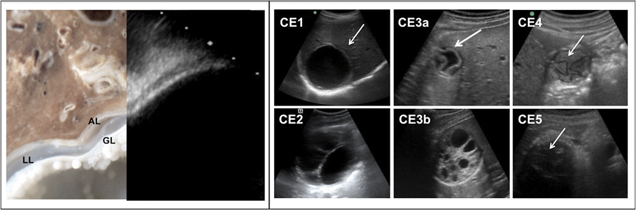
Figure 1. Left panel: ultrasonography showing CE1 cyst wall structures mirroring the cyst’s anatomical structure. Left: micrograph of liver with CE cyst wall, encompassing the adventitial layer (AL), the laminated layer (LL), and the germinal layer (GL). Right: on ultrasound the CE cyst wall’s structures are shown as an outer hypoechoic rim at the interface with normal liver and an inner hyperechoic rim at the interface with the cyst’s fluid content (double wall sign). Right panel: WHO-IWGE US stages of CE cysts. CE1 (unilocular fluid-filled cyst with double-wall [white arrow]), CE2 (fluid-filled cysts with daughter cysts), CE3a (unilocular fluid-filled cysts with detached parasitic layers [white arrow]) CE3b (daughter cysts in a solid matrix with folded hypoechoic parasitic layers), CE4 (solid content with folded hypoechoic parasitic layers [white arrow]), and CE5 (CE4 with evident egg-shell calcification [white arrow shows folded hypoechoic parasitic layers]).
 You have
Access
- 12
- Cited by
Introduction
Cystic echinococcosis (CE) is the infection with the larval stage (metacestode, named “echinococcal cyst” or “hydatid cyst”) of the cestode Echinococcus granulosus sensu lato, which develops its biological cycle mainly in a domestic environment between canids (definitive hosts; mainly dogs) and livestock (intermediate hosts; mainly sheep) (Romig et al. Reference Romig, Deplazes, Jenkins, Giraudoux, Massolo, Craig, Wassermann, Takahashi and de la Rue2017). Humans are accidental intermediate hosts who may develop the echinococcal cyst(s) mainly in the liver and lungs after ingestion of parasite eggs contaminating the environment. CE cysts present with variable morphology, which is classified in stages (CE1 to CE5 in the WHO Informal Working Group on Echinococcosis – IWGE – classification system; Brunetti et al. Reference Brunetti, Kern and Vuitton2010) that collectively reflect the biological viability of the metacestode (Hosch et al. Reference Hosch, Junghanss, Stojkovic, Brunetti, Heye, Kauffmann and Hull2008). This commentary will outline the importance of identifying hepatic CE cysts by ultrasonography (US), by which CE cyst stages were defined, and of staging for the diagnosis and clinical management of human CE.
Why imaging: “no cyst, no echinococcosis”
Because humans are intermediate hosts of E. granulosus harbouring the tissue larval stage of the parasite, classical direct parasitological diagnostic techniques such as coproparasitology are not adequate for its diagnosis and, currently, no molecular or antigen-detection test is able to reliably detect parasitic molecules in excreta or body fluids (Siles-Lucas et al. Reference Siles-Lucas, Casulli, Conraths and Müller2017; Siles-Lucas et al. Reference Siles-Lucas, Uchiumi and Tamarozzi2023). Diagnosis relies on visualization of the cyst by imaging and by detection of circulating antibodies against the parasite using seroassays. Although it might seem intuitive to use serology for screening of CE in asymptomatic subjects (i.e., in the context of screening) or in patients where the clinician might suspect CE (e.g., patients with abdominal symptoms or eosinophilia), current seroassays are not adequate for this purpose. First, seropositivity in population studies does not reflect high sensitivity but rather low specificity of serology when applied to an infection with low prevalence and therefore low pre-test probability such as CE (Torgerson and Deplazes Reference Torgerson and Deplazes2009). In other words, the overwhelming majority of seropositive cases in subjects with low pre-test probability of CE (e.g., in the general population or with very unspecific signs/symptoms such as abdominal pain or eosinophilia) will be false positives. Besides well-known causes of cross-reactivity of seroassays (E. multilocularis, Taenia solium/cysticercosis, and other less frequent conditions) (Hernandez-Gonzalez et al. Reference Hernández-González, Muro, Barrera, Ramos, Orduña and Siles-Lucas2008; Hernandez-Gonzalez et al. Reference Hernández-González, Santivañez, García, Rodríguez, Muñoz, Ramos, Orduña and Siles-Lucas2012), other causes of false positive serology results might derive from just “exposure” to the parasite in endemic areas (without evident risk of developing a CE cyst in the following months/years) (Hernandez et al. Reference Hernández, Cardozo, Dematteis, Baz, Trias, Nuñez, Barragué, López, Fuentes, López and Ferreira2005) and presence of poorly specific antibody isotypes (Mourglia-Ettlin et al. Reference Mourglia-Ettlin, Miles, Hernández and Dematteis2016).
Additionally, a negative serology cannot exclude the diagnosis of CE because in many conditions CE is associated with seronegative results. Low sensitivity of seroassays in confirmed CE cases is well-known to occur in the presence of “early” (CE1) and “inactive” (CE4–CE5) stage cysts (Tamarozzi et al. Reference Tamarozzi, Silva, Fittipaldo, Buonfrate, Gottstein and Siles-Lucas2021b), extra-hepatic cyst localizations (Rahimi et al. Reference Rahimi, Sadjjadi and Sarkari2011; Santivañez et al. Reference Santivañez, Arias, Portocarrero, Rodriguez, Gonzalez, Gilman, Gavidia and Garcia2012; Sanchez-Ovejero et al. Reference Sánchez-Ovejero, Akdur, Manzano-Román, Hernández-González, González-Sánchez, Becerro-Recio, González-Miguel, Akhan, Cretu, Vutova, Tamarozzi, Mariconti, Brunetti, Vola, Fabiani, Casulli and Siles-Lucas2020), and single, small, and uncomplicated CE cysts (Hernandez-Gonzalez et al. Reference Hernández-González, Santivañez, García, Rodríguez, Muñoz, Ramos, Orduña and Siles-Lucas2012; Santivañez et al. Reference Santivañez, Arias, Portocarrero, Rodriguez, Gonzalez, Gilman, Gavidia and Garcia2012; Lissandrin et al. Reference Lissandrin, Tamarozzi, Piccoli, Tinelli, De Silvestri, Mariconti, Meroni, Genco and Brunetti2016; Sanchez-Ovejero et al. Reference Sánchez-Ovejero, Akdur, Manzano-Román, Hernández-González, González-Sánchez, Becerro-Recio, González-Miguel, Akhan, Cretu, Vutova, Tamarozzi, Mariconti, Brunetti, Vola, Fabiani, Casulli and Siles-Lucas2020). Seropositivity in single, uncomplicated CE ranges from 64–85% in the presence of hepatic CE1 and from 53–90% in the presence of hepatic inactive CE4–CE5 cysts. In lung CE seropositivity ranges from 17%–80%, and it can be as low as 12% in cysts in uncommon sites (Siles-Lucas et al. Reference Siles-Lucas, Uchiumi and Tamarozzi2023). Therefore, the theoretical use of serology in screening campaigns for early diagnosis, for capturing people with active infection, and for capturing infection not detectable by portable imaging modalities (i.e., US) is thwarted by its low sensitivity especially in these conditions, and by the high rate of seropositive cases with eventually no detectable CE cysts.
The accuracy of seroassay results can be improved by performing serology only after a lesion compatible with CE is visualized on imaging because this strategy increases the pre-test probability of CE infection and, as a consequence, the post-test probability of the seroassay result (Vola et al. Reference Vola, Manciulli, De Silvestri, Lissandrin, Mariconti, Siles-Lucas, Brunetti and Tamarozzi2019; Manciulli et al. Reference Manciulli, Enríquez-Laurente, Tamarozzi, Lissandrin, Elizalde, Sedano, Bardales, Vola, De Silvestri, Tinelli, Brunetti, Santivanez and Mariconti2021; Tamarozzi et al. Reference Tamarozzi, Longoni, Vola, Degani, Tais, Rizzi, Prato, Scarso, Silva, Brunetti, Bisoffi and Perandin2021a)
Why ultrasound
The imaging features of CE cysts with different morphologies have been described based on US, resulting in the current standardized WHO-Informal Working Groups on Echinococcosis (WHO-IWGE) classification system (Brunetti et al. Reference Brunetti, Kern and Vuitton2010). This encompasses six stages (Figure 1) as follows: CE1 (unilocular fluid-filled cyst with double-wall), CE2 (fluid-filled cysts with daughter cysts), CE3a (unilocular fluid-filled cysts with detached parasitic layers), CE3b (daughter cysts in a solid matrix with folded hypoechoic parasitic layers), CE4 (solid content with folded hypoechoic parasitic layers), and CE5 (CE4 with evident egg-shell calcification). CE1, CE2, and CE3b cysts are biologically viable (active); CE3a may be biologically viable or not; CE4 and CE5 are biologically inactive (Hosch et al. Reference Hosch, Junghanss, Stojkovic, Brunetti, Heye, Kauffmann and Hull2008). Another still widely applied US cyst classification system is the one issued by Gharbi in 1981 (Gharbi et al. Reference Gharbi, Hassine, Brauner and Dupuch1981); however, some problems exist with this classification system due to its not univocal classification of Gharbi Type IV cysts, which could comprise both active (CE3b of the WHO-IWGE classification) and inactive (CE4) stages.
Left panel: ultrasonography showing CE1 cyst wall structures mirroring the cyst’s anatomical structure. Left: micrograph of liver with CE cyst wall, encompassing the adventitial layer (AL), the laminated layer (LL), and the germinal layer (GL). Right: on ultrasound the CE cyst wall’s structures are shown as an outer hypoechoic rim at the interface with normal liver and an inner hyperechoic rim at the interface with the cyst’s fluid content (double wall sign). Right panel: WHO-IWGE US stages of CE cysts. CE1 (unilocular fluid-filled cyst with double-wall [white arrow]), CE2 (fluid-filled cysts with daughter cysts), CE3a (unilocular fluid-filled cysts with detached parasitic layers [white arrow]) CE3b (daughter cysts in a solid matrix with folded hypoechoic parasitic layers), CE4 (solid content with folded hypoechoic parasitic layers [white arrow]), and CE5 (CE4 with evident egg-shell calcification [white arrow shows folded hypoechoic parasitic layers]).
Currently, US is the only imaging technique able to reliably depict the pathognomonic signs of CE cyst stages (Stojkovic et al. Reference Stojkovic, Rosenberger, Kauczor, Junghanss and Hosch2012) (Figure 1). The seminal study by Stojkovic et al. (Reference Stojkovic, Rosenberger, Kauczor, Junghanss and Hosch2012), comparing the performance of CT and MRI with US for the correct staging of hepatic CE cysts, showed very good agreement (Kappa = 0.83–1.00) between MRI (especially T2-weighted imaging) and US for CE1–CE4 cysts. In contrast, CT had only moderate agreement with US (Kappa = 0.62–0.72), although calcifications were shown better by CT than MRI. The unreliable performance of CT makes this technique less adequate than US and MRI for the work-up of suspect CE. While CT and to some extent MRI are less reliable for ruling in CE, together with contrast-enhanced US they can help to rule CE out, if they show clear features incompatible with CE, such as contrast enhancement of cysts’ components. Finally, studies published thus far on the usefulness of advanced imaging techniques, such as Diffusion Weighted MRI, for the differential diagnosis of CE cysts, had variable and overall unsatisfactory results (Siles-Lucas et al. Reference Siles-Lucas, Uchiumi and Tamarozzi2023).
Why staging
As mentioned before, cyst morphology, classified in stages, overall reflects some biological features of CE cysts, which have important epidemiological and clinical implications.
From results of observational population studies, it can be inferred that CE cysts progress from CE1 to CE4–CE5 stages, through the other stages (Rogan et al. Reference Rogan, Hai, Richardson, Zeyhle and Craig2006; Brunetti and Tamarozzi Reference Brunetti and Tamarozzi2023). Therefore, although cyst growth and evolution from stage to stage is uneven over time, the distribution of CE stages in a population arguably indicates transmission in the population, with presence of CE1 cysts indicating ongoing transmission.
From the clinical perspective, staging is pivotal for both diagnosis and clinical management of CE. When imaging alone is not conclusive, serology is often applied in the diagnostic workup of a lesion compatible with a CE cyst. As described above, negative serology cannot exclude the diagnosis of CE, while positive serology in a patient with a lesion compatible with CE may confirm the aetiological diagnosis (Siles-Lucas et al. Reference Siles-Lucas, Uchiumi and Tamarozzi2023). It must, however, be appreciated that seropositivity rate is associated with CE stage, as it is typically lower in the presence of CE1 and CE4–CE5 cysts and higher in the presence of CE2–CE3a–CE3b cysts (Tamarozzi et al. Reference Tamarozzi, Silva, Fittipaldo, Buonfrate, Gottstein and Siles-Lucas2021b). Therefore, in the presence of lesions such as simple cysts (in differential diagnosis with CE1) or solid lesions (in differential diagnosis with CE4–CE5), a high rate of negative serology can be expected, and differential diagnosis with CE can be more difficult to achieve using serology. On the contrary, in the presence of complex lesions such as cystic tumours (in differential diagnosis with CE2–CE3b cysts), a negative serology lowers the post-test probability of the lesion being CE (Manciulli et al. Reference Manciulli, Enríquez-Laurente, Tamarozzi, Lissandrin, Elizalde, Sedano, Bardales, Vola, De Silvestri, Tinelli, Brunetti, Santivanez and Mariconti2021; Tamarozzi et al. Reference Tamarozzi, Longoni, Vola, Degani, Tais, Rizzi, Prato, Scarso, Silva, Brunetti, Bisoffi and Perandin2021a).
Finally, no “one-size-fits-all” clinical management applies to CE, and current recommendations envisage a stage-specific approach to the management of hepatic uncomplicated CE (Brunetti et al. Reference Brunetti, Kern and Vuitton2010). In a very simplified synthesis, CE1 and CE3a cysts can be approached by medical treatment with albendazole or percutaneous interventions; CE4 and CE5 cysts should be only monitored with imaging (watch and wait approach); CE2 and CE3b cysts most often require surgical removal. It is therefore evident that the identification of CE cyst stage is strictly required for the correct clinical management of the patient.
Is WHO-IWGE staging classification reliable?
To be widely accepted and used, US classification must not only reliably support practical approaches to CE, as detailed above, but also be robust and easy to use. Solomon et al. (Reference Solomon, Fields, Tamarozzi, Brunetti and Macpherson2017) assessed the inter- and intra-observer reliability of the WHO-IWGE US classification system by presenting digitised US images to a panel of 11 experts in the diagnosis of CE who had different clinical backgrounds and were from different geographical origins. Inter-observer concordance ranged from 0.64–0.77, and intra-observer concordance ranged from 0.69–0.90. Agreement of experts’ performance with the image classification provided by the majority of experts was also significant. Altogether, these results showed that experts were able to consistently identify CE and stage CE cysts based on US features and that the WHO-IWGE classification system provides reliable staging for CE cysts.
In the context of the successful Rio Negro CE control programme in Argentina, US has been used since 1997 for CE screening in school children, and rural physicians without previous experience with imaging or US are provided with yearly brief focused training (Focused Assessment with Sonography for Echinococcosis) to implement the screening activity for CE, with suspect CE being referred for confirmation (Del Carpio et al. Reference Del Carpio, Mercapide, Salvitti, Uchiumi, Sustercic, Panomarenko, Moguilensky, Herrero, Talmon, Volpe, Araya, Mujica, Calabro, Mancini, Chiosso, Labanchi, Saad, Goblirsch, Brunetti and Larrieu2012).
Future perspectives
The diagnostic procedures and tools for the diagnosis of CE are heterogeneous, not standardized, and are still applied quite differently in different contexts. This hampers comparison of results of scientific studies and harmonized, appropriate clinical management of patients with CE.
The WHO is currently undertaking the writing of guidelines for the clinical management of CE (World Health Organization 2022), which will be flanked by diagnostic recommendations and workflow resulting from a Delphi study, all of which ongoing at the time of writing.
Ongoing research is attempting to improve the standardization of imaging and laboratory tools for the diagnosis of CE. The main limitations of imaging diagnosis of CE are the availability of US machines and the operator-dependency of the technique (and of the skills in recognizing pathognomonic features of CE). Recently, the dissemination of Artificial Intelligence has supported exploration of applying automatic classification algorithms to CE imaging. Although promising, results to date have unfortunately been inadequate for practical clinical application (Xin et al. Reference Xin, Shi, Jide, Zhu, Ma and Liao2020; Cheng et al. Reference Cheng, Wang, Li, Li, Zhou, Yang, Wang, Xiong, Fan, Wang, Li and Ni2022; Wu et al. Reference Wu, Yan, Wang, Liu, Liu and Song2022). At the laboratory level, diagnostic tests other than seroassays (e.g., cytokine release assays, antigen detection assays, molecular diagnosis on body fluids) have been applied to the diagnosis of CE, so far with unsatisfactory results (Siles-Lucas et al. Reference Siles-Lucas, Casulli, Conraths and Müller2017; Siles-Lucas et al. Reference Siles-Lucas, Uchiumi and Tamarozzi2023). The use of recombinant antigens for the detection of circulating antibodies, which has the advantage of scalability and standardization, has been more extensively explored, although assays’ sensitivities thus far have not overcome that of tests based on native antigenic preparations (Siles-Lucas et al. Reference Siles-Lucas, Casulli, Conraths and Müller2017).
Conclusions
The diagnosis of CE requires experience and skills, but tools currently available allow diagnosis and support clinical decision-making in the majority of cases. Unfortunately, they are too often not applied in the appropriate manner, as shown by the still frequent publication of prevalence studies using only serology (Siles-Lucas et al. Reference Siles-Lucas, Uchiumi and Tamarozzi2023) or misclassification of cysts due to use of inappropriate imaging tools. The publication of WHO guidelines will foster good clinical practice and should be widely disseminated outside the niche of scientists and physicians highly specialized in echinococcosis, including all relevant disciplines (radiology, microbiology, surgery, hepatology, etc).
Financial support
This work was supported by funding from the Italian Ministry of Health “Ricerca Corrente –L2” to IRCCS Sacro Cuore Don Calabria Hospital.
Competing interest
The author does not declare any conflict of interest.